first described in 1927 by german pathologist. Christian G, Schmori
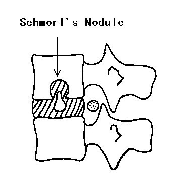
Hydrostatic loading of the Nucleus Pulposus (NP) of the IVD causes bulges of the nucleus into the Cartilagenous End Plate(CEP)
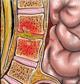
fractures of the CEP can occur if the compressive force is great enough.
CEP fracutures, also known as traumatic Schmorl's nodes, have been noted in postmoterm studies as features of disc digeneration
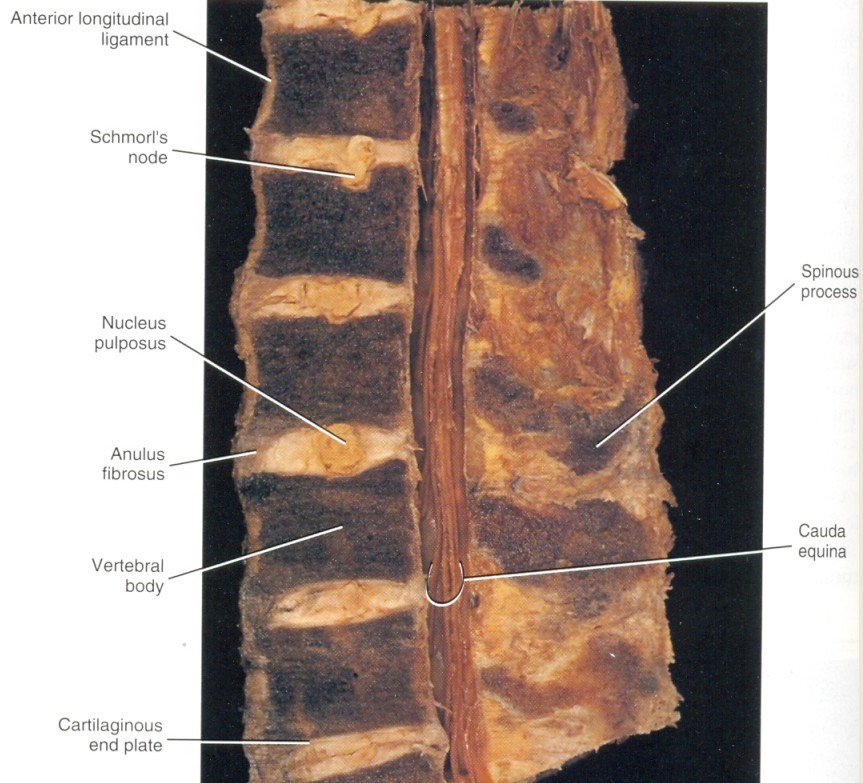
Schmorl's nodes are defined as heerniation of the IVD through the CEP and bony end flate.
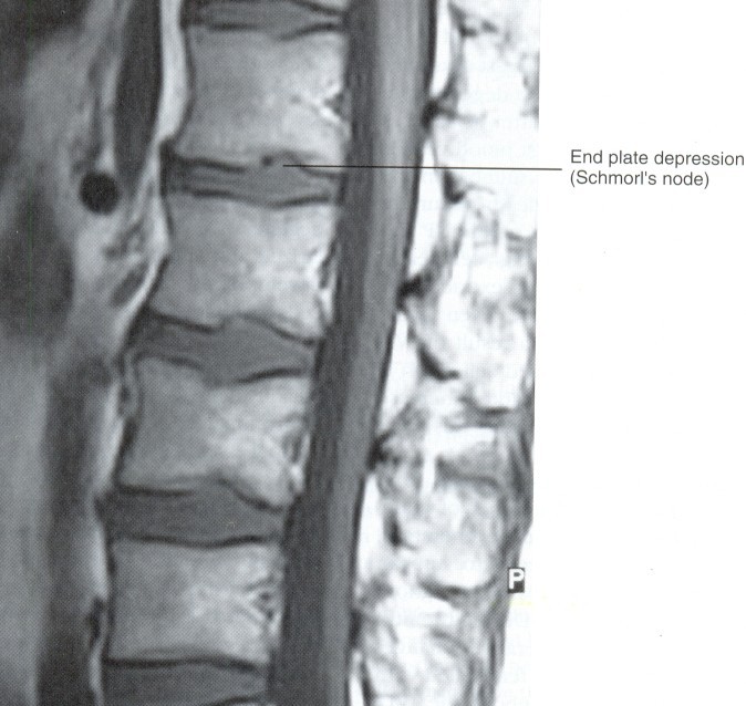
these lesions are believed to be associated with trauma and occur most frequently in the lower thoracic and lumbar region
Even though trauma is the most likely cause of Schmorl's node formation, a possible congenital origin, such as notochondrial cell "rests"(pockets of notochondral bone adjacent to the CEP, also been suggested
such congenital defects could predispose one to later CEP fractures
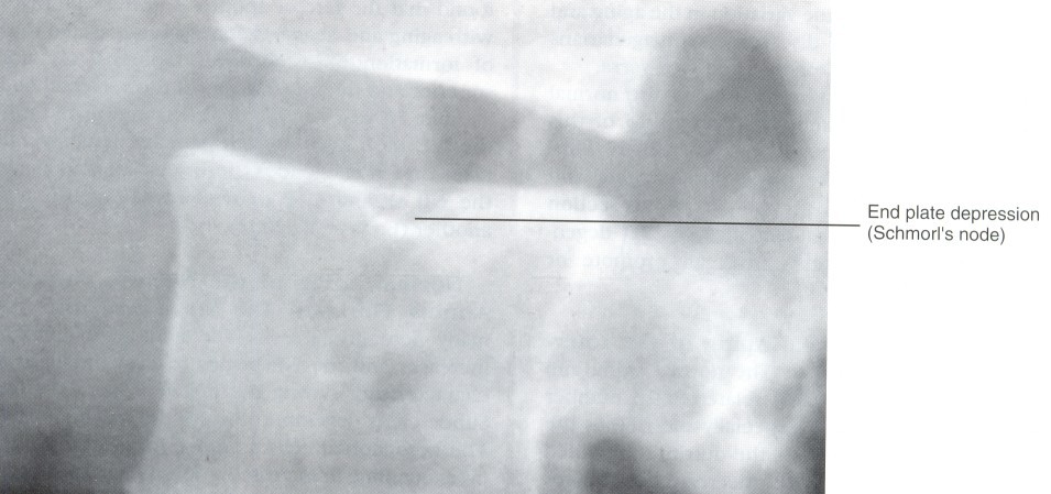
Reported Incidence: 38% in radiologic studies to 76% in postmortem studies
are thought to occur commonly between 20 and 40 when IVD has a relatively high fluid pressure
Schmorl's nodes probably predispose the IVD to early degenerative changes especially when observed in younger age group.
in fact a dorsolumbar kyphosis, seen in adolescents may be associated with Schmorl's nodes. therefore CEP fractures should be considered a possible etiologic cause when an active adolescent patient has back pain of the thoracolumbar region.
compression injury frequently results in an end plate fracure. this may completely resolve in som epatients, or in others, inflammatory repair processes may extend into the NP and result in disc degradation. such inflammatory disc degradation initiates internal disc disruption, which may become symptomatic, if the AF remains intact, isolated IVD resorption may follow, but if fissures and tears develop in the A, the degraded nuclear material may extrude
출저: Basic and Clinical Anatomy of the Spine, Spinal Cord and ANS 2nd edition, Gregory D. Cramer & Susan A, Darby 저
pp592 Part Ⅲ Spinal development, pediatric spine, and microscopic anatomy