통증의 간접경로를 이해하기 위해서는 PAG, RN, RF을 이해해야
panic bird..
간단히 정리하면
- PAG는 중뇌의 수도관과 같은 것으로 descending modulation of pain and in defensive behaviour 역할
- PAG가 자극되면 brain stem의 봉선핵(RN)에 연결된 엔케팔린 분비 뉴런이 활성화되어 봉선핵에서 5-TH(세로토닌)이 분비되어 척수의 dorsal horn으로 전해져 라미나 2(교양질)의 inhibitory interneuron에 자극을 전달
- inhibitory interneuron인 라미나2에서는 엔케팔린이나 다이놀핀을 분비하여 mu opioid 수용기에 붙어 말초에서 올라오는 통증전달을 차단하여 진통효과
- mu opioid 수용체의 활성은 물질 P를 억제.
- 4개의 opioid 수용체가 확인됨(mu, kappa, sigma, delta)
- 합성 opioid, opioid-derivative drugs(헤로인, 몰핀, pethidine, hydrocodone, oxycodone)등이 이 수용체를 활성화시킴
참고로 PAG활성은 defensive behavior, reproductive behavior를 활성화.
- Raphe nuclei의 주요기능은 세로토닌을 분비
- 항우울제 역할을 하는 Selective serotonin reuptake inhibitor (SSRI)는 봉선핵에서 작용함
- 봉선핵은 reticular formation의 내측부분이라고 받아들여지고 있음
- 뉴런의 상당부분이 serotonergic이므로 봉선핵은 중요
- 봉선핵에서 분비되는 세로토닌은 dorsal horn에 연결되어 엔케팔린 분비를 조절하기 때문에 통증 조절에 중요한 역할
- reticular formation은 awaking/sleeping cycle에 관여하는 대뇌의 일부
- reticular formation는 뇌간의 핵심으로 위로는 thalamus, hypothalamus, cortex에 연결되고, 아래로는 cerebellum과 sensory nerve에 연결됨
- reticular formation는 크게 5가지 기능을 함
1) somatic motor control - motor neuron이 하행성 pathway(reticulospinal tract)를 통해 운동신경에 작용하여 muscle tone, balance, posture에 중요한 역할, reticular formation은 눈, 귀의 신호를 소뇌에 전달하여 협응성에 관여함.
2) cardiovascular control - cardiac and vasomotor centers of the medulla oblongata.
3) pain modulation - reticular formation은 말초에서 대뇌로 통증을 전달하는 하나의 길, 또한 descending analgesic pathway임
4) sleep and consciousness - thalamus and cortex에 projection을 보내 의식, 수면에 관여
5) habituation
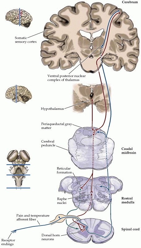
Periaqueductal gray (PAG; also called the "central gray") is the gray matter located around the cerebral aqueduct within the tegmentum of the midbrain. It plays a role in the descending modulation of pain and in defensive behaviour. The ascending pain and temperature fibers of the spinothalamic tract also send information to the PAG via the spinomesencephalic tract (so-named because the fibers originate in the spine and terminate in the PAG, in the mesencephalon or midbrain).
Role in analgesia
Stimulation of the periaqueductal gray matter of the midbrain activates enkephalin-releasing neurons that project to the raphe nuclei in the brainstem. 5-HT (serotonin) released from the raphe nuclei descends to the dorsal horn of the spinal cord where it forms excitatory connections with the "inhibitory interneurons" located in Laminae II (aka the substantia gelatinosa). When activated, these interneurons release either enkephalin or dynorphin (endogenous opioid neurotransmitters), which bind to mu opioid receptors* on the axons of incoming C and A-delta fibers carrying pain signals from nociceptors activated in the periphery. The activation of the mu-opioid receptor inhibits the release of substance P from these incoming first-order neurons and, in turn, inhibits the activation of the second-order neuron that is responsible for transmitting the pain signal up the spinothalamic tract to the ventroposteriolateral nucleus (VPL) of the thalamus. The nociceptive signal was inhibited before it was able to reach the cortical areas that interpret the signal as "pain" (such as the anterior cingulate). This is sometimes referred to as the Gate control theory of pain and is supported by the fact that electrical stimulation of the PAG results in immediate and profound analgesia.
- Four known kinds of opioid receptors have been identified: mu, kappa, sigma and delta. Synthetic opioid and opioid-derivative drugs activate these receptors (possibly by acting on the PAG directly, where these receptors are densely expressed) to produce analgesia. These drugs include heroin, morphine, pethidine, hydrocodone, oxycodone, and similar pain-reducing compounds.
Role in defensive behavior
Stimulation of the dorsal and lateral aspects of the PAG (in the rat) can provoke defensive responses characterised by freezing immobility, running, jumping, tachycardia, and increases in blood pressure and muscle tonus. In contrast, stimulation of the caudal ventrolateral PAG can result in an immobile, relaxed posture known as quiescence, whereas its inhibition leads to increased locomotor activity.
Lesions of the caudal ventrolateral PAG can greatly reduce conditioned freezing, whereas lesions of the dorsal aspect can reduce innate defensive behavior, virtually "taming" the animal.
Role in reproductive behavior
Neurons of the PAG are excited by endorphins and by opiate analgesics. It also plays a role in female copulatory behavior (see Lordosis behavior) via a pathway from the ventromedial nucleus of the hypothalamus.
Role in consciousness
If there is a lesion in the PAG, then consciousness is lost. This observation does not mean that the PAG, itself, is the center of consciousness, but rather that it is a critically-needed part of it.[1][2]
Raphe nuclei
The raphe nuclei ("raffe", Greek: ραφή = seam) are a moderate-size cluster of nuclei found in the brain stem. Their main function is to release serotonin to the rest of the brain.[1] Selective serotonin reuptake inhibitor (SSRI) antidepressants are believed to act in these nuclei, as well as at their targets.[2]
The raphe nuclei are traditionally considered to be the medial portion of the reticular formation, and they appear as a ridge of cells in the center and most medial portion of the brain stem.
Projections
These nuclei interact with almost every pertinent portion of the brain, but only a few of them have specifically independent interaction. These select nuclei are discussed as follows.
Overall, the caudal raphe nuclei, including the raphe magnus, pallidus and raphe obscurus, all project towards the spinal cord and brain stem. The more-rostral nuclei, including the raphe pontis, centralis (also called median), dorsal, tend to project towards the brain areas of higher function.[5]
Function
The raphe nuclei have a vast impact upon the central nervous system. Many of the neurons in the nuclei (but not the majority) are serotonergic; i.e., contain serotonin, a type of monoamine neurotransmitter. It is important to note that pharmacology traditionally affects global serotonin levels, while the actions of the raphe nuclei are dependent on the complex interplay between nuclei.[citation needed]
Projections from the raphe nuclei also terminate in the dorsal horn of spinal gray matter where they regulate the release of enkephalins, which inhibit pain sensation.
Reticular formation
The reticular formation is a part of the brain that is involved in actions such as awaking/sleeping cycle, and filtering incoming stimuli to discriminate irrelevant background stimuli.[1] It is essential for governing some of the basic functions of higher organisms, and is one of the phylogenetically oldest portions of the brain.
Location and relations
The reticular formation is a poorly-differentiated area of the brain stem, centered roughly in the pons. The reticular formation is the core of the brainstem running through the mid-brain, pons and medulla.[2] The ascending reticular activating system connects to areas in the thalamus, hypothalamus, and cortex, while the descending reticular activating system connects to the cerebellum and sensory nerves.
Functions
The reticular formation consists of more than 100 small neural networks, with varied functions including the following:
1. Somatic motor control - Some motor neurons send their axons to the reticular formation nuclei, giving rise to the reticulospinal tracts of the spinal cord. These tracts function in maintaining tone, balance, and posture--especially during body movements. The reticular formation also relays eye and ear signals to the cerebellum so that the cerebellum can integrate visual, auditory, and vestibular stimuli in motor coordination. Other motor nuclei include gaze centers, which enable the eyes to track and fixate objects, and central pattern generators, which produce rhythmic signals to the muscles of breathing and swallowing.
2. Cardiovascular control - The reticular formation includes the cardiac and vasomotor centers of the medulla oblongata.
3. Pain modulation - The reticular formation is one means by which pain signals from the lower body reach the cerebral cortex. It is also the origin of the descending analgesic pathways. The nerve fibers in these pathways act in the spinal cord to block the transmission of some pain signals to the brain.
4. Sleep and consciousness - The reticular formation has projections to the thalamus and cerebral cortex that allow it to exert some control over which sensory signals reach the cerebrum and come to our conscious attention. It plays a central role in states of consciousness like alertness and sleep. Injury to the reticular formation can result in irreversible coma.
5. Habituation - This is a process in which the brain learns to ignore repetitive, meaningless stimuli while remaining sensitive to others. A good example of this is when a person can sleep through loud traffic in a large city, but is awakened promptly due to the sound of an alarm or crying baby. Reticular formation nuclei that modulate activity of the cerebral cortex are called the reticular activating system or extrathalamic control modulatory system.
Pathology
Mass lesions in the brain stem cause severe alterations in level of consciousness such as coma due to their effects on the reticular formation.[5] Bilateral damage to the reticular formation of the midbrain may lead to a coma or death.[2]
Lesions in the reticular formation have been found in the brains of people who have post-polio syndrome, and some imaging studies have shown abnormal activity in the area in people with chronic fatigue syndrome, indicating a high likelihood that damage to the reticular formation is responsible for the fatigue experienced with these syndromes.
History and etymology
The term "reticular formation" was coined in the late 19th century, coinciding with Ramon y Cajal’s neuron doctrine. Allan Hobson states in his book The Reticular Formation revisited that he thought the name is an etymological vestige from the fallen era of the aggregate field theory in the neural sciences. The term reticulum means a netlike structure, which is what the Reticular Formation appears to be at first glance. It has been described as being either too complex to study or an undifferentiated part of the brain with no organization at all. Eric Kandel even describes the reticular formation as being organized in a similar manner to the intermediate gray matter of the spinal cord. This chaotic, loose, and intricate form of organization is what has turned off many researchers from looking farther into this mysterious area of the brain that seems to be at the crux of basic neurological and behavioral functions of the human being[citation needed]. The cells lack clear ganglionic boundaries, but do have clear functional organizations and distinct cell types.
The term reticular formation is seldom used any longer except to speak in generalities. Modern anatomy, or neuroscience articles, usually refer to the individual nuclei that comprise the reticular formation.
Structure
The reticular formation has been functionally cleaved both sagittally and coronally.
- The original functional differentiation was a division of caudal and rostral, this was based upon the observation that the lesioning of the rostral reticular formation induces a hypersomnia in the cat brain. In contrast, lesioning of the more caudal portion of the reticular formation produces insomnia in cats. This study has led to the idea that the caudal portion inhibits the rostral portion of the reticular formation.
- Sagittal division reveals more morphological distinctions. The raphe nuclei form a ridge in the middle of the reticular formation, and, directly to its periphery, there is a division called the medial reticular formation. The medial RF is large and has long ascending and descending fibers, and is surrounded by the lateral reticular formation. The lateral RF is close to the motor nuclei of the cranial nerves, and mostly mediates their function.
Medial and lateral reticular formation
The medial reticular formation and lateral reticular formation are two columns of neuronal nuclei with ill-defined boundaries, which go up through the medulla and into the mesencephalon (midbrain). The nuclei can only be teased out by function, cell type, and projections of efferent or afferent nature.