허리디스크의 치료적 맞춤운동은 참 어렵다.
살만운동을 통해 복압을 높이고, 골반의 전후방 움직임 속에서 조금만 잘못하면 nerve root가 압박(포착)되어 통증이 악화될 수 있기 때문이다.
그렇다고 다른 대안도 없다. 누워서 골반, 척추를 바닥에 붙여서 시행하는 치료적 운동조차 수행할 수 없다면 어떤 방법으로 core muscle을 강화할 수 있겠는가?
그 고민에 관련된 논문이다.
![]() A modern approach to abdominal training.pdf
A modern approach to abdominal training.pdf
![]() A modern approach to abdominal training. part2.pdf
A modern approach to abdominal training. part2.pdf
Introduction
Active care is a benchmark in the management of low back conditions. Unfortunately, voluntary exercise often perpetuates faulty movement patterns or abnormal motor control (AMC). This series of articles will describe how to train the abdominals and ensure that patients are performing their exercises without AMC.
The sit-up is a classic example of an exercise myth. The sit-up places high compressive load on the disc (McGill, 2006, 2007). It usually involves a posterior pelvic tilt which unnecessarily exacerbates disc load (Hickey and Hukins, 1980). Also, it is frequently performed early in the morning which is a time of high risk for annular injury (Adams and Hutton, 1985). Better and safer abdominal exercises exist which will be described in this series of papers.
Are specific patterns of muscle co-activation necessary?
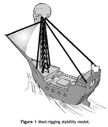
Whether the goal is preventive, injury recovery, rehabilitation, or performance enhancement good ‘‘core’’ fitness is a must. Muscles stabilize joints by stiffening like rigging on a ship (Fig. 1). According to Cholewicki and McGill (1996) spine stability is greatly enhanced by co-contraction (or co-activation) of antagonistic trunk muscles. Co-contractions increase spinal compressive load, as much as 12–18% or 440 N, but they increase spinal stability even more by 36–64% or 2925 N (Granata and Marras, 2000). They have been shown to occur during most daily activities (Marras and Mirka, 1990).
This mechanism is present to such an extent that without co-contractions the spinal column is unstable even in upright postures! (Gardner-Morse and Stokes, 1998). Co-contractions are most obvious during reactions to unexpected or sudden loading (Lavender et al., 1989). Involuntary coordination of ‘‘core’’ muscles has been found to be correlated to back pain. Researchers at Yale University have shown that a specific motor control signature of delayed agonist-antagonistic muscle activation predicts which asymptomatic people will later develop low back pain (LBP) (Cholewicki et al., 2005). In particular, what was found was longer muscle response latencies to perturbation in the ‘‘at risk’’ group than those in healthy control subjects.
Marras et al. (2005) have reported that there is a different pattern of antagonist muscle co-activation (kinematic ability) in LBP individuals than in asymptomatics. Patients were found to have greater spine load and greater kinematic compromise
during lifting tasks. Altered kinematics were strongly related to spine load, being able to predict 87% of the variability in compression, 61% in anteroposterior shear, and 65% in lateral shear. The
kinematic picture for the LBP individual showed
excessive levels of antagonistic muscle co-activation
which reduced trunk motion, but also increased
spine loading.
Ironically, when the spine is under load it is best
stabilized, but when ‘‘surprised’’ by trivial load at a
vulnerable time such as in the morning or after
prolonged sitting the spine stability system is most
dysfunctional (Adams and Dolan, 1995 ). Inappropriate
muscle activation patterns during seemingly
trivial tasks (only 60 N of force) such as bending
over to pick up a pencil can compromise spine
stability and potentiate buckling of the passive
ligamentous restraints (Andersson and Winters,
1990 ). This motor control skill has also been shown
to be more compromised under challenging aerobic
circumstances (McGill et al., 1995 ).
Australian researchers have demonstrated that a
delayed activation of the transverse abdominus
muscle during arm or leg movements has been
found to distinguish LBP patients from asymptomatic
individuals (Hodges and Richardson, 1998,
1999 ). However, according to Canadian scientists
focusing on a single muscle is like focusing on a
single guy wire (Kavcic et al., 2004 ). Research from
the University of Waterloo in Canada has found that
while certain muscles such as multifidus and
transverse abdominus may have special relevance
in distinguishing LBP subjects from asymptomatic
individuals that these muscles are part of a much
bigger orchestra responsible for spinal stability
(Kavcic et al., 2004 ). They demonstrated that
different muscles played greater or lesser roles
depending on the activity/exercise.
Sufficient stability, according to McGill, is defined
as the amount of muscle stiffness necessary for
stability along with a safety margin (McGill, 2006,
2007 ). Cholewicki et al. (1997) showed that modest
levels of co-activation are necessary, but if a joint
has lost its stiffness greater amounts of co-activation
are needed.
How effective is abdominal training?
Specific spinal stabilization exercises have been
shown to reduce future recurrences following an
acute LBP episode (Hides et al., 2001 ). Specific
spine stabilization exercises achieved superior
outcomes to isotonic exercises in chronic patients
with spondylolysthesis (O’Sullivan et al., 1997 ).
One study that compared McGill’s ‘‘general’’
stabilization exercise approach to the Australian
‘‘deep’’ local stabilization training demonstrated
that the ‘‘general’’ approach was superior
(Koumantakis et al., 2005 ).
Stuge et al. (2004) found that stabilizing exercises
were superior to traditional physical therapy for
pelvic girdle pain after pregnancy. The stabilization
group had lower pain intensity, disability, higher
quality of life, and less impairments. The results
persisted at 1-year check post-partum.
Yilmaz et al. (2003) administered an 8-week
stabilization program to post-operative lumbar
microdiscectomy patients. It was compared to
home exercise and to no exercise. At the 12th
week superior results were achieved in pain,
function, mobility, and lifting ability for the
stabilization group. Supervised stabilization training
was superior to home exercises which was
superior to no exercise.
Assessment of motor control during abdominal training
There are a few fundamental components necessary
to optimize motor control during abdominal
stabilization training. They are the abdominal
brace (AB), neutral spine posture, normal respiration,
and the sternal crunch. Avoiding AMC during
abdominal stabilization training is crucial to maintaining
‘‘sufficient stability’’.
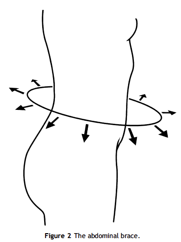
The abdominal brace
In assessing AMC the first criteria is achieving stiffness via pre-contraction or performing an AB. Co-contractions have been shown to occur automatically
in response to unexpected or sudden
loading (Lavender et al., 1989 ; Marras et al., 1987 ).
Stokes et al. (2000) has described how there are
basically two mechanisms by which this co-activation
occurs. One is a voluntary pre-contraction to
stiffen and thus dampen the spinal column when
faced with unexpected perturbations. The second
is an involuntary, reflex contraction of the muscles
quick enough to prevent excessive motion that
would lead to buckling following either expected or
unexpected perturbations (Cresswell et al., 1994 ;
Lavender et al., 1989 ; Marras et al., 1987 ; Stokes
et al., 2000 ; Wilder et al., 1996 ).
This can be achieved by having a patient perform a dead bug or bird dog and then ‘‘brace’’. While the patient performs an AB the clinician offers slow and then quick perturbations in different planes while asking the patient to maintain their posture. The patient can pretend they are about to be pushed or hit and they will ‘‘automatically’ brace (see Fig. 2 ).
Neutral spine posture
The second criteria for spine stability is maintainance of a ‘‘neutral spine’’ or normal lumbar lordosis. Many patients perform posterior pelvic tilts which actually places the lumbo-sacral spine in flexion and thus can potentially harm the disc via end-range loading in flexion. The ‘‘neutral zone is the inner region of a joint’s range of motion (ROM) where minimal resistance to motion is encountered (Panjabi, 1992 ).
Disc herniation has been shown to be related to
repeated flexion motion, especially (Callaghan and
McGill, 2001 ) if coupled with lateral bending and
twisting (Adams and Hutton, 1985 ). This was
supported by the work of Hickey and Hukins
(1980) who demonstrated the protective function
of lumbar lordosis on the disc. According to McGill
(2006) ‘‘Because ligaments are not recruited
when lordosis is preserved, nor is the disc bent, it
appears that the annulus is at low risk for failure.’’
Normal respiration
When performing the AB with neutral spine posture (e.g. slight lumbar lordosis) it is important that the normal respiration is maintained. The tendency when performing an AB and resisting perturbations is to hold the breath or chest breathe. Simple
cueing to continue breathing normally is usually all that is required. The best cue is to say ‘‘breathe and brace’’.
McGill et al. (1995) recommends that patients AB during both phases of respiration rather than only during exhalation. Typical gym training advice to exhale with exertion does not make sense in sports or work demands since stability must be ensured as endurance challenges are faced.
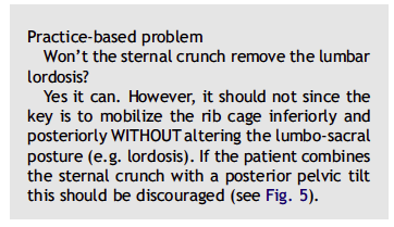
Sternal crunch
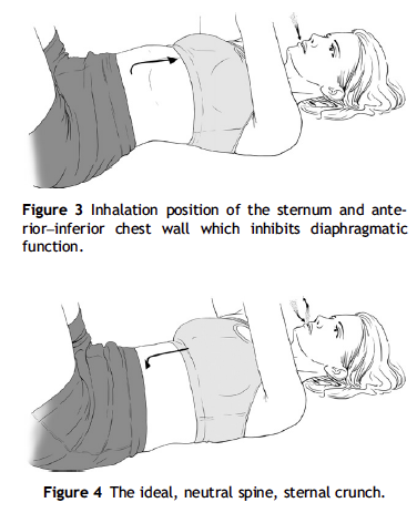
A novel approach to achieving stronger co-activation of all abdominal wall muscles is to observe the position of the anterior chest wall (Kolar, 2007 ). A cephalad position which can be thought of as an ‘‘inhalation’’ position is inhibitory of the normal
diaghragmatic function. It is noted that the thoraco– lumbar (T/L) junction is hyperlordotic and the diaphragm is oblique in this position (see Fig. 3 ). Ideally, a caudal anterior chest position is facilitated which is the ‘‘exhalation’’ position. In this case, the T/L junction is more neutral and the diaphragm is ‘‘centrated’’ in a horizontal position (see Fig. 4 ). The ‘‘exhalation’’ position is believed to be facilitory of the abdominal wall since active exhalation is produced by the abdominal muscles.
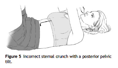

The overhead arm reach on a foam roll is an excellent training exercise for the abdominal wall. The patient should be able to maintain a sternal crunch while moving the arms overhead (see Fig. 6 ). This should be performed without T/L hyperlordosis and without a posterior pelvic tilt.
The clinician would initially cue the patient for
spinal stiffness, and thus an AB, by offering light
perturbations of the torso in a transverse plane
twisting direction. Last but not least, the patient
should be reminded to maintain normal respiration
throughout the duration of the exercise.
Conclusion
In a nutshell, abdominal exercises are commonly
prescribed to prevent and treat lower back pain as
well as to build overall fitness. Certain myths
should be dispelled about this subject regarding
sit-ups, morning exercise, the posterior pelvic tilt,
the transverse abdominis, exhaling with exertion,
etc. The role of maintaining a ‘‘neutral spine’’ and
performing an AB should be understood. The next two articles in this series will review more detail
about the basics of assessment and training of the
abdominal wall.
댓글
댓글 리스트-
작성자박건용 작성시간 13.09.08 abdominal stabilization training에 있어 기본 요소는 abdominal brace(dead bug or bird dog를 하면서 여러 방향에서의 자극에 자세를 유지함으로써 AB를 갖춤), neutral spine posture(골반 전후 tilt의 균형과 요추의 정상전만을 유지함으로써 척추, disc의 압박을 줄인다), normal respiration(neutral spine posture(slight lumbar lordosis)으로 AB를 유지할 때 정상호흡을 유지하는 것이 중요), and the sternal crunch(Ideally, a caudal anterior chest position is facilitated which is the ‘‘exhalation’’ position. without T/L hyperlordosis and without a posterior pelvic tilt.)이다.