motor unit와 근육의 수축, 그리고 motor unit plasticity, 신경발화빈도수, 신경동조의 개념 등- 대박 정리 완성 ㅎㅎㅎ
작성자문형철작성시간12.10.15조회수4,234 목록 댓글 1근육동원의 방법
motor unit, motor end plate, size principle, muscle recruitment에 대하여 엄밀한 학습이 필요하다.
정리하고 그림찾고
생각하고 또 생각하고
또 생각하라.
그리고 최적의 실행!!!
나부터 경험하자..........
벌써 3번째 다시 정리하는 중... 아직도 한눈에 안들어온다...
2014년 12월 13일 ...
이해하고 적용하고 설명하고
2017년 책쓰기를 위해 다시 생각 정리중!!
panic bird.......
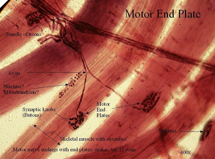
![]() lecture Notes, Neuromuscular Junction.pdf
lecture Notes, Neuromuscular Junction.pdf
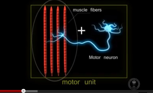
하나의 운동단위에는 5~1500개의 근섬유가 있다
한개의 neuron에 많은 운동단위가 있는 muscle with large ratio motor units는 강한 힘을 만들고
한개의 neuron에 적은 운동단위가 있는 muscle with small ratio motor unit는 세밀한 힘, 정확한 힘을 만든다.
참고) 1하나의 운동뉴런에 type 1섬유(slow twitch)는 100개의 근섬유, type 2 섬유는 1만개의 근섬유 지배...
그렇다면 대둔근과 같은 큰근육에는 하나의 운동단위에 1500개의 근섬유가 있고, 이 운동단위에는 50: 50의 type 1, type 2섬유가 있다고 가정할때, .....
750개의 type 1 섬유의 운동단위에는 하나의 운동단위당 100개의 근섬유(muscle fiber)가 연결되어 있으므로 7만5천개의 근섬유
750개의 type 2 섬유의 운동단위에는 하나의 운동단위당 1만개의 근섬유가 연결되어 있으므로 750만개의 근섬유가 있음.
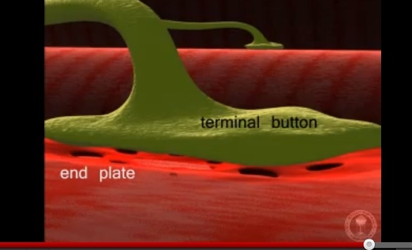
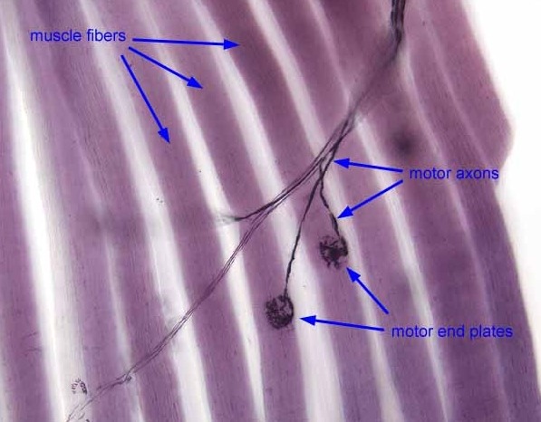
참고) 하나의 운동신경 axon에는 1~~160개의 운동종판에 신경근접합을 이룸.
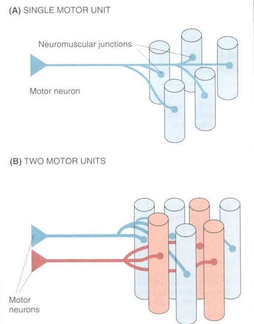
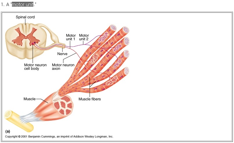
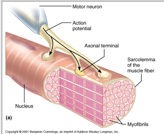
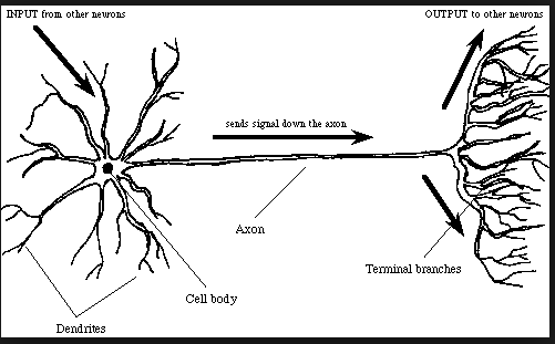
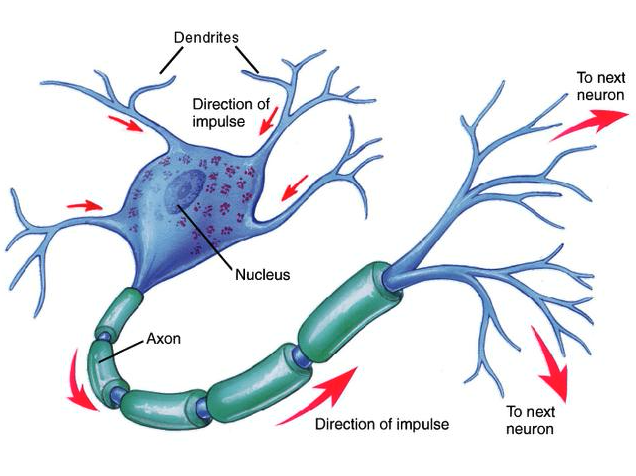
Each skeletal muscle fiber is innervated by a single motor axon. The same axon may also innervate other muscle fibers. All the fibers innervated by the same axon are called a motor unit.
각각의 골격근 섬유(muscle fiber)는 단일 신경축삭에 의해서 지배됨. 하나의 신경축삭은 다른 근섬유를 지배할 수 있음. 하나의 신경축삭에 의해서 지배받는 모든 근섬유를 motor unit라고 함.
motor unit
The functional unit of a skeletal muscle (organ), composed a voluntary motor neuron and the one or more skeletal muscle fibers which it innervates; muscles with large ratio motor units (1 neuron: many muscle fibers) can provide powerful contractions but cannot provide delicate control for precision movements while muscles with small ratio motor units (1 neuron: few muscle fibers) do not typically provide powerful contractions but can provide delicate control for very precise movements.
골격근의 기능적 단위는 자발적인 운동신경원과 하나 또는 그 이상의 근섬유가 있음.
운동단위비율이 많은 근육(1개의 근육에 많은 근섬유 지배)은 강한 힘을 내는 수축을 만들지만 정확하고 미세한 움직임을 조절하지 못함.
반면에 운동단위 비율이 적은 근육(1개의 근육에 적은 근섬유 지배)은 큰 힘을 만들지는 못하지만 매우 정확한 움직임을 위한 세밀한 조절을 함.
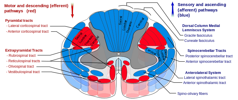
참고) spinal cord tract(상행로와 하행로)
1. 상행로(sensory and ascending pathway)
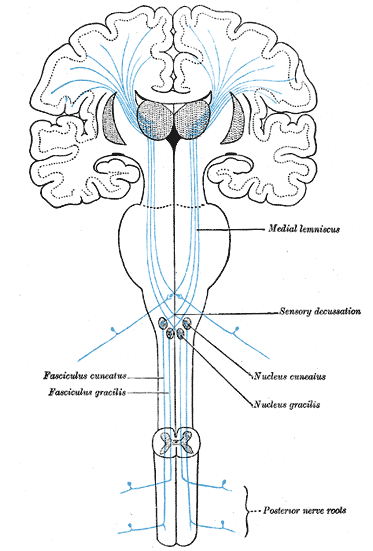
1) dorsal column medial lemniscus system
a) gracile fasciculus
b) cuneate faxciculus
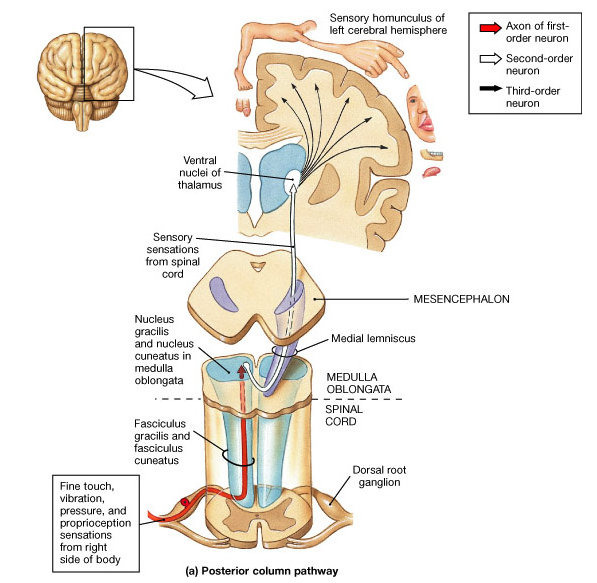
The posterior column-medial lemniscus pathway, PCML for short, (also known as the dorsal column, dorsal column-medial lemniscus pathway or the dorsal white column-medial lemniscus system) is thesensory pathway responsible for transmitting fine touch, vibration and conscious proprioceptive information from the body to the cerebral cortex;[1] as well as tactile pressure, barognosis, graphesthesia, stereognosis, recognition of texture, kinesthesia and two-point discrimination.[2]
The name comes from the two structures that the sensation travels up: the posterior (or dorsal) columns of the spinal cord, and the medial lemniscus in the brainstem. Because the posterior columns are also called dorsal columns, the pathway is often called the dorsal column-medial lemniscus system, or DCML for short. The PCML pathway is composed of rapidly conducting, large, myelinated fibers.[2]
First neuron[edit]
This fine sensation is detected by Meissner's corpuscles that lie in the dermis of the skin close to the epidermis. When these structures are stimulated by slight pressure, an action potential is started. Alternatively, proprioceptive muscle spindles and other skin surface touch receptors such as merkel cells, Ruffini endings, Pacinian corpuscles, and hair follicle receptors (Peritrichial endings) may involve the first neuron in this pathway.
The action potential travels up an axon (the cell body of the neuron will be in a dorsal root ganglion). (The neurons are classified as pseudo-unipolar, so they are regarded as having just one long process, which includes both a peripheral branch dendrite and central branch axon.) So the sensation travels from the skin, along the axon, past the neuronal cell body, and into the dorsal column of the spinal cord.

The axons continue inside the spinal cord, running up the posterior (dorsal) column. Axons from the lower body are most medial (closer to the midline), and run in the gracile tract of the spinal column. Sensory axons from the upper body enter the spinal cord later, specifically from T6 on up, so are more lateral and travel up the cuneate tract. At the level of the closed medulla oblongata, these axons synapse with neurons in the gracile nucleus and cuneate nucleus.
Second neuron[edit]
The secondary neurons (that start in the nuclei) cross over to the other side of the medulla (as internal arcuate fibres) to form the medial lemniscus. This crossing over is commonly referred to as the sensory decussation.
At the medulla, the medial lemniscus is orientated perpendicular to the way the fibres travelled in the posterior columns. For example, in the columns, lower limb is medial, upper limb is more lateral. At the medial leminiscus, axons from the leg are more ventral, and axons from the arm are more dorsal. Fibres from the trigeminal nerve (supplying the head) come in dorsal to the arm fibres, and travel up the lemniscus too.
The medial lemniscus rotates 90 degrees at the pons. The secondary axons from neurons giving sensation to the head, stay at around the same place, while the leg axons move outwards.
The axons travel up the rest of the brainstem, and synapse at the thalamus (at the ventral posterolateral nucleus for sensation from the neck, trunk, and extremities, and at the ventral posteromedial nucleus for sensation from the head).
Third neuron[edit]
Neurons starting in the thalamus travel up the posterior limb of the internal capsule, and again head and leg swap relative positions. The axons synapse in the primary sensory cortex, with lower body sensation most medial (e.g., the paracentral lobule) and upper body more lateral.
2) spinocerebellar tract
a) posterior spinocerebellar tract
b) anterior spinocerebellar tract
The spinocerebellar tract is a set of axonal fibers originating in the spinal cord and terminating in the ipsilateral cerebellum. This tract conveys information to the cerebellum about limb and joint position (proprioception).
Proprioceptive information is obtained by Golgi tendon organs and muscle spindles.
- Golgi tendon organs consist of a fibrous capsule enclosing tendon fascicles and bare nerve endings that respond to tension in the tendon by causing action potentials in Ib afferent neurons (relatively large, myelinated, quickly conducting).
- Muscle spindles fibers are complicated systems of length monitoring within muscles which result in information being carried via Ia neurons (larger and faster than Ib) (from both nuclear bag fibers and nuclear chain fibers) and II neurons (solely from nuclear chain fibers).
All of these neurons are "first order" or "primary", are sensory (and thus have their cell bodies in the dorsal root ganglion) and pass through Rexed laminae layers I-VI of the dorsal horn, to form synapses with "second order" or "secondary" neurons in the layer just beneath the dorsal horn (layer VII)
3) anterolateral system
a) lateral spinothalamic tract
b) anterior spinothalamic tract
The spinothalamic tract is a sensory pathway originating in the spinal cord. It is one component of the anterolateral system. It transmits information to the thalamus about pain, temperature, itch and crude touch. The pathway decussates at the level of the spinal cord, rather than in the brainstem like the posterior column-medial lemniscus pathway and corticospinal tract.
There are two main parts of the spinothalamic tract (STT):
- The lateral spinothalamic tract transmits pain and temperature.
- The anterior spinothalamic tract (or ventral spinothalamic tract) transmits crude touch and pressure.
# spino-olivary fiber
2. 하행로(motor and descending pathways)
1) pyramidal tracts
a) lateral corticospinal tract
b) anterior corticospinal tract
2) extrapyramidal tract
a) rubrospinal tract
The rubrospinal tract is a part of the nervous system. It is a part of the lateral indirect extra-pyramidal tract.
In humans, the rubrospinal tract is one of several major motor control pathways. It is smaller and has fewer axons than the corticospinal tract, suggesting that it is less important in motor control. It is one of the pathways for the mediation of voluntary movement. The tract is responsible for large muscle movement as well as fine motor control, and it terminates primarily in the cervical spinal cord, suggesting that it functions in upper limb but not in lower limb control. It primarily facilitates flexion in the upper extremities (see decorticate posture).
It is small and rudimentary in humans. In some other primates, however, experiments have shown that over time, the rubrospinal tract can assume almost all the duties of the corticospinal tract when the corticospinal tract is lesioned.
b) reticulospinal tract
The reticulospinal tract (or anterior reticulospinal tract) is an extrapyramidal motor tract that descends from the reticular formation in two tracts to act on the motor neurons supplying the trunk and proximal limb muscles. It is involved mainly in locomotion and postural control, although it does have other effects as well.
1. Integrates information from the motor systems to coordinate automatic movements of locomotion and posture
2. Facilitates and inhibits voluntary movement; influences muscle tone
3. Mediates autonomic functions
4. Modulates pain impulses
5. Influences blood flow to lateral geniculate nucleus of the thalamus.
The tract is divided into two parts, the medial (or pontine) and lateral (or medullary) reticulospinal tracts (MRST and LRST).
- The MRST is responsible for exciting anti-gravity, extensor muscles. The fibers of this tract arise from the caudal pontine reticular nucleus and the oral pontine reticular nucleus and project to the lamina VII and lamina VIII of the spinal cord (BrainInfo)
- The LRST is responsible for inhibiting excitatory axial extensor muscles of movement. The fibers of this tract arise from the medullary reticular formation, mostly from the gigantocellular nucleus, and descend the length of the spinal cord in the anterior part of the lateral column. The tract terminates in lamina VII mostly with some fibers terminating in lamina IX of the spinal cord.
The sensory tract conveying information in the opposite direction is known as the "spinoreticular tract".
If the superior colliculus is damaged, it is called decerebration and causes decerebrate rigidity.
The reticulospinal tracts also provide a pathway by which the hypothalamus can control sympathetic thoracolumbar outflow and parasympathetic sacral outflow.
c) olivospinal tract
The olivospinal fasciculus (Helweg) arises in the vicinity of the inferior olivary nucleus in the medulla oblongata, and is seen only in the cervical region of the medulla spinalis, where it forms a small triangular area at the periphery, close to the most lateral of the anterior nerve roots.
d) vestibulospinal tract
The vestibulospinal tract is a neural tract in the central nervous system. Specifically, it is a component of the extrapyramidal system and is classified as a component of the medial pathway. Like other descending motor pathways, the vestibulospinal fibers of the tract relay information from nuclei to motor neurons.[1] The vestibular nuclei receive information through the vestibulocochlear nerve about changes in the orientation of the head. The nuclei relay motor commands through the vestibulospinal tract. The function of these motors commands are to alter muscle tone, extend, and change the position of the limbs and head with the goal of supporting posture and maintaining balance of the body and head.[1]
The vestibulospinal tract is part of the "extrapyramidal system" of the central nervous system. In human anatomy, the extrapyramidal system is a neural network located in the brain that is part of the motor systeminvolved in the coordination of movement.[2] The system is called "extrapyramidal" to distinguish it from the tracts of the motor cortex that reach their targets by traveling through the "pyramids" of the medulla. Thepyramidal pathways, such as corticospinal and some corticobulbar tracts, may directly innervate motor neurons of the spinal cord or brainstem. This is seen in anterior (ventral) horn cells or certain cranial nerve nuclei. Whereas the extrapyramidal system centers around the modulation and regulation through indirect control of anterior (ventral) horn cells. The extrapyramidal subcortical nuclei include the substantia nigra, caudate, putamen, globus pallidus, thalamus, red nucleus and subthalamic nucleus.[3]
The traditional thought was that the extrapyramidal system operated entirely independently of the pyramidal system. However, more recent research has provided a greater understanding of the integration of motor control. Motor control from both the pyramidal and extrapyramidal systems have extensive feedback loops and are heavily interconnected with each other.[1] A more appropriate classification of motor nuclei and tracts would be by their functions. When broken down by function there are two major pathways: medial and lateral. The medial pathway helps control gross movements of the proximal limbs and trunk. The lateral pathway helps control precise movement of the distal portion of limbs.[1] The vestibulospinal tract, as well as tectospinal and reticulospinal tracts are examples of components of the medial pathway.[1]
The vestibulospinal tract is part of the vestibular system in the CNS. The primary role of the vestibular system is to maintain head and eye coordination, upright posture and balance, and conscious realization of spatial orientation and motion. The vestibular system is able to respond correctly by recording sensory information from hairs cells in the labyrinth of the inner ear. Then the nuclei receiving these signals project out to the extraocular muscles, spinal cord, and cerebral cortex to execute these functions.[4]
One of these projections, the vestibulospinal tract, is responsible for upright posture and head stabilization. When the vestibular sensory neurons detect small movements of the body, the vestibulospinal tract commands motor signals to specific muscles to counteract these movements and re-stabilize the body.
The vestibulospinal tract is an upper motor neuron tract consisting of two sub-pathways:
- The medial vestibulospinal tract projects bilaterally from the medial vestibular nucleus within the medial longitudinal fasciculus to the ventral horns in the upper cervical cord (C6 vertebra).[5] It promotes stabilization of head position by innervating the neck muscles, which helps with head coordination and eye movement.
- The lateral vestibulospinal tract provides excitatory signals to interneurons, which relay the signal to the motor neurons in antigravity muscles.[6] These antigravity muscles are extensor muscles in the legs that help maintain upright and balanced posture.
Lateral vestibulospinal tract[edit]
The lateral vestibulospinal tract is a group of descending extrapyramidal motor neurons, or efferent fibers.[2] This tract is found in the lateral funiculus, a bundle of nerve roots in the spinal cord. Thelateral vestibulospinal tract originates in the lateral vestibular nucleus or Deiters’ nucleus in the pons.[2] The Deiters' nucleus extends from pontomedullary junction to the level of abducens nervenucleus in the pons.[2]
Lateral vestibulospinal fibers descend uncrossed, or ipsilateral, in the anterior portion of the lateral funiculus of the spinal cord.[2][7] Fibers run down the total length of the spinal cord and terminate at the interneurons of laminae VII and VIII. Additionally, some neurons terminate directly on the dendrites of alpha motor neurons in the same laminae.[2]
Medial vestibulospinal tract[edit]
The medial vestibulospinal tract is a group of descending extrapyramidal motor neurons, or efferent fibers found in the anterior funiculus, a bundle of nerve roots in the spinal cord. The medial vestibulospinal tract originates in the medial vestibular nucleus or Schwalbe's nucleus.[2] The Schwalbe's nucleus extends from the rostral end of the inferior olivary nucleus of the medulla oblongata to the caudal portion of the pons.[2]
Medial vestibulospinal fibers join with the ipsilateral and contralateral medial longitudinal fasciculus, and descend in the anterior funiculus of the spinal cord.[2][7] Fibers run down to the anterior funiculus to the cervical spinal cord segments and terminate on neurons of laminae VII and VIII. Unlike the lateral vestibulospinal tract, the medial vestibulospinal tract innervates muscles that support the head. As a result, medial vestibulospinal fibers run down only to the cervical segments of the cord.[2]
The vestibulospinal reflex uses the vestibular organs as well as skeletal muscle in order to maintain balance, posture, and stability in an environment with gravity. These reflexes can be further broken down by timing into a dynamic reflex, static reflex or tonic reflex. It can also be categorized by the sensory input as either canals, otolith, or both. The term vesitbulospinal reflex, is most commonly used when the sensory input evokes a response from the muscular system below the neck. These reflexes are important in the maintenance of homeostasis.[8]
Example of vestibulospinal reflex[edit]
- The head is tilted to one side which stimulates both the canals and the otoliths.
- This movement stimulates the vestibular nerve as well as the vestibular nucleus.
- These impulses are transmitted down both the lateral and medial vestibulospinal tracts to the spinal cord.
- The spinal cord induces extensor effects in the muscle on the side of the neck to which the head is bent, and flexor effects in the muscle in the side of the neck away from the direction of the displaced head.
Tonic labyrinthine reflex[edit]
The tonic labyrinthine reflex (TLR) is a reflex that is present in newborn babies directly after birth and should be fully inhibited by 3.5 years.[9] This reflex helps the baby master head and neck movements outside of the womb as well as the concept of gravity. Increased muscle tone, development of the proprioceptive and vestibular senses and opportunities to practice with balance are all consequences of this reflex. During early childhood, the TLR matures into more developed vestibulospinal reflexes to help with posture, head alignment and balance.[10]
The tonic labyrinthine reflex is found in two forms.
- Forward: When the head bends forward, the whole body, arms, legs and torso curl together to form the fetal position.
- Backwards: When the head is bent backward, the whole body, arms, legs and torso straighten and extend.
Righting reflex[edit]
The righting reflex is another type of reflex. This reflex positions the head or body back into its "normal" position, in response to a change in head or body position. A common example of this reflex is the cat righting reflex, which allows them to orient themselves in order to land on their feet. This reflex is initiated by sensory information from the vestibular, visual, and the somatosensory systems and is therefore not only a vestibulospinal reflex.[8]
A typical person sways from side to side when the eyes are closed. This is the result of the vestibulospinal reflex working correctly. When an individual sways to the left side, the left lateral vestibulospinal tract is activated to bring the body back to midline.[7] Generally damage to the vestibulospinal system results in ataxia and postural instability.[11] For example, if unilateral damage occurs to the vestibulocochlear nerve, lateral vestibular nucleus, semicircular canals or lateral vestibulospinal tract, the person will likely sway to that side and fall when walking. This occurs because the healthy side "over powers" the weak side in a way that will cause the person to veer and fall towards the injured side.[6] Potential early onset of damage can be witnessed through a positive Romberg's test.[6] Patients with bilateral or unilateral vestibular system damage will likely regain postural stability over weeks and months through a process called vestibular compensation.[11] This process is likely related to a greater reliance on other sensory information.
- Recent research has shown that damage to the medial vestibulospinal tract alters vestibular evoked myogenic potential in the sternocleidomastoid muscle (SCM),[12][13] which are involved in head rotation. The vestibular evoked myogenic potential is an assessment of the sacculo-collic reflex and a test of function in otolithic organs. Also, lesions to the tract impair ascending efferent fiber signaling, which led to nystagmus.[12][13]
- There has also been recent research to determine if there is a difference in vestibulospinal function when there is damage to the superior vestibular nerve as opposed to the inferior vestibular nerve and vice versa. They defined vestibulospinal function by ability to have proper posture, as well as by self reported dizziness. The results were determined by using the Sensory Organization Test (SOT) of the computerized dynamic posturography (CDP) as well as the dizziness handicap inventory (DHI). It was determined that subjects with damaged inferior spinal nerve performed worse on the posture test than the control group, but performed better than patients with superior vestibulo nerve damage. With this they determined that the superior vestibular nerve plays a larger role in balance than the inferior vestibulo nerve but that they both play a role. In terms of the DHI it was concluded that there was no difference between the patients with the two different impairments.[14]
- Vestibular compensation after unilateral or bilateral vestibular system damage can be accomplished by sensory addition and sensory substitution. Sensory substitution occurs when any remaining vestibular function, vision, or light touch of a stable surface substitute for the lost function. Postural sway and gait ataxia can be reduced by augmenting sensory information for balance control. Recent research has shown that as little as 100 grams of light touch of a fingertip can provide enough sensory reference to reduce sway and ataxia during gait.[11]
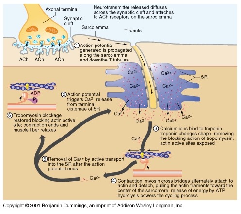
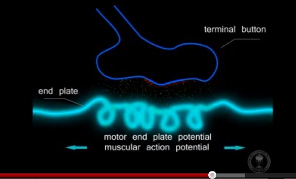
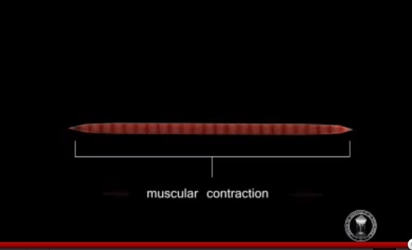
1) 생성된 활동전위가 근초와 T 튜블로 들어감.
2) 활동전위는 SR의 terminal cisternae로부터 칼슘을 분비하도록 자극함
3) 칼슘이온이 트로포닌에 부착함. 트로포닌은 형태를 바꾸고 트로포마이오신의 액틴차단을 제거하고 액틴 활성위치가 열림
4) 근수축 : 마이오신이 액틴에 교차다리형태로 부착하여 액틴필라멘트를 끌어당김.
5) 활동전위가 끝나고 SR로 능동수동에 의해서 칼슘이 제거됨.
6) 트로포마이오신 블록은 액틴활성부위 차단을 회복하면 근수축이 끝나고 근육이 이완됨.
Motor unit plasticity
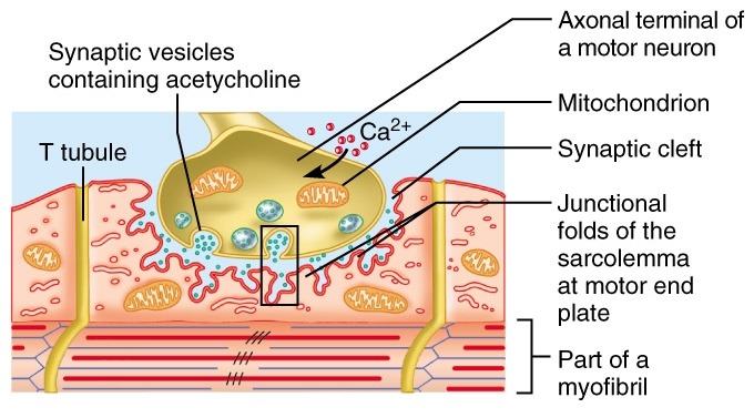
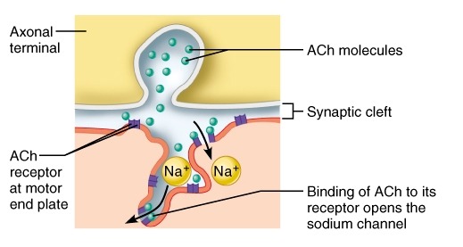
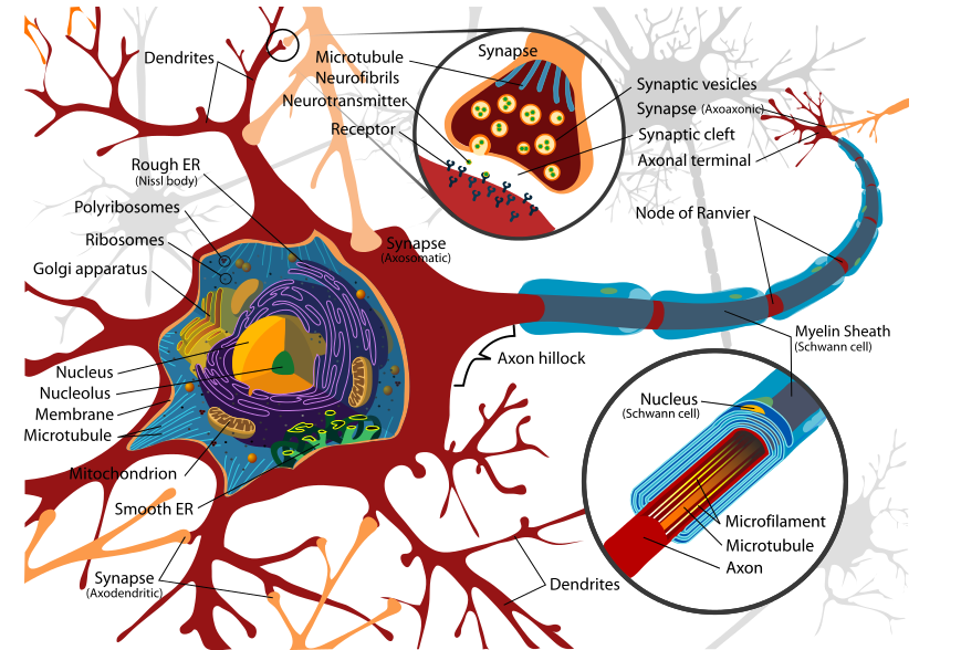
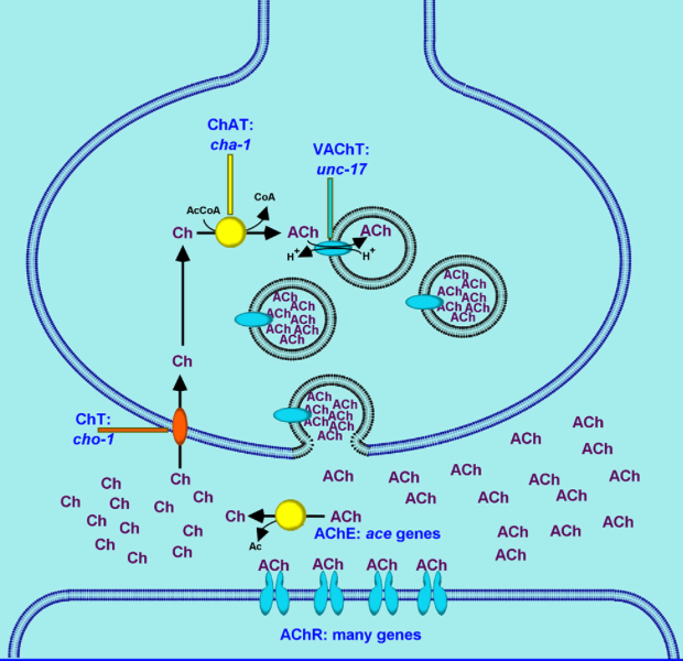
The motor unit consists of a voluntary alpha motoneuron and all of the collective muscle fibers that it controls, known as the effector muscle. The alpha motoneuron communicates with acetylcholine receptors on the motor end plate of the effector muscle. Reception of acetylcholine neurotransmitters on the motor end plate causes contraction of that effector muscle.
- 운동단위는 자발적 알파운동신경원으로 구성되어 있고, 지배하고 있는 모든 근육의 움직임을 조절
- 알파운동신경원은 작용근육의 운동종판에 아세틸콜린 수용체와 교통함.
- 운동종판에서 아세틸콜린 신경전달물질의 reception은 효과기 근육의 수축을 일으킴.
Motor unit plasticity is defined as the ability of motoneurons and their respective effector muscles to physically and functionally change as a result of activity, age, and other factors. Motor unit plasticity has implications for improved athletic performance and resistance to immobility as a result of age. Recent advanced training techniques and physical therapy techniques that focus on improving neural function in addition to muscular function show promising results to improving athletic performances and extending mobility for the elderly.[1]
- 운동단위 가소성은 운동신경원과 연관 효과기 근육이 운동, 나이, 기타 요소에 의해 물리적, 기능적으로 그 기능이 달라져 지속적 변화가 유지되는 능력임.
- 운동단위 가소성은 엘리트 스포츠 선수들이 기능이 향상되고, 나이에 따른 기능 쇠퇴에 저항하여 기능이 향상됨을 의미
- 최신의 트레이닝은 신경기능을 증진시키는 것과 함께 근육기능을 증진시키는 것에 초점(운동선수, 노인들 모두)
Plasticity due to Resistance Training
Resistance training has been shown to drastically increase performance of motor units of the larger muscle groups.[2] Motor unit plasticity of the larger muscle groups is extremely important for athletes, especially those participating in high impact and fast pace sports such as track and field, martial arts, and American football.
저항 트레이닝은 큰 근육그룹의 운동단위의 수행능력을 과감하게 증가시킴.
큰 근육그룹의 운동단위 가소성은 육상 선수에게 매우 중요함.
Training that focuses on improving muscle strength and neural function via resistance training, or more commonly known as plyometrics, is currently incorporated into many professional and collegiate training regiments. Motor unit plasticity can be measured in many ways, the most important of which being neural firing frequency, EMG amplitude, muscle force output, pre-synaptic inhibition, and synchronization.
저항 트레이닝 또는 플라이오메트릭스 운동을 통한 근력증가와 신경기능 증진에 초점을 맞춘 운동은 많은 전문가와 대학교육으로 편입됨.
운동단위 가소성은 다양한 방식으로 측정가능함.
신경발화 빈도수, 근전도 amplitude, 근육힘 발산, 시냅스전 억제, 동조화 등이 중요한 운동신경 가소성 측정방식임.
[edit]1. Neural Firing Frequency
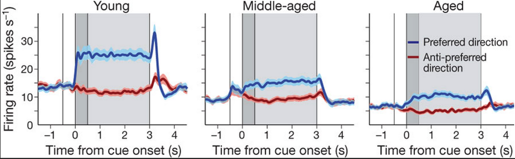
- 최대발화빈도수는 100~200Hz, 최대 40%까지 증진
- reaction time의 증가
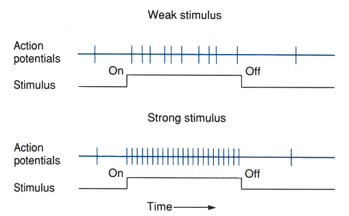
Firing frequency is defined as the number of neuronal signals sent per second on one motoneuron. This frequency is measured in Hertz. Maximum firing frequency in humans typically ranges from 100–200 Hz. Studies incorporating electromyography have proven that adaptation mechanisms occurring as a result of resistance training have can drastically increase the maximal firing frequency of a motoneuron.
발화 빈도수는 하나의 운동신경원에 초당 신경 신호(neuronal signal)의 숫자로 정의됨. 이 빈도수는 Hz로 측정됨.
인류의 경우 최대발화빈도수는 100~200Hz임. 근전도검사는 저항운동의 결과로서 적응기전을 증명해왔는데, 저항운동은 운동신경원의 최대 발화빈도수를 과감하게 증가시킬 수 있음.
Firing frequency has been shown to increase by as much as 40 percent in professional athletes as a result of resistance training.[2] Increases in firing frequency improve athletic force by decreasing the time to maximum muscle contraction (also known as reaction time) rather than increasing the maximum force output.
발화빈도수는 저항 트레이닝의 결과 전문 운동선수에서 40%까지 증가되어지는 것을 볼 수 있음. 발화빈도수의 증가는 최대힘의 분출증가보다는 반응속도(최대 근수축시간)의 감소에 의해서 운동선수의 힘을 증가시킴.
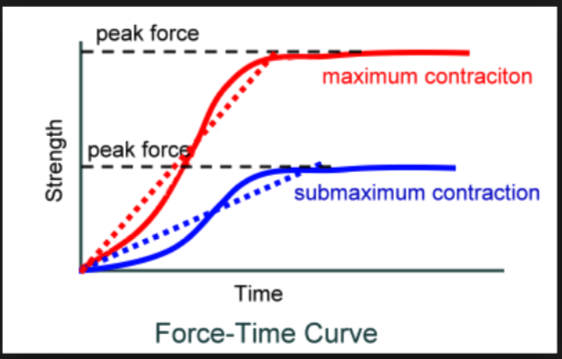
참고) 힘-시간 그래프
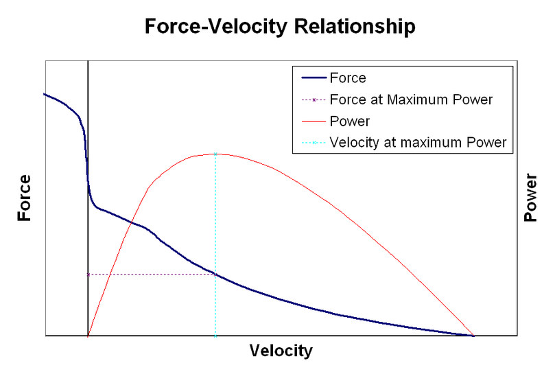
참고) 힘과 속도관계 그래프
[edit]2. EMG amplitude
- maximal neuronal output을 측정.
- 1개월 저항운동으로 50%증진가능.
EMG, or electromyography, amplitude is the measure of the electric potential of motor units. Maximum EMG amplitude is more commonly referred to as maximum neuronal output. Studies have shown that maximum EMG amplitude increases with continual resistance training. These increases can range from minuscule to as much as a 50 percent increase after as little as a one-month training period.
On average, at the end of a one-month period of consistent and repetitive resistance training, EMG amplitude reaches a plateau. Further increases in amplitude after this one-month period occur if the training regimen is varied.[3] This plateau effect is assumed to occur as a result of sufficient neuronal adaptation to the resistance load. Variance in load or repetition causes the neuron to increase its output, and therefore EMG amplitude, to allow for increased muscular force of contraction after this plateau occurs.
근전도 진폭은 운동단위의 전기활동의 수치임. 최대 근전도 진폭은 최대신경 분출로 인정됨.
최대 근전도 진폭은 지속적인 저항트레이닝과 함께 증가함.
1개월 트레이닝을 통해서 근전도 진폭의 증가량은 아주 적은 증가부터 50%까지 증가할 수 있음.
참고) Maximum voluntary isometric contraction (MVIC)
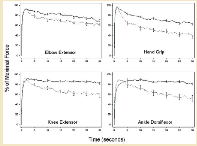
Figure 2. Mean percent of maximal voluntary isometric contraction force plotted against time during 30-s sustained contractions of elbow extensors, hand grip, knee extensors, and ankle dorsiflexors on dominant side in MS patients (dotted lines) and healthy controls (solid lines). Error bars represent one standard error of the mean and are only shown at certain time points to improve visualization of the curves. MS patients had greater decrements in contraction force than controls in all muscles tested. Source: See Schwid (70).
[edit]3. Force Output
- 저항운동으로 최대 1천% 증가 가능.
Force output undergoes much the same effects as EMG amplitude in response to resistance training. Force output significantly increases with resistance training and plateaus, on average, after about a month or less of consistent and repetitive training.[4] Increases in force output after this plateau can only occur as a result of variation in training load or repetition.
힘분출은 근전도 진폭과 같은 의미임. 힘분출은 저항 트레이닝과 함께 의미심장하게 증가함. 고원에 도달한 후 최대 힘분출의 증가는 다양한 부하와 반복 트레이닝을 다양하게 시행한 결과로 얻어짐.
Increases in force output occur as a result of an increase in muscle fiber diameter. It is unknown exactly how much force output of a single motor unit can increase as it varies incredibly from person to person. For example, Olympic weight lifters have been able to increase their total force output (the output produced a group of muscles) by as much as one thousand percent from the start of training.
힘분출의 증가는 근섬유 직경 증가의 결과와 함께 일어남. 단일 운동단위의 얼마만큼의 힘이 증가하는지 잘 모름.
올림픽 역도선수는 트레이닝 시작부터 1천 퍼센트만큼 그들의 최대힘 증가가 보일 수 있음 .....
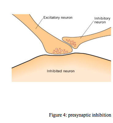
[edit]4. Pre-synaptic Inhibition 시냅스 전억제
- 시냅스 전 억제는 하나의 뉴런에서 이어지는 뉴런으로 신경신호 전달이 제한되는 것을 말함.
This form of inhibition commonly refers to the limiting of neural signals to transfer from one neuron to a subsequent neuron. Pre-synaptic inhibition is known to decrease in response to resistance training. This decrease occurs as a result of increased excitability of the motoneuron and decreased activity of inhibitory pathways. However, changes in pre-synaptic inhibition do not have as large an impact on motor unit performance as changes in force output, EMG amplitude, or firing frequency.
시냅스 전 억제는 하나 신경원에서 이어지는 신경원으로 신호전달되는 한계에 대한 언급임.
시냅스전 억제는 저항트레이닝에 반응하여 감소되는 것으로 알려짐. 이 감소는 운동신경원흥분 증가의 결과와 억제 전달로의 활성감소에 의함.
하지만 시냅스전 억제에서 변화는 힘 분출, 근전도 진폭, 발화빈도수 측면에서 운동단위 수행만큼 큰 영향을 주지 못함.
[edit]5. Neural Synchronization 신경동조화
- 신경동조화는 효과적인 운동단위 동원임.
Neural synchronization is the simultaneous firing of motoneurons. Synchronization leads to more efficient motor unit recruitment. Neural synchronization is important for muscle performance because the more motoneurons that are activated at once, the more muscle fibers that contract at once and hence the stronger the total force of contraction.
신경동조화는 운동신경원의 동시발화임. 신경동조화는 운동단위 동원을 좀더 효과적으로 이끌어냄. 신경동조화는 근육활동 수행에 중요한 역할을 함. 왜냐하면 한번에 활성화되는 좀더 많은 운동신경원 많으면 좀더 많은 근섬유가 한번에 수축하고 그 결과 총수축의 강한 힘을 발휘하기 때문임.
Synchronization is known to slightly increase as a result of resistance training.[5] Motor unit recruitment is frequently associated with synchronization and is defined as the order and number of neurons that are needed to perform a movement. Recruitment is not known to change in response to training or age.[5]
신경동조화는 저항트레이닝으로 약간 증가한다고 알려짐. 운동단위 동원은 신경동조화와 밀접하게 연관되고 운동신경원의 숫자와 질서에 의해서 움직임을 수행한다고 정의됨. 동원은 트레이닝이나 노화에 반응하여 변화하지 않는다고 알려짐.
[edit]Plasticity due to Age and Inactivity 노화와 비활동에 의한 가소성
일반적으로 저항트레이닝의 반대 효과는 노화와 운동단위의 비활성으로 알려짐. 발화빈도수, 근전도 진폭, 힘분출은 노화와 비활동에 의해서 심각하게 감소함. 하지만 노화와 비활동은 운동단위에서 신경동조화에서 심각한 감소를 유발하지는 않음.
노화와 비활동으로 신경동조화가 감소되지 않는 이유는 알려지지 않음.
[edit]Plasticity according to Muscle Type
Fast twitch muscle units and slow twitch muscle units differ in their ability to produce force and resist fatigue. Fast twitch muscle units have the ability to produce great amounts of force but they do not resist fatigue for long periods of time whereas slow twitch muscle units do not produce great amounts of force but can resist fatigue for very long periods of time.[8]
빠른 연축 섬유단위와 느린 연축섬유단위는 힘을 생성하고 피로에 저항하는 능력이 다름. 빠른 연축을 일으키는 근육운동단위는 큰힘을 생성하는 능력이 있으나 오랫동안 피로에 저항하지는 못함. 반면에 느린 연축근육단위는 큰힘을 생성하지 못하지만 오랫동안 근피로에 저항함.
Fast twitch muscles include large muscle groups such as the upper thigh and upper arm muscles whereas slow twitch muscles include high endurance muscles such as those used for posture. However, despite their drastic differences in structure and function, studies have shown that these types of muscle show the same trends in plasticity as a result of training and aging.[9]
빠른 연축근육은 대퇴와 윗팔 근육과 같은 큰 근육그룹을 포함하고, 느린 연축그육은 자세를 유지하는 높은 지구력을 가진 근육임. 하지만 그들의 구조와 기능의 이러한 많은 차이에도 불구하고 트레이닝과 노화의 결과로 나타나는 가소성은 비슷한 경향을 가짐.
빠른 연축섬유가 더 빨리 노화된다는 논문
참고) 빠른 연축섬유의 위성세포가 노화로 인해 근육재생이 느려짐 - 클릭
[edit]Structural Changes of the Motor Unit
[edit]Nerve Terminal Branching and Acetylcholine Receptor Plasticity
Studies have shown that both activity and inactivity of motor unit affect the pre-synaptic and post-synaptic relationship of the neuromuscular junction of the motor unit. The ability of a motoneuron to branch at the neuromuscular junction represents the pre-synaptic plasticity of the motor unit.
운동단위의 활동과 비활동은 운동단위의 신경근 접합의 시냅스전과 시냅스 후 관계에 영향을 미친다는 연구
The ability of acetylcholine receptors to increase and decrease in number on the motor end plate of the effector muscle represents the post-synaptic plasticity of the motor unit.
아세틸콜린 수용체가 효과기 근육종판의 숫자 감소와 증가시켜 하는 역할은 운동단위의 시냅스 후 가소성을 표현함.
Studies have shown that with increased physical activity (resistance training or otherwise), volume of nerve terminal branching significantly increases. However, physical activity did not seem to increase the amount of acetylcholine receptors on the effector muscle. On the contrary, inactivity of the motor unit proved to significantly decrease the amount of acetylcholine receptors on the effector muscle and have no effect on nerve terminal branching.[10]
연구는 저항트레이닝으로 증가된 물리적 활성, 신경말단 brancing의 양은 의미심장하게 증가함. 하지만 물리적 활성은 효과기 근육의 아세틸 콜린 수용체의 양을 증가시키지는 않는 듯함. 반대로 운동단위의 비활동은 효과기 근육에 아세틸콜린 수용체의 양을 의미심장하게 감소시키고 신경말단 가지에는 영향을 미치지 않음.
It is assumed that this loss of acetylcholine receptors due to inactivity is a result of a decrease in muscle fiber size. However, it is not known why the number of acetylcholine receptors does not increase as a result of activity or why nerve terminal branching does not decrease as a result of inactivity.
비활동때문에 발생하는 아세틸콜린 수용체의 소실은 근섬유크기의 감소 결과임. 하지만 아세틸콜린 수용체의 숫자가 활동의 결과 증가하지 않는 이유는 설명하지 못함. 또한 신경말단 가지가 비활동의 결과 감소되지 않는 이유는 밝혀지지 않음.
Prior to activity based research having been done on the motor unit, it perhaps would have been logical to assume that the amount of acetylcholine receptors nerve terminal branching would both increase or decrease as a result of activity or inactivity, respectively. The cause of the apparent incongruence between the effects of inactivity and activity on the motor unit is not yet known.
- 연구에 의하면 아세틸콜린 수용체 신경말단 가지는 활동과 비활동의 결과로 감소되거나 증가함.
- 운동단위에서 활동과 비활동의 효과사이 불일치는 설명되지 않음.
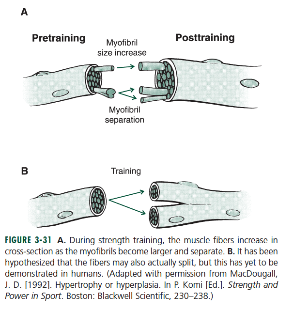
[edit]Effector Muscle Size
Muscle fibers have the ability to drastically increase in size as a result of all types of training. Muscle size is not directly related to muscle strength (force output) as would most likely be assumed.[11] Endurance training can increase the size of low force-producing slow twitch muscle by as much as resistance training can increase the size of high force-producing fast twitch muscle. These situations have the ability to result in two muscles of equal size, but the slow twitch muscle produces only a small fraction of the maximum contractile strength produced by the fast twitch muscle.[12]
- 근섬유는 모든 트레이닝의 결과 크기가 증가함.
- 근육크기는 직접적으로 근력과 연관을 갖지는 않음.
근력저항 트레이닝이 큰힘을 생성하는 빠른 연축섬유 크기를 증가시킬수 있는 것 만큼 지구력 트레이닝은 작은 힘을 내는 느린연축근섬유의 크기를 증가시킬 수 있음.
[edit]Force Output due to Muscle Growth in Comparison to Neuronal Plasticity
Neurons adapt much quicker to training than muscle. The drastic increase in initial changes in force output at the start of physical training are due mostly to increases in firing frequency and EMG amplitude of the neuron. After this initial period of increase in force output, firing frequency and maximum EMG amplitude plateau.
Only very small subsequent increases in these neural functions are seen after this plateau effect. This phenomenon of neurons adapting to training much quicker than muscle likely explains the frequently observed increase in strength at the very beginning of training occurring prior to muscle growth. Increases in force output after the plateau of neural functions are due almost entirely to muscle growth.[2]
- 신경원은 근육보다 트레이닝에 더 빨리 적응함.
- 신경원의 근전도 진폭과 발화빈도수에서 증가때문에 초기 운동을 시작하면 힘의 증가는 바로 나타남. 초기 힘분출 증가기간이 지난 후, 발화빈도수와 최대 근전도 진폭은 고원에 도달함.
- 이러한 신경기능의 작은 연속적인 개선은 고원에 도달한 후에 보임. 이러한 운동에 대한 신경적응현상은 근육보다 빨리 일어남. 신경기능의 고원후에 힘 증가는 근육증진에 의존함.