스쿼트, 달리기를 한다고 할때..
느린연축섬유, 안정화 근육, 지근섬유 type 1 섬유가 지구력과 안정성을 만들고
빠른연축섬유, 속근섬유, type 2 섬유가 빠른 움직임을 만든다. ...
어떤 운동을 통해 근육의 무엇을, 어떻게 바꿀 수 있는가?
운동을 해도 아래 그림에서 보듯이 type 1의 비율은 바뀌지 않는다. type 2a가 증가함.
panic bird..
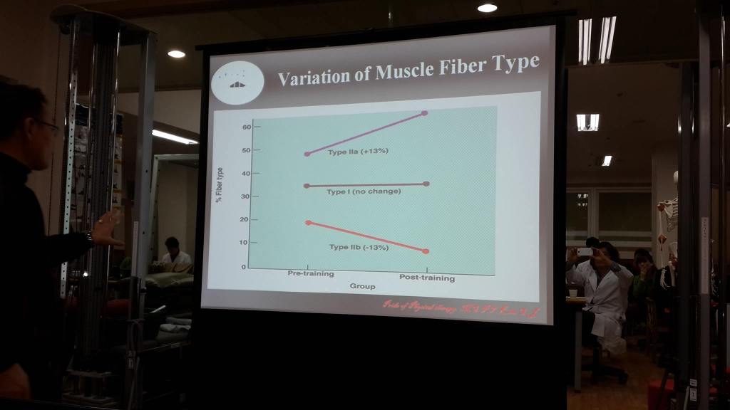
항중력근, 짧아지는 특성, 지근, 적근, type 1, 산소에너지 사용하는 섬유는 그 숫자가 변하지 않는다.
운동을 통해서 무엇이 바뀌는 것인가?
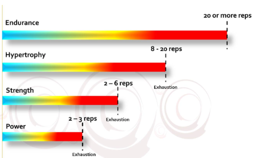
근지구력운동 - 1RM의 60% 힘으로 15~25회반복 운동을 할때, Type 1 섬유가 트레이닝된다. 근지구력이 길러지고
근력운동 - 1RM의 80%의 힘으로 3~7회 반복운동을 할때, Type 2섬유가 트레이닝된다. 근력이 길러지고
그런데 근지구력트레이닝을 해도 type 1섬유의 숫자는 변하지 않는다.
그럼 무엇이 바뀌어 무엇이 개선되는 것인가?
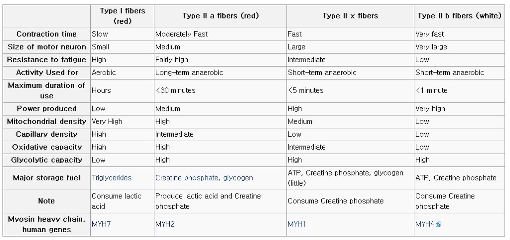
type 1 섬유, 적색, 느린근수축, 크기가 작은 운동단위, 근피로 저항성 높음, 산소이용, 상대적으로 약한 힘의 오랜 지속(근지구력), 미토콘드리아 많음, 모세혈관많음, 산소용적 높음, glycolytic capacity 낮음...
What is a motor unit?
- A motor unit is the number of muscle fibres triggered by the firing of a single alpha motor neurone unit in the anterior horn of the spinal cord.
- Each motor unit has the same type of muscle fibre - either Type-1's, the various intermediate fibre types, Type-IIA's or Type-IIB's.
- Slow-twitch [Type-1's] usually have small numbers of muscle fibres - approximately 100 fibres per unit17.
- A fast-twitch motor unit [Type-IIBs] may have approximately 10,000 fibres17.
- Each muscle in the average human has about a 50:50 ratio of slow and fast twitch muscle fibres.
- When an impulse travels down the nerve to its motor unit, all the fibres within that unit whether it is a Type-1, Intermediate or Type-IIB motor unit --- will contract simultaneously. This is the law of "all or none" in muscle physiology17.
대둔근은 주로 지근이므로 느린연축 근섬유 비율이 약간 더 많다는 의미임.
What is the orderly [sequential] recruitment of muscle fibres?
- With the right load, ie one that is modest enough to allow you to reach exhaustion after all the motor units are called into play will result in the weakest endurance Type-1 fibres firing first and then when these fibres are exhausted, the intermediate fibre motor units will fire and towards the end of the set, when your muscle is becoming exhausted and you can barely lift this load, the Type-IIB's which are the strongest fibres will contract.
- The Type-1's are the weakest fibres but can contract for long periods of time if the load is light enough.
- The Type-IIB muscle fibres are the strongest fibres but exhaust easily - however, these muscle fibres are the 'Holy Grail' of the bodybuilders because these fibres have the greatest potential to grow.
What about loads and muscle fibre recruitment?
- Power Training [1- 3 reps to exhaustion] If the load is high, for example a 1-3 RM where you reach exhaustion in 1 to 3 repetitions, this load is heavy enough to simultaneously fire all muscle types with the Type-1's exhausting rapidly leaving only the strongest Type-IIA and mainly Type-IIB to finish the last repetition. This type of training is used for powerlifters where explosive but short-lived force is needed by the strongest muscle fibres. Myofibrillar hypertrophy occurs. Hypertrophy is due to true protein increase in the muscle due to satellite cells helping to increase the number and size of the contractile proteins [Actin and Myosin]. The number of muscle fibres stays the same in humans but the satellite cells fuse to existing cells and donate their nuclei and DNA to the existing fibres helping them to increase in size. To find out more click on 'The Mystery of Skeletal Muscle Hypertrophy
- Strength Training [2 - 6 reps to exhaustion] - this load will once again exhaust the Type-1 motor units rapidly leaving only the higher intermediates and Type-IIBs to complete the repetitions. Strength training is for those who want strength but also anaerobic endurance where resistance is being met relatively frequently such as football. Myofibrillar hypertrophy occurs due to satellite cells increasing the contractile proteins actin and myosin [see above]
 Hypertrophy Training [8 - 20 reps to exhaustion]-This load and the number of repetitions will ensure an orderly firing of the Type-1, intermediates and Type-IIB motor units. Hypertrophy occurs significantly in Type-1 fibres due to resistance and aerobic training40but more hypertrophy is possible with Type-IIB fibres which have the greatest potential to grow. Unlike power and strength training, hypertrophy is not due to myofibrillar hypertrophy but due to sarcoplasmic hypertrophy where the sarcoplasma volume increases. The number of reps and the load used to enable this number of repetitions to be done will ensure all fibre types have the potential to grow.
- Endurance Training [20 or more repetitions] - Type-1 fibres are endurance fibres and recover rapidly compared with fast-twitch fibres. Thse Type-1 fibres are ready to contract again in 90 seconds. Ideally with endurance training, a weight should be used that will allow repetitive lifting or pushing of that load without exhaustion occuring within 90 seconds. The reason for this is that if a light enough load is used theoretically you can train just the Type-1 muscle fibres without progressing to the intermediates or fast-twitch fibres. Importantly, those that want to progress to hypertrophy should ensure that they never lift or push a load beyond 90 seconds to reach exhaustion as this will allow the Type-1 fibres to recover and 'kick-in' once again giving you a 'second wind'17. This will mean you will not trigger off the stronger intermediate and fast-twitch muscle fibres.
The direct exhaustion and adaptation of a muscle fiber was only one benefit of the program. The second was that you develop both types of hypertrophy which lead to a greater cross sectional area within a muscle. These two types of hypertrophy are termed myofibril hypertrophy and sarcoplasmic hypertrophy and both types can be seen from fig 2.
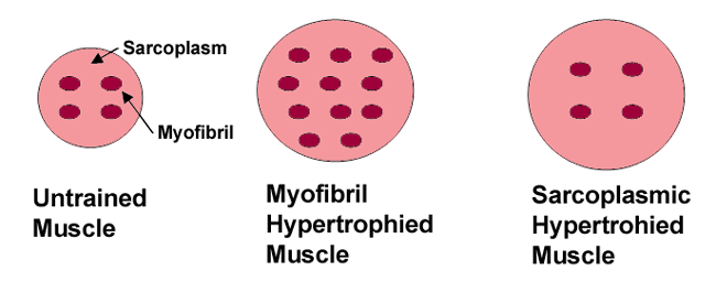
Fig 2 Cross Sectional View Of A Muscle Fiber.
 Click To Enlarge.
When a muscle grows it is either because the muscle has added additional myofibrils which are strands of protein within the muscle or due to increased anaerobic energy demands there is an increase in the cellular fluid between the myofibrils.
Heavy strength training tends to develop the amount of myofibrils due to reasons explained later and higher volume training tends to promote sarcoplasmic hypertrophy. The holistic hypertrophy program gives you the best of both worlds.
One set to failure
- It has been dogma that many sets need to be done to elicit strength or hypertrophy.
- A study out of Adelphi University looked a a large number of research papers on single vs multiple-set resistance training. Their conclusion:
- "Perhaps the most controversial element of any strength training programme is the number of sets required to increase muscular strength and hypertrophy. There is a prevalent belief that at least 3 sets of each exercise are required to elicit optimal increases in strength and hypertrophy. However, most of the studies that reported the results of training with single versus multiple sets do not substantiate this tenet. In fact, the preponderance of evidence suggests that for training durations of 4 to 25 weeks there is no significant difference in the increase in strength or hypertrophy as a result of training with single versus multiple sets42."
- Dr Doug McGuff and John Little in their book 'Body by Science' also state that one set to failure is all that is required. They stress the point that once exhaustion is reached and the Type-IIB motor units have been engaged, it becomes redundant to perform any more repetitions as you have worked your way through the orderly recruitment of all the fibres to these top-level fast-twitch fibres. If you do one set well with the right load and perfect form, you will have reached your 'Holy Grail' if you want strength and hypertrophy. They also point out that it will take 4 - 10 days for the Type-IIB muscle fibres to recuperate and that in any given week, it would once again be pointless to exercise that particular muscle. It would be far better to perform split routines exercising various muscle groups, taking them to exhaustion in one set and then resting that muscle group for one week. Remember that famous aphorism that you grow out of the gym and not in the gym. Also to grow you need to take advantage of the all important immediate post-exercise metabolic window.
Skeletal muscle is a form of striated muscle tissue which is under the control of the somatic nervous system; that is to say, it is voluntarily controlled. It is one of three major muscle types, the others being cardiac and smooth muscle. As their name suggests, most skeletal muscles are attached to bones by bundles of collagen fibers known as tendons.
골격근은 횡문근으로 자발적 근수축을을 일으키는 체신경계의 조절을 받음.
대부분의 골격근은 콜라겐 섬유 다발인 건에 의해 뼈에 부착함.
Skeletal muscle is made up of individual components known as myocytes, or "muscle cells", sometimes colloquially called "muscle fibers". They are formed from the fusion of developmental myoblasts (a type of embryonic progenitor cell that gives rise to a muscle cell) in a process known as myogenesis. These long, cylindrical, multinucleated cells are also called myofibers.
골격근은 근섬유로 이루어져 있음. myofiber = muscle fiber
The myofibers are in turn composed of myofibrils. The myofibrils are composed of actin and myosin filaments repeated as a sarcomere, the basic functional unit of the muscle fiber and responsible for skeletal muscle's striated appearance and forming the basic machinery necessary for muscle contraction. The term muscle refers to multiple bundles of muscle fibers held together by connective tissue.
근섬유는 근원섬유의 연결이고, 근원섬유은 근절의 반복연결인 액틴과 마이오신 필라멘트로 구성됨.
근섬유(muscle fiber) - 근수축을 일으키는 단위
Individual muscle fibers are formed during development from the fusion of several undifferentiated immature cells known as myoblasts into long, cylindrical, multi-nucleated cells. Differentiation into this state is primarily completed before birth with the cells continuing to grow in size thereafter. Skeletal muscle exhibits a distinctive banding pattern when viewed under the microscope due to the arrangement of cytoskeletal elements in the cytoplasm of the muscle fibers. The principal cytoplasmic proteins are myosin and actin (also known as "thick" and "thin" filaments, respectively) which are arranged in a repeating unit called a sarcomere. The interaction of myosin and actin is responsible for muscle contraction.
근섬유는 결국 마이오신과 액틴으로 구성되고, 근절을 이루어 근수축함.
근섬유 분류의 근원
1) type of myosin percent - fast or slow
2) degree of oxidative phosphorylation
There are two principal ways to categorize muscle fibers: the type of myosin (fast or slow) present, and the degree of oxidative phosphorylation that the fiber undergoes. Skeletal muscle can thus be broken down into two broad categories: Type I and Type II. Type I fibers appear red due to the presence of the oxygen binding protein myoglobin. These fibers are suited for endurance and are slow to fatigue because they use oxidative metabolism to generate ATP (adenosine triphosphate). Type II fibers are white due to the absence of myoglobin and a reliance on glycolytic enzymes. These fibers are efficient for short bursts of speed and power and use both oxidative metabolism and anaerobic metabolism depending on the particular sub-type. These fibers are quicker to fatigue.
근섬유 타입 두가지로 나뉘는데, 마이오신의 형태(fast or slow), oxidative phosphorylation 의 정도에 따라서 결정됨.
type 1 fiber는 oxygen binding protein myoglobin때문에 붉은 색. 지구력 움직임에 적합하고 근피로가 느린이유는 ATP를 만드는데 산소적 대사를 사용하기 때문.
type 2 fiber는 myoglobin이 적어서 흰색. 포도당효소를 사용하여 ATP를 만듬. 산소대사와 무산소대사를 동시에 이용하여 빠르고 강한 힘을 폭발적으로 냄. 쉽게 근피로에 빠짐.
Not all skeletal muscle fibers are the same. Traditionally, they were categorized depending on their varying color, which is a reflection of myoglobin content.
Red Fibers: Those containing high levels of myoglobin and oxygen storing proteins had a red appearance. Red muscle fibers tend to have more mitochondria and greater local capillary density.
White Fibers: Those with a low content had a white appearance.
As more was learned about the functional differences between skeletal muscle fibers, they were also classified, depending on their twitch capabilities, into fast and slow twitch, traits that largely, but not completely, overlap the previous classification based on color.
Fast Twitch: Some authors define a fast twitch fiber as one in which the myosin can split ATP very quickly.
However, fast twitch fibers also demonstrate a higher capability for electrochemical transmission of action potentials and a rapid level of calcium release and uptake by the sarcoplasmic reticulum. The fast twitch fibers rely on a well-developed, short term, glycolytic system for energy transfer and can contract and develop tension at 2-3 times the rate of slow twitch fibers. Fast twitch muscles are much better at generating short bursts of strength or speed than slow muscles, and so fatigue more quickly.[1]
Slow Twitch: The slow twitch fibers generate energy for ATP re-synthesis by means of a long term system of aerobic energy transfer. They tend to have a low activity level of ATPase, a slower speed of contraction with a less well developed glycolytic capacity. They contain large and numerous mitochondria and with the high levels of myoglobin that gives them a red pigmentation. They have been demonstrated to have high concentration of mitochondrial enzymes, thus they are fatigue resistant. Slow twitch muscles fire more slowly than fast twitch fibers and so are able to fire for a longer time before fatiguing.[2]
The 2 main categories of muscle fibers become several, when further differentiating type II into several subtypes, based on myosin isoforms and denoted with letters of the alphabet. In humans the two subtypes are IIa and IIx; IIx is often referred to as IIb because earlier classification had clumped together two different types. Later on, further research recognized these subtypes as distinct, but the use of the name IIb remained entrenched.[3] Non human fiber types include true IIb fibers, IIc, IId and so on.
Type I Red fibers. Slow oxidative (also called slow twitch or fatigue resistant fibers). Contain:
- Large amounts of myoglobin.
- Many mitochondria.
- Many blood capillaries.
- Generate ATP by the aerobic system, hence the term oxidative fibers.
- Split ATP at a slow rate.
- Slow contraction velocity.
- Resistant to fatigue.
- Found in large numbers in postural muscles.
- Needed for aerobic activities like long distance running.
Type IIa Red fibers. Fast oxidative (also called fast twitch A or fatigue resistant fibers). Contain:
- Large amounts of myoglobin.
- Many mitochondria.
- Many blood capillaries.
- Large amount of glycogen.
- High capacity for generating ATP by oxidation. Split ATP at a very rapid rate and, hence, high contraction velocity
- Resistant to fatigue but not as much as slow oxidative fibers.
- Needed for sports such as middle distance running and swimming.
Type IIx / IIb (dependent upon species) White. Fast glycolytic (also called fast twitch B or fatigable fibers). Contain:
- Low myoglobin content.
- Few mitochondria.
- Few blood capillaries.
- Large amount of Creatine phosphate.
- Split ATP very quickly.
- Fatigue easily.
- Needed for sports like sprinting.
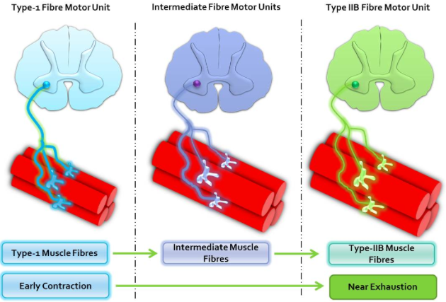
Individual muscles are a mixture of 3 types of muscle fibers (type 1, type 2a and type 2b), but their proportions vary depending on the action of that muscle. It must be remembered that skeletal muscles, although a mixture, can only have one type of muscle fiber within a motor unit.
하나의 운동단위내에는 같은 type 섬유를 가짐.
This is demonstrated if we look at contractions. E.g. If a weak contraction is needed only the type 1 motor units will be activated. These fibers are used mainly for endurance activities. If a stronger contraction is required the type 2a fibers will be activated or used to assist the type 1 fibers.
약한 근수축은 오직 type1 운동단위가 활성화되는 것이 필요함. 근지구력 활동
강한 근수축은 type2a 섬유가 활성화되거나 type 1 섬유가 도와주는 것이 필요함.
Maximal contractions facilitate the use of type 2b fibers which are always activated last. These fibers are used during ballistic activities but tire easily. With advanced EMG techniques it is possible to look at which muscle fibers are recruited when performing an exercise/test. The total number of skeletal muscle fibers has traditionally been thought not to change. It is believed there are no sex or age differences in fiber distribution, however, relative fiber types vary considerably from muscle to muscle and person to person. Sedentary men and women (as well as young children) have 45% type 2 and 55% type 1 fibers.[citation needed]
최대 근수축은 type2b 섬유를 사용는데 항상 마지막 단계에서 활성화됨. 이 섬유는 탄도활성과 같은 강한 움직임동안 사용되지만 쉽게 지침.
앉아서만 일하는 사람은 남녀차이없이 type 2 : type 1=45 : 55%
People at the higher end of any sport tend to demonstrate patterns of fiber distribution e.g. endurance athletes show a higher level of type 1 fibers. Sprint athletes, on the other hand, require large numbers of type 2 b fibers. Middle distance event athletes show approximately equal distribution of the 2 types.
- 근지구력 운동선수는 type 1 섬유비율이 높음.
- 단거리 달리기 선수는 반면에 type 2 섬유비율이 많음.
This is also often the case for power athletes such as throwers and jumpers. It has been suggested that various types of exercise can induce changes in the fibers of a skeletal muscle.[4] It is thought that if you perform endurance type events for a sustained period of time, some of the type 2b fibers transform into type 2a fibers. However, there is no consensus on the subject. It may well be that the type 2b fibers show enhancements of the oxidative capacity after high intensity endurance training which brings them to a level at which they are able to perform oxidative metabolism as effectively as slow twitch fibers of untrained subjects. This would be brought about by an increase in mitochondrial size and number and the associated related changes not a change in fiber type.
- 만약 당신이 지구력 타입의 운동을 시행한다면 type2b 섬유의 어떤 것은 type 2a 섬유로 변할 것이라고 생각됨.
- 높은 강도의 근지구력 운동을 시행하면 type 2b의 산소저장능력의 증진이 일어나 산소대사능력이 증가하는 것임.
- 미토콘드리아 크기와 숫자는 모든 섬유에서 모두 증가함.
근섬유 구조
Structure of skeletal muscle fiber[edit]
Every organelle and macromolecule of a muscle fiber are arranged to ensure form meets function. The plasma membrane is called the sarcolemma with the cytoplasm known as the sarcoplasm. In the sarcoplasm are the myofibrils. The myofibrils are long protein bundles about 1 micrometer in diameter each containing myofilaments. Pressed against the inside of the sarcolemma are the unusual flattened nuclei. Between the myofibrils are the mitochondria. While the muscle fiber does not have a smooth endoplasmic reticulum, it contains a sarcoplasmic reticulum. The sarcoplasmic reticulum surrounds the myofibrils and holds a reserve of the calcium ions needed to cause a muscle contraction. Periodically, it has dilated end sacs known as terminal cisternae. These cross the muscle fiber from one side to the other. In between two terminal cisternae is a tubular infolding called a transverse tubule (T tubule). T tubules are the pathways for action potentials to signal the sarcoplasmic reticulum to release calcium, causing a muscle contraction. Together, two terminal cisternae and a transverse tubule form a triad.[5]
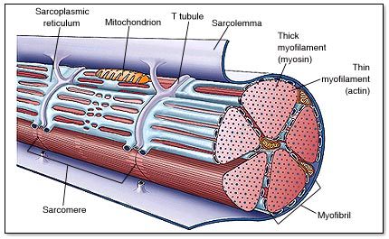
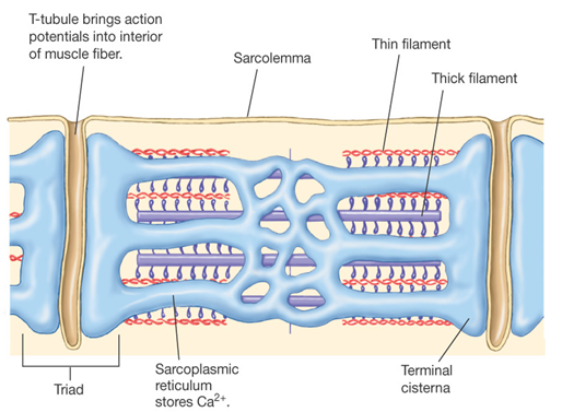
- muscle fiber, myofibril, sarcoplasmic reticulum, terminal cisternae, t-tubule 등의 구조.
Organization of skeletal muscle fibers[edit]
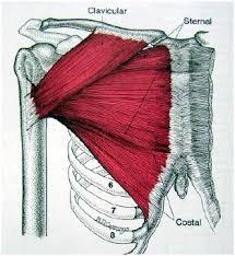
While the muscle fibers of the fascicles lie parallel to one another, the fascicles themselves can vary in their relationship to one another and to their tendons.[6] The different patterns of arrangement of the fascicles produce four different types of skeletal muscles: parallel muscles, convergent muscles, pennate muscles, and sphincter muscles.[6]
- 4가지 형태의 골격근 : parallel muscles, convergent muscles, pennate muscles, and sphincter muscles
- 1. Parallel muscles
The fascicles of parallel muscles run parallel to the direction of the muscle, thus these muscles on a whole function similarly to a single muscle fiber.[6]Most skeletal muscles in the body are parallel muscles; although they can be seen in a variety of shapes such as flat bands, spindle shaped, and some can have large protrusions in their middle known as the belly of the muscle.[6]
- 2. Convergent muscles
The fibers in convergent muscles fan out from a common point of attachment.[6] Covering a broad surface these fibers allow for more versatile types of movement.[6] These muscles do not pull as hard on their corresponding tendons as their parallel muscle counterparts however due to the fibers not all pulling in the same direction, even pulling in different directions at opposite ends.[6]
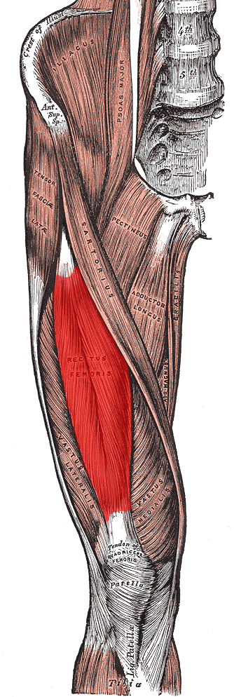
- 3. Pennate muscles
In a pennate muscle one or more tendons run through the body of the muscle with the fascicles forming an oblique angle to the tendons.[6] Because the fascicles pull on the tendons at an angle they do not move the tendon as far as their parallel muscle counterparts. Despite this they generate greater tension due to their possessing a greater amount of muscle fibers than similarly sized parallel muscles.[6]
- ex: Rectus femoris
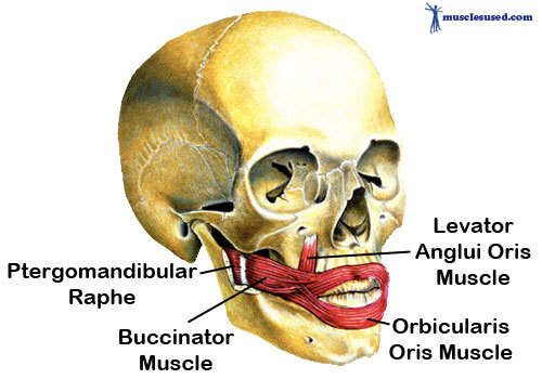
- 4. Sphincter muscles
The fibers of the sphincter or circular muscles are arranged concentrically around an opening or recess.[6] As the muscle contracts, the opening it circumvents gets smaller, for this reason these muscles are often found at the entrances and exits of external and internal passage ways.[6]
ex: Orbicularis oris
Cellular physiology and contraction
In addition to the actin and myosin components that constitute the sarcomere, skeletal muscle fibers also contain two other important regulatory proteins,troponin and tropomyosin, that are necessary for muscle contraction to occur. These proteins are associated with actin and cooperate to prevent its interaction with myosin. Skeletal muscle cells are excitable and are subject to depolarization by the neurotransmitter acetylcholine, released at the neuromuscular junction by motor neurons.[7] Once a cell is sufficiently stimulated, the cell's sarcoplasmic reticulum releases ionic calcium (Ca2+), which then interacts with the regulatory protein troponin. Calcium-bound troponin undergoes a conformational change that leads to the movement of tropomyosin, subsequently exposing the myosin-binding sites on actin. This allows for myosin and actin ATP-dependent cross-bridge cycling and shortening of the muscle.
- 액틴과 마이오신이 근절을 구성함. 트로포닌과 트로포마이오신, 아세틸콜린, 신경근접합, 운동신경 등의 근수축개념 설명.
physics - 근육역학
Muscle force is proportional to physiologic cross-sectional area (PCSA), and muscle velocity is proportional to muscle fiber length.[8] The strength of a joint, however, is determined by a number of biomechanical parameters, including the distance between muscle insertions and pivot points and muscle size. Muscles are normally arranged in opposition so that as one group of muscles contract, another group relaxes or lengthens. Antagonism in the transmission of nerve impulses to the muscles means that it is impossible to fully stimulate the contraction of two antagonistic muscles at any one time. During ballistic motions such as throwing, the antagonist muscles act to 'brake' the agonist muscles throughout the contraction, particularly at the end of the motion. In the example of throwing, the chest and front of the shoulder (anterior Deltoid) contract to pull the arm forward, while the muscles in the back and rear of the shoulder (posterior Deltoid) also contract and undergo eccentric contraction to slow the motion down to avoid injury. Part of the training process is learning to relax the antagonist muscles to increase the force input of the chest and anterior shoulder.
Contracting muscles produce vibration and sound.[9] Slow twitch fibers produce 10 to 30 contractions per second (10 to 30 Hz). Fast twitch fibers produce 30 to 70 contractions per second (30 to 70 Hz).[10] The vibration can be witnessed and felt by highly tensing one's muscles, as when making a firm fist. The sound can be heard by pressing a highly tensed muscle against the ear, again a firm fist is a good example. The sound is usually described as a rumbling sound. Some individuals can voluntarily produce this rumbling sound by contracting the tensor tympani muscle of the middle ear. The rumbling sound can also be heard when the neck or jaw muscles are highly tensed.
Signal transduction pathways
Skeletal muscle fiber-type phenotype in adult animals is regulated by several independent signaling pathways. These include pathways involved with theRas/mitogen-activated protein kinase (MAPK) pathway, calcineurin, calcium/calmodulin-dependent protein kinase IV, and the peroxisome proliferator γ coactivator 1 (PGC-1). The Ras/MAPK signaling pathway links the motor neurons and signaling systems, coupling excitation and transcription regulation to promote the nerve-dependent induction of the slow program in regenerating muscle. Calcineurin, a Ca2+/calmodulin-activated phosphatase implicated in nerve activity-dependent fiber-type specification in skeletal muscle, directly controls the phosphorylation state of the transcription factor NFAT, allowing for its translocation to the nucleus and leading to the activation of slow-type muscle proteins in cooperation with myocyte enhancer factor 2 (MEF2) proteins and other regulatory proteins. Ca2+/calmodulin-dependent protein kinase activity is also upregulated by slow motor neuron activity, possibly because it amplifies the slow-type calcineurin-generated responses by promoting MEF2 transactivator functions and enhancing oxidative capacity through stimulation of mitochondrial biogenesis.
Contraction-induced changes in intracellular calcium or reactive oxygen species provide signals to diverse pathways that include the MAPKs, calcineurin and calcium/calmodulin-dependent protein kinase IV to activate transcription factors that regulate gene expression and enzyme activity in skeletal muscle.

PGC1-α (PPARGC1A), a transcriptional coactivator of nuclear receptors important to the regulation of a number of mitochondrial genes involved in oxidative metabolism, directly interacts with MEF2 to synergistically activate selective ST muscle genes and also serves as a target for calcineurin signaling. A peroxisome proliferator-activated receptor δ (PPARδ)-mediated transcriptional pathway is involved in the regulation of the skeletal muscle fiber phenotype. Mice that harbor an activated form of PPARd display an “endurance” phenotype, with a coordinated increase in oxidative enzymes and mitochondrial biogenesis and an increased proportion of ST fibers. Thus—through functional genomics—calcineurin, calmodulin-dependent kinase, PGC-1α, and activated PPARδ form the basis of a signaling network that controls skeletal muscle fiber-type transformation and metabolic profiles that protect against insulin resistance and obesity.
The transition from aerobic to anaerobic metabolism during intense work requires that several systems are rapidly activated to ensure a constant supply of ATP for the working muscles. These include a switch from fat-based to carbohydrate-based fuels, a redistribution of blood flow from nonworking to exercising muscles, and the removal of several of the by-products of anaerobic metabolism, such as carbon dioxide and lactic acid. Some of these responses are governed by transcriptional control of the FT glycolytic phenotype. For example, skeletal muscle reprogramming from an ST glycolytic phenotype to an FT glycolytic phenotype involves the Six1/Eya1 complex, composed of members of the Six protein family. Moreover, the hypoxia-inducible factor 1-α (HIF1A) has been identified as a master regulator for the expression of genes involved in essential hypoxic responses that maintain ATP levels in cells.Ablation of HIF-1α in skeletal muscle was associated with an increase in the activity of rate-limiting enzymes of the mitochondria, indicating that the citric acid cycle and increased fatty acid oxidation may be compensating for decreased flow through the glycolytic pathway in these animals. However, hypoxia-mediated HIF-1α responses are also linked to the regulation of mitochondrial dysfunction through the formation of excessive reactive oxygen species in mitochondria.
Other pathways also influence adult muscle character. For example, physical force inside a muscle fiber may release the transcription factor serum response factor (SRF) from the structural protein titin, leading to altered muscle growth.