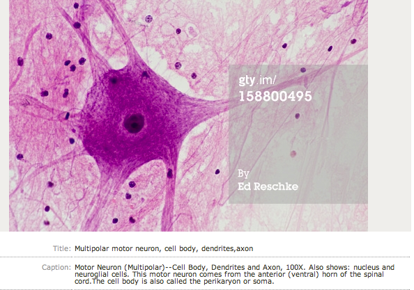
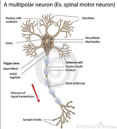
In neurology, the term motor neuron (or motoneuron) classically applies to neurons located in the central nervous system (CNS) that project their axons outside the CNS to directly or indirectly control muscles. Motor neurons are efferent nerves also called effector neurons, that carry signals from the spinal cord to the muscles to produce (effect) movement.[1]Examples of motor neurons are primary motor neurons, alpha motor neurons, beta motor neurons and gamma motor neurons.
신경과학에서 운동신경원은 중추신경계에 위치하는 신경원임.
운동신경원은 그것의 축삭을 중추신경계 바깥쪽으로 방사하여 직접적으로 또는 간접적으로 근육을 조절함.
운동신경원은 원심성 신경으로 효과기 신경원이라고 하여 척수로부터 전달되는 신호를 근육으로 전달하여 움직임을 생성함.
예를 들어 일차신경원, 알파운동신경원, 베타운동신경원, 감마운동신경원
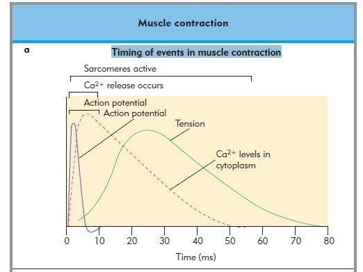
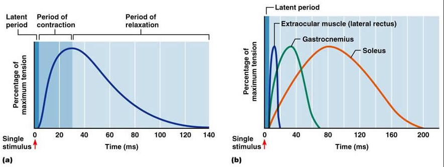
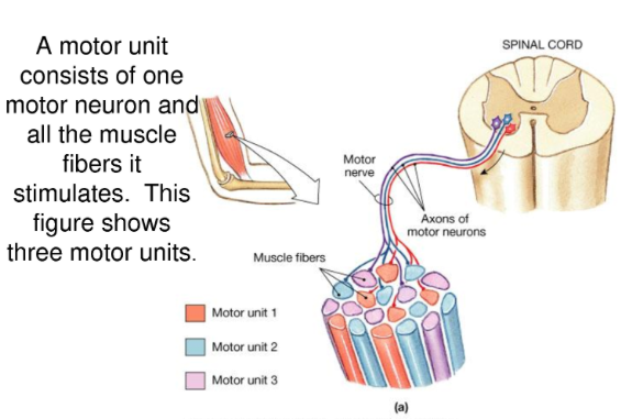
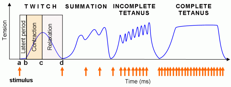
A single motor neuron may innervate many muscle fibres (muscle cells), and a muscle fibre can undergo many action potentials in the time taken for a single muscle twitch (fasciculation). As a result, if an action potential arrives before a twitch has completed, the twitches can superimpose on one another, either through summation or tetanus.
하나의 운동신경원은 많은 근섬유를 지배할 수 있고, 근섬유는 많은 활동전위아래서 단일근육연축을 일으킬 수 있음. 그 결과 활동전위가 근연축이 완성되기 전에 도착하면, 근육연축은 다른 근연축과 겹처서 summation을 만들어져 가중(summation) 또는 근경축이 발생함.
In summation, the muscle is stimulated repetitively such that additional action potentials coming from the somatic nervous system arrive before the end of the twitch. The twitches thus superimpose on one another, leading to a force greater than that of a single twitch. On the other hand, tetanus is caused by constant, very high frequency stimulation - the action potentials come at such a rapid rate that individual twitches are indistinguishable, and tension rises smoothly eventually reaching a plateau.[2]
가중에서, 근육은 한번의 연축반응이 끝나기 전에 체신경으로부터 다시 추가적인 활동전위가 발생, 축적하는 것을 말함.
근육 연축은 단일 연축에 힘이 추가되는 것을 말함.
근 경축(tetanus)은 고빈도의 가중이 지속되어 빠른 근연축이 발생하고 고원에 이르는 것을 말함.
motor neuron의 해부, 생리
According to their targets, motor neurons are classified into three broad categories:
Somatic motor neurons, which originate in the central nervous system, project their axons to the target tissues, which are always skeletal muscles.[3] Skeletal muscles are involved in locomotion (such as the muscles of the limbs, abdominal, and intercostal muscles).
체신경 운동신경원은 중추신경계에서 시작한 자극이 축삭을 따라 근육조직에 방사되는 것을 말함.
Special visceral motor neurons, also called branchial motor neurons, which directly innervate branchial muscles (that motorize the gills in fish and the face and neck in land vertebrates).
특수 내장운동신경원은 아가미 운동신경원으로 직접적으로 아가미 근육에 신경지배함.
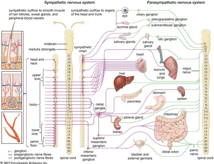
General visceral motor neurons (visceral motor neurons for short) which indirectly innervate cardiac muscle and smooth muscles of the viscera ( the muscles of the arteries): they synapse onto neurons located in ganglia of the autonomic nervous system (sympathetic and parasympathetic), located in the peripheral nervous system (PNS), which themselves directly innervate visceral muscles (and also some gland cells).
일반내장운동신경원은 심장, 내장근육, 혈관근육에 간접적으로 신경지배하여 운동신경원에 시냅스하여 ....
In other words:
- the motor command of skeletal and branchial muscles is monosynaptic (involving only one motor neuron, respectively, somatic and branchial, which synapses onto the muscle).
- the command of visceral muscles is disynaptic (involving two neurons: the general visceral motor neuron located in the CNS, which synapses onto a ganglionic neuron, located in the PNS, which synapses onto the muscle).
골격근육과 아가미근육은 단일시냅스
내장근육은 disynaptic으로 두개의 신경원이 관여함. 일반적 내장운동신경원은 중추신경계에 위치하고, 말초신경계에 존재하는 강글리온 신경원과 시냅스하여 근육으로 연결됨.
It could be argued that, in the command of visceral muscles, the ganglionic neuron, parasympathetic or sympathetic, is the real motor neuron, being the one that directly innervates the muscle (whereas the general visceral motor neuron is, strictly speaking, a preganglionic neuron).[citation needed] But, for historical reasons, the term motor neuron is reserved for the CNS neuron.
내장근육, 강글리오닉 신경원, 부교감신경 또는 교감신경은 진짜 운동신경원으로 근육에 직접적으로 신경지배함.
All vertebrate motor neurons are cholinergic, that is, they release the neurotransmitter acetylcholine. Parasympathetic ganglionic neurons are also cholinergic, whereas most sympathetic ganglionic neurons are noradrenergic, that is, they release the neurotransmitter noradrenaline. (see Table)
모든 척추운동신경원은 콜리너직 신경원으로 아세틸콜린이라는 신경전달물질을 분비함. 부교감신경 강긍리오닉 신경원은 또한 콜리너직이고, 대부분의 교감신경 강글리오닉 신경원은 노르아드레너직 신경원으로 노르아드레날린 신경물질을 분비함.
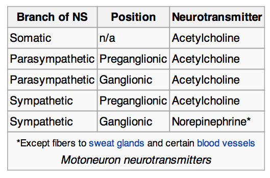
motor neuron의 기능
The interface between a motor neuron and muscle fiber is a specialized synapse called the neuromuscular junction. Upon adequate stimulation, the motor neuron releases a flood of neurotransmitters that bind to postsynapticreceptors and triggers a response in the muscle fiber which leads to muscle movement.
운동신경원과 근육의 관련성은 특수한 시냅스를 통해 신경근접합을 이루고 있음. 정확한 자극을 통해 운동신경원은 신견전달물질을 분비하여 시냅스후수용기와 부착하여 근육섬유를 자극하고 움직임을 만들어냄.
- In invertebrates, depending on the neurotransmitter released and the type of receptor it binds, the response in the muscle fiber could be either excitatory or inhibitory.
- For vertebrates, however, the response of a muscle fiber to a neurotransmitter can only be excitatory, in other words, contractile. Muscle relaxation and inhibition of muscle contraction in vertebrates is obtained only by inhibition of the motor neuron itself. Although muscle innervation may eventually play a role in the maturation of motor activity. This is how muscle relaxants work by acting on the motoneurons that innervate muscles (by decreasing their electrophysiological activity) or on cholinergic neuromuscular junctions, rather than on the muscles themselves.
Somatic motoneurons are the alpha efferent neurons, beta efferent neurons, and gamma efferent neurons. (They are called efferent to indicate the flow of information from the central nervous system (CNS) to the periphery.)
- Alpha motor neurons innervate extrafusal muscle fibers (a type of slow switch fiber) located throughout the muscle. Their cell bodies are in the ventral horn of the spinal cord and they are sometimes called ventral horn cells.
- Beta motor neurons innervate intrafusal muscle fibers of muscle spindles, with collaterals to extrafusal fibres.
- Gamma motor neurons innervate intrafusal muscle fibers found within the muscle spindle.
In addition to voluntary skeletal muscle contraction, alpha motor neurons also contribute to muscle tone, the continuous force generated by noncontracting muscle to oppose stretching. When a muscle is stretched, sensory neurons within the muscle spindle detect the degree of stretch and send a signal to the CNS. The CNS activates alpha motoneurons in the spinal cord, which cause extrafusal muscle fibers to contract and thereby resist further stretching. This process is also called the stretch reflex.
Gamma motoneurons regulate the sensitivity of the spindle to muscle stretching. With activation of gamma neurons, intrafusal muscle fibers contract so that only a small stretch is required to activate spindle sensory neurons and the stretch reflex.
motor unit
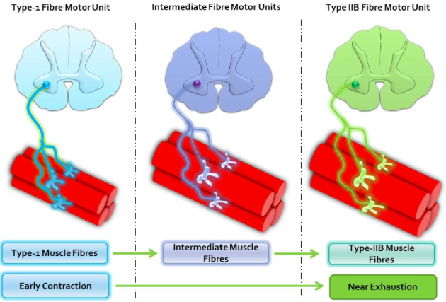
According to Purves D, et al, a single motor neuron may synapse with one or more muscle fibers. The motor neuron and all of the muscle fibers to which it connects is a motor unit. Motor units are split up into 3 categories: slow motor units, fast fatiguing motor units, and fast fatigue-resistant motor units.
- Slow motor units are used to stimulate small muscle fibres which contract very slowly and provide small amounts of energy but are very resistant to fatigue, so they are used to sustain muscular contraction such as keeping the body upright.
- Fast fatiguing motor units are used to stimulate larger muscle groups which apply large amounts of force but fatigue very quickly. They are used for tasks that requires large brief bursts on energy such as jumping or running.
- Fast fatigue-resistant motor units stimulate moderate-sized muscles groups that don't react as fast as the FF motor units, but can be sustained much longer, as implied by the name, and provide more force than S motor units.[4]
ㅌㅌ