Article
18 December 2024
Pilot Study on the Relationship Between Different Lower Limb Raising Velocities and Trunk Muscle Contraction in Active Straight Leg Raise
Kohei Yoshikawa1,2,
Noriyuki Kida3,*,
Takumi Jiroumaru4,
Yuta Murata2,5 and
Shinichi Noguchi6
1
Kanazawa Orthopaedic and Sports Medicine Clinic, 881 Ono, Ritto 520-3016, Japan
2
Graduate School of Science and Technology, Kyoto Institute of Technology, Kyoto 606-8585, Japan
3
Faculty of Arts and Sciences, Kyoto Institute of Technology, Kyoto 606-8585, Japan
4
Department of Physical Therapy, School of Health Science, Bukkyo University, Kyoto 604-8418, Japan
Show more
J. Funct. Morphol. Kinesiol.2024, 9(4), 276;https://doi.org/10.3390/jfmk9040276
This article belongs to the Section Functional Anatomy and Musculoskeletal System
Abstract
Background/Objectives: The active straight leg raise requires intricate coordination between the hip, knee, pelvis, and spine. Despite its complexity, limited research has explored the relationship between lower limb raising velocity and trunk muscle motor control during an active straight leg raise in healthy individuals. This study aimed to explore the potential effects of increased lower limb raising velocity on core muscle contractions during active straight leg raises.
Methods: Six healthy adult men (mean age: 24.5 ± 2.5 years) participated in this study. Electromyography signals were recorded using surface electrodes placed on the rectus abdominis, external oblique, and internal oblique/transverse abdominis muscles. The participants performed active straight leg raises at three different velocities: 3 s, 2 s, and as fast as possible (max). The electromyography data were analyzed from 250 ms before to 1000 ms after movement initiation, with muscle activity expressed as a percentage of the maximal voluntary isometric contraction. Statistical analyses were conducted using non-parametric tests, including the Friedman test for overall differences, followed by pairwise Wilcoxon signed-rank tests with Bonferroni correction for multiple comparisons (p < 0.05).
Results: During the 250 ms before movement initiation, the internal oblique/transverse abdominis, external oblique, and rectus abdominis muscles showed greater activity in the max condition compared to the 3 s and 2 s conditions (Friedman test, p < 0.05), but no significant differences were found in pairwise comparisons (Wilcoxon test, p > 0.05). Similarly, during the 500 ms after movement initiation, internal oblique/transverse abdominis activity was higher in the max condition, with no significant pairwise differences observed.
Conclusions: Faster lower limb raising velocities during active straight leg raise may enhance core stability by activating anticipatory and sustained internal oblique/transverse abdominis, external oblique, and rectus abdominis activity on the raised limb side. Training to promote this activation could improve dynamic stability in rapid or asymmetric movements.
초록
배경/목적:
능동적 직각 다리 들기 운동은
고관절, 무릎, 골반, 척추 간의 정교한 협응을 요구한다.
이러한 복잡성에도 불구하고,
건강한 개인에서 능동적 직각 다리 들기 운동 중
하지 들기 속도와 체간 근육 운동 조절 간의 관계를 탐구한 연구는 제한적이다.
본 연구는 능동적 직각 다리 들기 운동 중
하지 들기 속도 증가가 코어 근육 수축에 미치는 잠재적 영향을 탐구하고자 하였다.
방법:
건강한 성인 남성 6명(평균 연령: 24.5 ± 2.5세)이 본 연구에 참여하였다.
복직근, 외복사근, 내복사근/횡복근에 부착된 표면 전극을 사용하여 근전도 신호를 기록하였다.
참가자들은 3초, 2초, 그리고 가능한 한 빠르게(최대)라는 세 가지 다른 속도로 능동적 직각 다리 들어올리기 운동을 수행하였다.
근전도 데이터는
동작 시작 250밀리초 전부터 1000밀리초 후까지 분석되었으며,
근육 활동은 최대 자발적 등척성 수축의 백분율로 표현되었다.
통계 분석은 전체 차이에 대한 프리드먼 검정을 포함한 비모수 검정과 다중 비교를 위한 본페로니 보정을 적용한 쌍별 윌콕슨 부호순위 검정(p < 0.05)으로 수행되었다.
결과:
동작 시작 250밀리초 전 구간에서,
내복사근/횡복근, 외복사근, 복직근은
최대 조건에서 3초 및 2초 조건 대비 더 높은 활동을 보였으나(프리드먼 검정, p < 0.05),
쌍간 비교에서는 유의한 차이가 관찰되지 않았다(윌콕슨 검정, p > 0.05).
마찬가지로,
동작 시작 후 500밀리초 동안에도 최대 조건에서 내복사근/횡복근 활동이 더 높았으며,
유의한 쌍간 차이는 관찰되지 않았다.
결론:
능동적 직각 다리 들기 시 하체 들어 올리는 속도를 높이면,
들어 올린 다리 쪽의 내측복사근/횡복근, 외측복사근, 복직근의 예측적·지속적 활성화를 통해
코어 안정성이 향상될 수 있다.
이러한 활성화를 촉진하는 훈련은
신속하거나 비대칭적인 동작에서의 동적 안정성을 개선할 수 있다.
Keywords:
active straight leg raise; lower limb raising velocities; surface electromyography
1. Introduction
Core stability plays a crucial role in athletic performance, daily movements, and injury prevention; however, its definition remains subject to diverse interpretations. Core stability has no absolute definition because it comprises various interacting factors [1]. Generally, it is categorized into “static stability” and “dynamic stability.” Static stability is supported by structural components such as bones, joints, and ligaments [2], whereas dynamic stability is controlled by muscles and the nervous system [3,4]. Dynamic stability involves the coordination of passive elements such as bones and ligaments, active elements such as muscles and tendons, and neural elements to maintain spinal stability, allowing safe movement [5]. In sports contexts, core stability is essential for the efficient transmission and control of force and movement to the extremities. In sports, core stability is defined as the ability to control the position and movement of the trunk over the pelvis, allowing for optimal generation, transfer, and control of forces and movements to the extremities [5]. In addition, core stability requires adaptive control during movement in response to different situations [1]. Although core stability in sports has no unified definition, this study defines dynamic core stability as a state in which passive, active, and neural elements work together to maintain movement within a certain range.
The core typically refers to the muscles of the abdomen, spinal muscles, diaphragm, pelvic floor muscles, and the muscles surrounding the hips, which together form a cylindrical structure supporting the spine. Core muscles play a critical role in maintaining posture and transferring force. Deep muscles, such as the transverse abdominis (TrA), internal obliques (IO), and multifidus (MF), are considered more suitable than superficial muscles for controlling the segmental movements of the spine [6]. These deep muscles coordinate with superficial muscles depending on the velocity and type of movement [7]. For example, deep muscles are reportedly activated before movements in tasks such as single-leg standing and the active straight leg raise (ASLR) test [8,9,10]. Although deep muscles finely tune the segmental spinal stability, superficial muscles stabilize the entire spine through kinetic chains.
1. 서론
코어 안정성은
운동 능력, 일상 동작, 부상 예방에 중요한 역할을 하지만,
그 정의는 여전히 다양한 해석의 대상이다.
코어 안정성은
상호작용하는 여러 요소를 포함하기 때문에
절대적인 정의가 존재하지 않는다[1].
일반적으로
코어 안정성은 “정적 안정성”과 “동적 안정성”으로 분류된다.
정적 안정성은
뼈, 관절, 인대 등의 구조적 구성요소에 의해 유지되는 반면[2],
동적 안정성은 근육과 신경계에 의해 제어된다[3,4].
동적 안정성은
척추 안정성을 유지하여 안전한 움직임을 가능하게 하기 위해
뼈와 인대 같은 수동적 요소, 근육과 힘줄 같은 능동적 요소, 신경적 요소의 조화를 포함한다[5].
스포츠 맥락에서 코어 안정성은
사지로의 힘과 움직임의 효율적인 전달 및 제어에 필수적이다.
스포츠에서 코어 안정성은
골반 위에서 몸통의 위치와 움직임을 제어하여
사지로의 힘과 움직임을 최적으로 생성, 전달, 제어할 수 있는 능력으로 정의된다[5].
또한 코어 안정성은
다양한 상황에 대응하여 움직임 중 적응적 제어를 요구한다[1].
스포츠에서의 코어 안정성에 대한 통일된 정의는 없으나,
본 연구에서는 동적 코어 안정성을 수동적, 능동적, 신경적 요소가 협력하여
특정 범위 내에서의 움직임을 유지하는 상태로 정의한다.
코어는 일반적으로 복부 근육, 척추 근육, 횡격막, 골반저근, 고관절 주변 근육을 지칭하며,
이들은 함께 척추를 지지하는 원통형 구조를 형성한다.
코어 근육은 자세 유지와 힘 전달에 핵심적인 역할을 한다.
횡복근(TrA), 내복사근(IO), 다열근(MF)과 같은 심부 근육은
척추의 분절적 움직임을 제어하는 데 표층 근육보다 더 적합한 것으로 간주된다[6].
이러한 심부 근육은 움직임의 속도와 유형에 따라 표층 근육과 협조한다[7].
예를 들어,
한쪽 다리 서기나 능동적 직각 다리 들어올리기(ASLR) 테스트와 같은 과제에서
심부 근육이 동작 전에 활성화된다고 보고되었습니다[8,9,10].
심부 근육이 척추 분절 안정성을 미세하게 조절하는
반면,
표층 근육은 운동 사슬을 통해 척추 전체를 안정화시킵니다.
Several studies have demonstrated the impact of movement velocity on core muscle activity. During shoulder flexion movements in a standing position, faster movement velocities promote greater anticipatory muscle activity of the TrA [11]. TrA activation has also been observed during hip flexion and extension, suggesting that movement velocity influences core muscle activity [12]. Anticipatory contraction of the TrA during rapid limb movements is critical for maintaining core stability. Thus, core muscle function may vary depending on movement velocity and posture.
Various methods have been used to assess core stability. Static assessments such as the plank and side-bridge tests are commonly used to evaluate core muscle endurance and strength [13,14,15]. However, these tests do not reflect the stability or muscle coordination during dynamic movements. Methods for evaluating dynamic stability include the Sherman core stability test, which assesses core stability during movement by evaluating the response of core muscles to external forces and movements, as well as balance abilities [16,17]. The star excursion balance test (SEBT) is used to assess the dynamic stability of the core and lower limbs in single-leg standing by measuring the reach in multiple directions [17,18,19,20]. Furthermore, the functional movement screen (FMS) is used to evaluate basic movement patterns, assessing overall stability, flexibility, and coordination [21,22,23]. Although the FMS and SEBT involve whole-body movements, they have limitations in directly assessing the core muscles. The ASLR test, which evaluates the coordination of the lower limbs and core muscles while raising one leg in the supine position, is effective for assessing dynamic core stability [24,25].
여러 연구에서
운동 속도가 코어 근육 활동에 미치는 영향을 입증하였다.
서 있는 자세에서 어깨 굴곡 운동 시, 더 빠른 운동 속도는
복횡근(TrA)의 예측적 근육 활동을 증가시킨다[11].
고관절 굴곡 및 신전 시에도 복횡근 활성화가 관찰되어 운동 속도가
코어 근육 활동에 영향을 미친다는 점을 시사한다[12].
신속한 사지 운동 중 TrA의 예측 수축은
코어 안정성 유지에 핵심적이다.
따라서 코어 근육 기능은 운동 속도와 자세에 따라 달라질 수 있다.
코어 안정성 평가에는 다양한 방법이 활용된다.
플랭크 및 사이드 브리지 테스트와 같은 정적 평가는
코어 근육의 지구력과 근력을 평가하는 데 흔히 사용된다[13,14,15].
그러나 이러한 테스트는
동적 운동 중 안정성이나 근육 협응력을 반영하지 못한다.
동적 안정성 평가 방법에는
외부 힘과 움직임에 대한 코어 근육의 반응 및 균형 능력을 평가하여
동작 중 코어 안정성을 측정하는 셔먼 코어 안정성 검사(Sherman core stability test)가 포함된다[16,17].
스타 익스커션 밸런스 테스트(SEBT)는
단일 다리 서기 자세에서 다방향으로의 도달 거리를 측정하여
코어와 하지의 동적 안정성을 평가하는 데 사용된다[17,18,19,20].
또한 기능적 동작 검사(FMS)는
기본 동작 패턴을 평가하여 전반적인 안정성, 유연성 및 조화를 측정하는 데 사용됩니다[21,22,23].
FMS와 SEBT는 전신 동작을 포함하지만
코어 근육을 직접 평가하는 데 한계가 있습니다.
ASLR 검사는
누운 자세에서 한쪽 다리를 들어 올리는 동안 하지와 코어 근육의 협응력을 평가하며,
동적 코어 안정성 평가에 효과적이다[24,25].
The ASLR is a standard test for assessing the coordination between the lower limb and core muscles by flexing the hip while lying supine with the knee extended. This test can effectively measure dynamic core stability [26]. The ASLR is well suited for electromyographic (EMG) analysis, allowing the evaluation of muscle onset time and contraction intensity, which provides a more detailed understanding of dynamic core stability. Studies on healthy individuals have reported that the IO and TrA are activated during the later stages of ASLR, whereas the external oblique (EO) and rectus abdominis (RA) muscles are delayed compared with the psoas major and IO muscles when the leg is lifted [27]. In patients with lumbopelvic pain, TrA and IO activation during ASLR was lower than that in pain-free groups [28]. Although these studies have indicated that muscle activity during ASLR contributes to core stability and postural control, the effect of movement velocity on muscle activity remains unclear. Different movement velocities may significantly affect the timing and intensity of muscle contraction. Faster movements require faster muscle contractions and greater explosive force, whereas slower movements require sustained muscle activity. Understanding the relationship between movement velocity and muscle activity is crucial [29]. Even for the same movement, the effect of movement velocity on the activity patterns, timing, and intensity of core muscles remains unclear. Further research is needed, particularly on how the deep and superficial core muscles work together to ensure stability in response to different movement velocities during tasks such as ASLR.
It is generally accepted that faster movements prompt anticipatory TrA activation to maintain dynamic core stability. This pilot study aimed to explore the effects of increased lower limb raising velocity during ASLR on the contraction of core muscles, particularly the IO/TrA, EO, and RA, to provide preliminary data and a methodological foundation for future comprehensive studies.
ASLR은
무릎을 펴고 누운 상태에서 고관절을 굴곡시키며
하지와 코어 근육 간의 협응력을 평가하는 표준 검사이다.
이 검사는
동적 코어 안정성을 효과적으로 측정할 수 있다[26].
ASLR은
근전도(EMG) 분석에 적합하여 근육 발현 시간과 수축 강도를 평가할 수 있으며,
이는 동적 코어 안정성에 대한 보다 상세한 이해를 제공한다.
건강한 대상자를 대상으로 한 연구에 따르면,
다리를 들어 올릴 때 외복사근(EO)과 복직근(RA)은
대요근과 내복사근(IO)에 비해 활성화가 지연되는 반면,
내복사근(IO)과 복횡근(TrA)은 ASLR 후기 단계에서 활성화된다[27].
요추골반 통증 환자군에서는
ASLR 수행 중 TrA와 IO 활성화가 통증 없는 대조군보다 낮았다[28].
이러한 연구들은
ASLR 중 근육 활동이 코어 안정성과 자세 제어에 기여함을 시사하지만,
운동 속도가 근육 활동에 미치는 영향은 여전히 불분명하다.
서로 다른 운동 속도는
근육 수축의 시기와 강도에 상당한 영향을 미칠 수 있다.
빠른 동작은 더 빠른 근육 수축과 더 큰 폭발력을 요구하는 반면,
느린 동작은 지속적인 근육 활동을 필요로 한다.
운동 속도와 근육 활동 간의 관계를 이해하는 것은
매우 중요하다[29].
동일한 동작이라 하더라도 운동 속도가
코어 근육의 활동 패턴, 시기, 강도에 미치는 영향은 여전히 불분명하다.
특히
ASLR과 같은 작업 중 다양한 운동 속도에 대응하여 안정성을 확보하기 위해
심부 및 표층 코어 근육이 어떻게 협력하는지에 대한 추가 연구가 필요하다.
일반적으로
빠른 동작은 동적 코어 안정성 유지를 위해
TrA의 예측적 활성화를 유발한다고 알려져 있다.
본 예비 연구는 ASLR 수행 중 하체 들어올리기 속도 증가가 코어 근육,
특히 IO/TrA, EO, RA의 수축에 미치는 영향을 탐구하여
향후 포괄적 연구를 위한 기초 자료와 방법론적 토대를 마련하는 것을 목표로 한다.
2. Materials and Methods
2.1. Participants
Six healthy adult men (24.5 ± 2.5 years, 170.5 ± 5.1 cm, 68.2 ± 9.0 kg) participated in this experiment. Participants with a history of pain, significant postural abnormalities, or serious neurological or respiratory conditions were excluded. Informed consent was obtained from all the participants. This study was approved by the Ethics Committee of Kanazawa Orthopedic Surgery Clinic (Kanazawa-OSMC-2024-003) and conducted in accordance with the Declaration of Helsinki.
2.2. Electromyography Recording
The skin was carefully prepared by shaving excess hair and reducing the skin impedance to below 5 kΩ. After drying the skin, pairs of Ag/AgCl surface electrodes (SMP-300, METS Co., LTD., Tokyo, Japan) with a size of 19 mm per side were placed 20 mm apart at the following locations: rectus abdominis (RA), 1 cm above and 2 cm lateral to the umbilicus [30]; external oblique (EO), 1 cm above the horizontal line passing through the umbilicus and 1 cm lateral to the RA boundary [31]; and IO/TrA, 1 cm medial to the anterior superior iliac spine (ASIS) and 0.5 cm below the line connecting the ASIS [32]. This placement captures the combined activity of the IO and TrA muscles, as demonstrated in a previous study. Additionally, the muscle fibers of the IO/TrA were identified under ultrasound guidance, and it was confirmed that there was no overlap with the EO. The electrodes were placed bilaterally at each site for a total of six locations. To minimize cross-talk, careful attention was paid to electrode placement, inter-electrode distance, and orientation along the muscle fibers [33]. EMG signals were sampled at 1000 Hz, amplified, and collected using an EMG analysis software (VitalRecorder2 ver. 3.8.4. 1403, KISSEI COMTEC Co., Ltd., Nagano, Japan).
2.3. Kinematics
Kinematic data were collected by attaching a foot sensor to the heel, which defined the movement initiation point.
2.4. Experimental Task
EMG and kinematic data were synchronously recorded during ASLR. The participants performed the ASLR task in the supine position with their legs straight, raising their right lower limb to 45° of hip flexion at three different velocities. A bar was set up such that the participant’s patella would touch it at 45° of hip flexion (Figure 1).
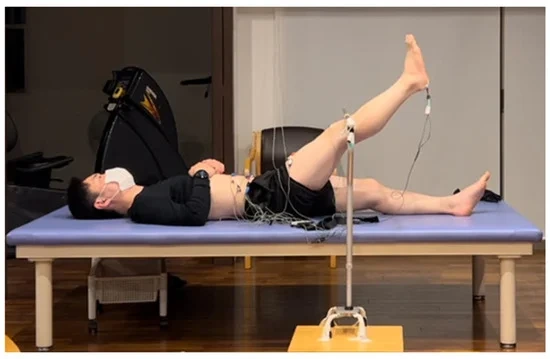
Figure 1. Experimental design of the active straight leg raise task.
The three conditions were grouped based on the speed of raising the leg to 45°. In the 3 s group (3 s) (slow; angular velocity ω = 15°/s), the leg was raised at a slow speed, taking exactly 3 s. In the 2 s group (2 s) (moderate; ω = 22.5°/s), the leg was raised at a moderate speed, taking 2 s. Finally, in the max group (max) (as fast as possible; ω = approximately 45°/s), the leg was raised at the fastest possible speed, typically in about 1 s. Each condition was performed six times with the order of the trials randomized, and the movements were initiated following verbal commands. The participants performed MVICs for 5 s in the supine against manual resistance for each muscle. Two MVIC trials were performed for each muscle with sufficient rest between trials, and the higher value from the two trials was used.
2.5. Data Analysis
EMG data were analyzed based on kinematic data using movement initiation as the reference point. The analysis focused on the period from 250 ms before movement initiation to 1000 ms after initiation. First, the raw data were bandpass-filtered at 20–500 Hz and then full-wave rectified. The root mean square of the EMG amplitude was calculated using a 50 ms window for each ASLR trial and MVIC.
For the data 250 ms before movement initiation (pre-250 ms), the average was calculated using a 50 ms window. The data 1000 ms after movement initiation were divided into four intervals as follows: 0–250 ms (post-250 ms), 251–500 ms (post-500 ms), 501–750 ms (post-750 ms), and 751–1000 ms (post-1000 ms). The average value for each 50 ms interval was calculated. Muscle activity levels are expressed as a percentage of MVIC (%MVIC) (MATLAB R2024a, MathWorks, Inc., Natick, MA, USA).
2.6. Statistical Analysis
Due to the small sample size and the results of the normality tests, non-parametric statistical methods were employed. The Friedman test was used to assess differences among the three velocities (3 s, 2 s, and max). When the Friedman test indicated significant differences, post-hoc pairwise comparisons were conducted using the Wilcoxon signed-rank test with Bonferroni correction to adjust for multiple comparisons. Statistical significance was set at p < 0.05. All statistical analyses were performed using the SPSS ver. 29.0.2 (IBM Corp., Armonk, NY, USA).
3. Results
During the 250 ms before movement initiation, the IO/TrA, EO, and RA muscles on the same side as the raised lower limb tended to be higher compared to the 3 s and 2 s conditions, as indicated by the Friedman test. For the IO/TrA, the activity in the max condition (7.5 ± 4.8%) tended to be higher compared to the 3 s (3.2 ± 1.8%) and 2 s (3.2 ± 1.8%) conditions (χ2 = 9.3, p = 0.009, Kendall’s W = 0.78). For EO, the activity in the max condition (18.5 ± 3.6%) tended to be higher compared to the 3 s (10.9 ± 3.9%) and 2 s (11.2 ± 2.7%) conditions (χ2 = 7.0, p = 0.030, Kendall’s W = 0.58). For RA, the activity in the max condition (6.6 ± 3.3) tended to be higher compared to the 3 s (3.6 ± 1.1%) and 2 s (3.8 ± 1.1%) conditions (χ2 = 8.3, p = 0.016, Kendall’s W = 0.69) (Table 1).

Table 1. EMG (%MVIC) values for each muscle, with the mean ± SD, median (IQR), and Friedman test results across trials.
However, subsequent pairwise comparisons using the Wilcoxon test did not reveal significant differences between any of the conditions (p > 0.05 for all comparisons) (Table 2, Table 3 and Table 4).

Table 2. Results of the Wilcoxon test following the Friedman test for right IO/TrA in pre-250 ms.

Table 3. Results of the Wilcoxon test following the Friedman test for right EO in pre-250 ms.

Table 4. Results of the Wilcoxon test following the Friedman test for RA in pre-250 ms.
During the 500 ms after movement initiation, the IO/TrA tended to be higher in the max condition compared to the 3 s and 2 s conditions, as indicated by the Friedman test (χ2 = 9.0, p = 0.011, Kendall’s W = 0.75). For the IO/TrA, the activity in the max condition (8.8 ± 7.0%) tended to be higher compared to the 3 s (4.0 ± 2.3%) and 2 s (4.2 ± 2.6%) conditions. However, subsequent pairwise comparisons using the Wilcoxon test did not reveal significant differences between any of the conditions during this period (p > 0.05 for all comparisons) (Table 5).

Table 5. Results of the Wilcoxon test following the Friedman test for right IO/TrA in post-500 ms.
4. Discussion
This pilot study aimed to investigate how differences in lower limb raising velocities during ASLR influence the contraction of core muscles, particularly the IO/TrA, EO, and RA. During the 250 ms before movement initiation, the muscle activities of the IO/TrA, EO, and RA on the same side as the raised lower limb tended to be higher compared to the 3 s and 2 s conditions, as indicated by the Friedman test. Similarly, during the 500 ms after movement initiation, the IO/TrA also tended to be higher compared to the 3 s and 2 s conditions. However, pairwise comparisons using the Wilcoxon test did not reveal significant differences between conditions during either period.
These findings suggest that both anticipatory and early post-movement contractions of the IO/TrA, EO, and RA may play a role in maintaining core stability during fast lower limb raising in the supine position.
Unlike previous studies that investigated postural control during task performance in standing positions [7,8,10], this study examined postural control in the supine position. When standing, gravity exerts a greater effect, requiring more active stabilization by the core muscles [11]. In the supine position, core stability is partially supported by passive elements, such as bones and ligaments, reducing the demand for strong core muscle contractions [3]. However, as movement velocity increases, maintaining core stability requires anticipatory contractions of both deep muscles, such as the IO/TrA, and superficial muscles, including the EO and RA. During the max condition, the coordinated contraction of the EO, RA, and IO/TrA on the same side as the raised lower limb is essential for ensuring core stability, whereas, during slower movements (2 s and 3 s conditions), anticipatory muscle contractions may not be as necessary, allowing stability to be maintained more easily. The notable finding that the IO/TrA, EO, and RA on the same side as the raised lower limb exhibited increased activity before movement initiation aligns with the concept of “feedforward postural control”, where core muscles activate in anticipation of limb movement to ensure core stability [34]. This result highlights the importance of preparatory muscle activation across both deep and superficial muscles, particularly during rapid movements like those observed in the max condition, to counteract the increased demands for core stabilization.
The findings partially align with previous studies on movements in the standing position. For instance, during fast shoulder flexion in standing, anticipatory contraction of the deep muscle TrA is promoted regardless of the direction of preparatory trunk movement, and superficial muscles like the IO and EO exhibit direction-specific activity. However, in standing, the activity of deep muscles (TrA and MF) is more critical for trunk support [11].
Similarly, our study revealed that, in the relatively stable supine position, the deep muscle IO/TrA on the same side as the raised lower limb showed a trend toward greater activation, along with a tendency for higher activity in the superficial muscles EO and RA on the same side during fast movements. This suggests that both deep and superficial core muscles are actively involved in maintaining core stability during fast lower limb raising in the supine position. These results further support the notion that posture significantly influences muscle activity patterns and that the muscles involved in maintaining core stability adapt depending on the posture.
The unilateral dominance of the IO/TrA, EO, and RA on the side of the raised lower limb can be explained as an adaptive response of the core muscles to asymmetric movements. Previous studies have reported that unilateral limb movements result in asymmetric activity of the core muscles to ensure stability on that side [5]. This finding aligns with the increased activity of the IO/TrA, EO, and RA on the lower limb raising side in this study, suggesting that unilateral core stability contributes to lower limb movement control.
The effect of movement velocity on core muscle activity tended to be evident, with the EO and RA showing greater activity in the fast velocity condition. Additionally, the deep muscle IO/TrA also exhibited a trend toward greater activation in the fast condition. Faster movements generate greater rotational forces (moments) around the hip joint, requiring both superficial muscles like the EO and RA and deep muscles such as the IO/TrA to contract more rapidly to maintain core stability. As movement velocity increases, the moment at which movement initiation increases and the activities of the EO, RA, and IO/TrA increase accordingly to counteract these forces. This suggests that both superficial and deep core muscles are actively involved in stabilizing the core during fast movements.
Deep and superficial muscles play different roles in core stability. Deep muscles like the TrA, IO, and MF contribute to segmental stabilization of the spine, whereas superficial muscles like the EO, RA, and IO support overall spinal stabilization [34,35]. Previous studies have reported a muscle-activation pattern during dynamic movements in which deep muscles activate first, followed by superficial muscles [34]. In this study, conducted in a relatively stable supine position, it was observed that the deep muscle IO/TrA showed a trend toward higher activity before movement initiation. Similarly, the superficial muscles EO and RA also demonstrated a trend toward higher activity before movement initiation during fast movements. This coordinated activation of deep and superficial muscles suggests that these muscle groups work together to maintain core stability before and during fast lower-limb raising in the supine position. These findings highlight the influence of posture on muscle activity and suggest that the muscles involved in maintaining core stability adapt their activation patterns according to the posture. Previous studies have focused on movements performed in standing positions, where trunk stabilization is critical [12]. In contrast, ASLR involves lower limb raising in a supine position with a relatively light load, suggesting that the demands on core stability are lower. However, even under light loads, the IO/TrA, EO, and RA on the side of the raised lower limb may contribute to overall movement stability by controlling trunk rotation and lateral flexion during lower limb raising.
Furthermore, the IO/TrA showed a trend toward higher activity even during the 500 ms after movement initiation. This suggests that the IO/TrA plays a crucial role in maintaining core stability not only before but also during movement. On the other hand, the EO and RA were primarily active before movement initiation, indicating that their role may be limited to ensuring core stability before movement. These findings suggest that deep and superficial muscles perform distinct roles and function at different times in maintaining core stability, with the IO/TrA playing a central role in sustaining core stability throughout the movement.
Additionally, the lack of differences in EO and RA activity after movement initiation suggests that, under the maximal velocity condition, significant muscle exertion to counteract a large moment at movement initiation was followed by the leg being raised by inertia. Consequently, the need for muscle exertion decreased after movement initiation. In ASLR with a light load, the EO and RA are most active before or at the onset of movement, after which the movement requires only the maintenance of muscle strength. This indicates that high muscle activity was not necessary after movement initiation, allowing for stable motion.
The results of this study provide valuable insights into core training in sports and rehabilitation. Eliciting the coordinated activity of both deep and superficial core muscles, such as the IO/TrA, EO, and RA, on the same side as the raised lower limb before movement initiation may play a crucial role in enhancing dynamic core stability. This coordinated muscle activation is particularly important during fast movements, as it helps stabilize the core in anticipation of increased demands. For athletes, training programs that focus on improving the anticipatory and cooperative activation of these core muscles could be essential in activities that require rapid and explosive movements.
This study had several limitations. The small sample size and lack of statistical power limit the ability to detect significant differences, potentially leading to false negatives. However, as a pilot study, these findings hold significant value in laying the groundwork for hypothesis-driven experimental research. The insights gained from this exploratory study are critical for designing future experiments with refined methodologies and larger, more diverse sample populations. Moreover, the results are specific to the studied population and conditions, which may limit their generalizability. Future research is required to validate these findings, expand on the observed trends, and provide robust evidence to confirm the role of lower limb raising velocity in enhancing core muscle activity and stability. Additionally, the results are specific to the studied population and conditions, which may limit their generalizability. Future research is required to validate these findings with a larger sample size, more diverse populations, and more refined methodologies to confirm the observed trends and provide robust evidence. In this study, we assessed muscle activity of the IO/TrA and EO using surface electromyography. We acknowledge that recording sEMG signals from deep muscles like the TrA has inherent limitations, and it is difficult to completely eliminate the influence of crosstalk [35]. However, we took measures to minimize these effects through careful electrode placement. These limitations should be considered when interpreting the results.
4. 논의
본 예비 연구는 ASLR 수행 중 하지 들어올리기 속도 차이가 코어 근육, 특히 IO/TrA, EO, RA의 수축에 미치는 영향을 조사하고자 하였다. 동작 시작 전 250ms 동안 들어올린 하지와 같은 측의 IO/TrA, EO, RA 근육 활동은 프리드먼 검정 결과 3초 및 2초 조건에 비해 높은 경향을 보였다. 마찬가지로, 동작 시작 후 500ms 동안에도 IO/TrA의 활동은 3초 및 2초 조건에 비해 높은 경향을 보였다. 그러나 윌콕슨 검정을 이용한 쌍간 비교에서는 두 기간 모두 조건 간 유의미한 차이를 확인하지 못했다.
이러한 결과는 IO/TrA, EO 및 RA의 예측 수축과 초기 운동 후 수축이 모두 누운 자세에서 빠른 하지 들어 올리기 동안 코어 안정성을 유지하는 데 중요한 역할을 할 수 있음을 시사한다.
이 연구는 서 있는 자세에서 작업 수행 중 자세 조절을 조사한 기존 연구[7,8,10]와 달리 누운 자세에서 자세 조절을 조사했다. 서 있는 자세에서는 중력이 더 큰 영향을 미치므로 코어 근육에 의한 능동적 안정화가 더 많이 요구된다[11]. 누운 자세에서는 골격과 인대 같은 수동적 요소가 코어 안정성을 부분적으로 지원하므로 강한 코어 근육 수축에 대한 요구가 감소한다[3]. 그러나 운동 속도가 증가함에 따라 코어 안정성 유지를 위해서는 IO/TrA 같은 심부 근육과 EO 및 RA 같은 표층 근육의 예측 수축이 모두 필요하다. 최대 조건에서는 들어 올린 하지와 같은 쪽의 EO, RA, IO/TrA의 조화된 수축이 코어 안정성 확보에 필수적인 반면, 느린 동작(2초 및 3초 조건)에서는 예측적 근육 수축이 그다지 필요하지 않아 안정성을 더 쉽게 유지할 수 있습니다. 들어 올린 하지와 같은 쪽의 IO/TrA, EO 및 RA가 동작 시작 전에 활동이 증가했다는 주목할 만한 발견은 “피드포워드 자세 제어” 개념과 일치하는데, 이는 코어 근육이 사지 동작을 예상하여 코어 안정성을 보장하기 위해 활성화된다는 개념이다 [34]. 이 결과는 특히 최대 조건에서 관찰된 것과 같은 빠른 동작 동안 심부 및 표층 근육 전반에 걸친 준비적 근육 활성화가 코어 안정화에 대한 증가된 요구를 상쇄하는 데 중요함을 강조한다.
이 결과는 서 있는 자세에서의 동작에 관한 기존 연구와 부분적으로 일치한다. 예를 들어, 서 있는 상태에서 빠른 어깨 굴곡 시, 준비적 몸통 움직임의 방향과 무관하게 깊은 근육 TrA의 예측 수축이 촉진되며, IO 및 EO와 같은 표층 근육은 방향 특이적 활성을 보인다. 그러나 서 있는 자세에서는 깊은 근육(TrA 및 MF)의 활성이 몸통 지지력에 더 중요하다[11].
마찬가지로 본 연구에서는 상대적으로 안정된 앙와위 자세에서, 빠르게 움직일 때 들어 올린 하지와 같은 쪽의 깊은 근육(IO/TrA)이 더 큰 활성화 경향을 보였으며, 같은 쪽의 표층 근육(EO 및 RA)도 더 높은 활동 경향을 보인다는 사실을 밝혀냈다. 이는 앙와위 자세에서 빠른 하지 들어올리기 동안 심부 및 표층 코어 근육 모두 코어 안정성 유지에 적극적으로 관여함을 시사한다. 이러한 결과는 자세가 근육 활동 패턴에 상당한 영향을 미치며, 코어 안정성 유지에 관여하는 근육이 자세에 따라 적응한다는 개념을 더욱 뒷받침한다.
들어올린 하지 측의 IO/TrA, EO, RA 근육의 일측성 우세는 비대칭 운동에 대한 코어 근육의 적응적 반응으로 설명될 수 있다. 기존 연구에서는 일측 사지 운동 시 해당 측 안정성 확보를 위해 코어 근육의 비대칭적 활동이 발생한다고 보고된 바 있다[5]. 본 연구에서 하지 들어 올림 측의 IO/TrA, EO, RA 활동 증가가 관찰된 것은 이러한 결과와 부합하며, 일측 코어 안정성이 하지 운동 제어에 기여함을 시사한다.
운동 속도가 코어 근육 활동에 미치는 영향은 속도가 빠를수록 뚜렷한 경향을 보였으며, EO와 RA는 고속 조건에서 더 큰 활동을 나타냈다. 또한 심부 근육인 IO/TrA 역시 고속 조건에서 활성화 증가 경향을 보였다. 빠른 운동은 고관절 주위 회전력(모멘트)을 증가시켜 코어 안정성 유지를 위해 EO, RA와 같은 표층근과 IO/TrA와 같은 심부근 모두의 더 빠른 수축을 요구한다. 운동 속도가 증가함에 따라 운동 시작 시점의 모멘트가 증가하고, 이에 대응하여 EO, RA, IO/TrA의 활동도 증가한다. 이는 빠른 운동 중 코어 안정화에 표층 및 심층 코어 근육이 모두 적극적으로 관여함을 시사한다.
심층근과 표층근은 코어 안정성에서 서로 다른 역할을 수행합니다. TrA, IO, MF와 같은 심층근은 척추의 분절적 안정화에 기여하는 반면, EO, RA, IO와 같은 표층근은 전반적인 척추 안정화를 지원합니다[34,35]. 기존 연구들은 동적 동작 중 심층근이 먼저 활성화되고 표층근이 뒤따르는 근육 활성화 패턴을 보고한 바 있습니다[34]. 상대적으로 안정된 앙와위 자세에서 수행된 본 연구에서는 깊은 근육인 IO/TrA가 동작 시작 전에 더 높은 활성화를 보이는 경향이 관찰되었다. 마찬가지로 표층 근육인 EO와 RA 역시 빠른 동작 중 동작 시작 전에 더 높은 활성화를 보이는 경향을 나타냈다. 이러한 깊은 근육과 표층 근육의 조화된 활성화는 앙와위 자세에서 빠른 하지 들어올리기 동작 전과 동작 중에 이 근육군들이 함께 작용하여 코어 안정성을 유지함을 시사한다. 이러한 결과는 자세가 근육 활동에 미치는 영향을 강조하며, 코어 안정성 유지에 관여하는 근육들이 자세에 따라 활성화 패턴을 조정함을 시사한다. 기존 연구들은 체간 안정화가 중요한 서 있는 자세에서의 동작에 집중해왔다[12]. 반면 ASLR은 상대적으로 가벼운 하중으로 누운 자세에서 하지 들어올리기를 수행하므로 코어 안정성에 대한 요구가 낮다고 볼 수 있다. 그러나 가벼운 하중 하에서도, 들어 올린 하지 측의 내회전근/내전근(IO/TrA), 외회전근(EO), 외전근(RA)은 하지 들어 올리기 동안 몸통 회전과 측면 굴곡을 제어함으로써 전체적인 동작 안정성에 기여할 수 있습니다.
또한, IO/TrA는 동작 시작 후 500밀리초 동안에도 더 높은 활동 경향을 보였습니다. 이는 IO/TrA가 동작 전뿐만 아니라 동작 중에도 코어 안정성 유지에 중요한 역할을 한다는 것을 시사합니다. 반면, EO와 RA는 주로 동작 시작 전에 활성화되었는데, 이는 이들의 역할이 동작 전 코어 안정성 확보에 국한될 수 있음을 시사한다. 이러한 결과는 심층근과 표층근이 코어 안정성 유지에 있어 서로 다른 시점에 상이한 역할을 수행하며, IO/TrA가 동작 전반에 걸쳐 코어 안정성을 유지하는 중심적 역할을 한다는 점을 시사한다.
또한 동작 시작 후 EO와 RA 활동에 차이가 없었던 점은 최대 속도 조건에서 동작 시작 시 큰 모멘트를 상쇄하기 위한 상당한 근육 발력이 이루어진 후 관성으로 다리가 들어 올려졌음을 시사한다. 결과적으로 동작 시작 후 근육 발력의 필요성은 감소했다. 경량 부하를 적용한 ASLR에서는 EO와 RA가 동작 시작 전 또는 시작 시점에 가장 활발히 활동하며, 이후 동작은 근력 유지만 필요로 한다. 이는 동작 시작 후 높은 근육 활동이 필요하지 않으며 안정적인 움직임을 가능케 함을 시사한다.
본 연구 결과는 스포츠 및 재활 분야의 코어 훈련에 대한 귀중한 통찰력을 제공한다. 동작 시작 전 들어 올린 하지와 같은 측의 IO/TrA, EO, RA와 같은 심부 및 표층 코어 근육의 조화된 활동을 유도하는 것이 동적 코어 안정성 향상에 중요한 역할을 할 수 있다. 이러한 조화된 근육 활성화는 특히 빠른 동작 중 증가된 요구를 예상하여 코어를 안정화하는 데 도움이 되므로 중요합니다. 운동선수에게 있어, 이러한 코어 근육들의 예측적 및 협력적 활성화를 개선하는 데 초점을 맞춘 훈련 프로그램은 신속하고 폭발적인 동작이 필요한 활동에서 필수적일 수 있습니다.
본 연구에는 몇 가지 한계가 존재한다. 적은 표본 크기와 통계적 검정력의 부족은 유의미한 차이를 발견하는 능력을 제한하여 잠재적으로 위음성 결과를 초래할 수 있다. 그러나 예비 연구로서, 이러한 발견은 가설 기반 실험 연구의 토대를 마련하는 데 상당한 가치를 지닌다. 이 탐색적 연구에서 얻은 통찰은 정교화된 방법론과 더 크고 다양한 표본 집단을 대상으로 한 향후 실험 설계를 위해 중요하다. 또한 결과는 연구된 집단과 조건에 특이적이어서 일반화 가능성을 제한할 수 있다. 향후 연구에서는 이러한 결과를 검증하고 관찰된 경향을 확장하며, 하체 들어올리기 속도가 코어 근육 활동과 안정성 향상에 미치는 역할을 확인하기 위한 강력한 증거를 제시해야 한다. 또한 결과는 연구 대상 집단과 조건에 특이적이어서 일반화 가능성을 제한할 수 있다. 향후 연구에서는 더 큰 표본 규모, 더 다양한 집단, 더 정교한 방법론을 통해 이러한 결과를 검증하고 관찰된 경향을 확인하며 강력한 증거를 제공해야 한다. 본 연구에서는 표면근전도(sEMG)를 사용하여 IO/TrA 및 EO 근육의 활동을 평가했습니다. TrA와 같은 심부 근육의 sEMG 신호 기록에는 본질적인 한계가 있으며, 간섭(crosstalk)의 영향을 완전히 제거하기 어렵다는 점을 인정합니다[35]. 그러나 신중한 전극 배치로 이러한 영향을 최소화하기 위한 조치를 취했습니다. 결과 해석 시 이러한 한계를 고려해야 합니다.
5. Conclusions
This study provides preliminary evidence suggesting that the coordinated activity of both deep and superficial core muscles, including the IO/TrA, EO, and RA, plays crucial roles in maintaining core stability during high-velocity lower limb raising in ASLR. The IO/TrA exhibited activity both before and during movement, suggesting its continuous contribution to core stabilization across different phases of the movement. Meanwhile, the EO and RA were primarily active before movement initiation, highlighting their anticipatory roles in ensuring core stability. Practically, incorporating training programs that promote both the anticipatory activation of the IO/TrA, EO, and RA and the sustained activation of the IO/TrA during lower limb raising or unilateral movements in the supine position may be beneficial for enhancing dynamic core stability. Strengthening these muscles could improve performance in sports and daily activities, particularly in movements that require rapid or asymmetric core stabilization.
5. 결론
본 연구는 IO/TrA, EO, RA를 포함한 심부 및 표층 코어 근육의 조화된 활동이 ASLR에서 고속 하지 들어올리기 동작 중 코어 안정성 유지에 핵심적인 역할을 한다는 예비적 증거를 제시한다. IO/TrA는 동작 전과 동작 중 모두에서 활성을 보였으며, 이는 동작의 다양한 단계에 걸쳐 코어 안정화에 지속적으로 기여함을 시사한다. 한편, EO와 RA는 주로 동작 시작 전에 활성화되어 코어 안정성 확보를 위한 예측적 역할을 강조한다. 실용적으로, IO/TrA, EO, RA의 예측적 활성화와 하반신 들어올리기 또는 앙와위 자세에서의 일측성 동작 중 IO/TrA의 지속적 활성화를 모두 촉진하는 훈련 프로그램을 도입하는 것이 동적 코어 안정성 향상에 도움이 될 수 있다. 이러한 근육을 강화하면 특히 신속하거나 비대칭적인 코어 안정화가 필요한 동작에서 스포츠 및 일상 활동 수행 능력이 향상될 수 있다.
Author Contributions
Conceptualization, K.Y.; methodology, K.Y. and N.K.; validation, K.Y. and N.K.; formal analysis, K.Y. and T.J.; Investigation, K.Y., Y.M. and S.N.; data curation, K.Y. and N.K.; writing—original draft, K.Y.; writing—review and editing, K.Y.; visualization, K.Y. and N.K. All authors have read and agreed to the published version of the manuscript.
Funding
This research received no external funding.
Institutional Review Board Statement
This study was conducted in accordance with the Declaration of Helsinki, and approved by the Institutional Review Board (Ethics Committee) of Kanazawa Orthopedic Surgery Clinic (protocol code Kanazawa-OSMC-2024-003).
Informed Consent Statement
Informed consent was obtained from all subjects involved in this study.
Data Availability Statement
The original contributions presented in the study are included in the article, further inquiries can be directed to the corresponding author.
Acknowledgments
We appreciate the contributions of all those who provided data and resources for this study.
Conflicts of Interest
The authors declare no conflicts of interest.
References