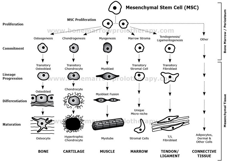
Structure of the dermis에 관한 그림들..
epidermis보다 dermis는 15배에서 40배가 두껍고 많은 콜라겐으로 구성되어 있다. 근막이완의 메카니즘을 잘 이해하기위해서는 dermis를 잘 이해해야 할듯...
http://www.derm-hokudai.jp/shimizu-dermatology/ch01/index.html
122페이지 skin layer mechanics 논문
a. Structure of the dermis
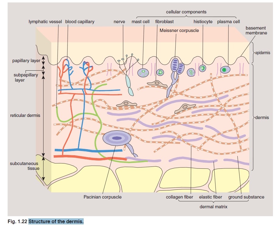
The dermis is the structure beneath the epidermis, and the two are separated by the basal membrane (Fig. 1.3). The dermis is approximately 15 to 40 times as thick as the epidermis. It consists of three layers.
1) Papillary layer: The dermal area that projects into the intervals between the epidermal ridges. The fiber components are thin and richly supplied with capillaries, sensory nerve endings and cytoplasm.
2) Subpapillary layer: The area underlying the epidermis, containing the same components as the papillary layer.
3) Reticular layer: Accounts for the largest part of the dermis and has dense connective tissue comprising fiber components. The lower part comes into contact with the subcutaneous fatty tissue.
There are blood vessels and nerves in some parts. The components of the dermis comprise the fibrous tissue and
the dermal matrix formed by cells in the interstitial components (Fig. 1.22). The major components mainly consist of collagen fibers (mainly types I and III), with smaller amounts of elastic fibers, reticular fibers and matrix. This matrix generally comprises the extra-cellular matrix and ground substance made up of proteoglycans and gelatin. Fibroblasts, macrophages, mast cells, plasma cells, vascular channels and nerves are common cellular components.
b. Interstitial components
1. Collagen fibers
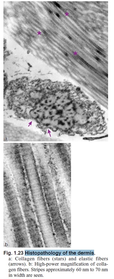
Collagen fibers account for 70% of the weight of dry dermis (Fig. 1.23) and appear white to the naked eye. The collagen
fibrous component is poorly extensible; however, it is extremely tough and especially resistant to tension parallel to the fibers. This characteristic is important in maintaining the dynamic strength of skin.
Collagen fibers form from aggregations of thin fibrils. The more fibrils there are in the fiber, the thicker and stronger the
fiber. Thin collagen fibers are sparsely seen in the papillary layers and subpapillary layers; however, collagen bundles, with fully developed thick collagen fibers, are densely distributed in the reticular dermal upper layers.
In light microscopy, the proteinaceous collagen fibers stain well in eosin solution; they stain red in Van Gieson and blue after Mallory staining. The fibril is observed by electron microscopy as being very long, 100 nm to 500 nm in diameter with cross striations that repeat at intervals of 60 nm to 70 nm (Figs. 1.23 and 1.24). Fibrils become collagen fibers by aggregating with glycoproteins. A thick collagen bundle can reach 2 mm to 15 mm in diameter.
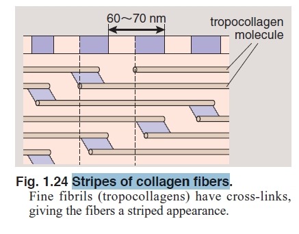
Collagen fiber molecules are produced in the rough endoplasmic reticulum of fibroblasts. Helical procollagens with three a-chains are secreted and the molecular ends are cut by procollagen peptidase to become tropocollagens. The molecules are crosslinked to each other with a regular gap that forms the striped collagen fiber (Fig. 1.23b).
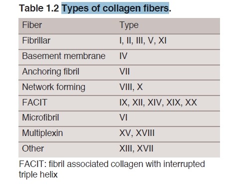
Twenty subtypes of collagen molecules with a-chains of different molecular structures are known to exist (Table 1.2); however, type I collagen accounts for 80% of the collagen fibers that make up the dermis. Reticular fibers, which distribute in the perivascular regions as thin argyrophilic fibers and do not form thick fiber bundles, are type III collagen, and these account for about 15% of all fibers. Most of the remainder is thought to be type V collagen. Types IV, VII and XVII are mainly found in the basal membranes, associated with epidermal keratinocytes.
2. Elastic fibers
The elastic fiber is not as tough as the collagen fiber; however, it is extremely elastic and found abundantly in the dermis of the scalp, face and the extensible organs such as arteries and tendons.
In the dermis, the deeper the elastic fiber, the thicker it is. In the reticular layers, elastic fibers are scattered among collagen bundles running parallel to the skin surface. The closer the elastic fiber is to the papillary layer, the thinner it is and the more perpendicular it is to the skin surface. It forms an arch shape in the papillary layer from which thin fibers are produced that perpendicularly reach the lamina densa.
Elastic fibers are also connected to the lamina densa of glands, sweat ducts, smooth muscle, nerves and blood vessels.
Elastic fibers are 1 mm to 3 mm in diameter. They cannot be differentiated from collagen fibers by HE staining. Elastic fibers stain dark blue to black in Weigert resorcin fuchsin, red-violet in aldehyde fuchsin, and brownish black in orcein. In elastic fibers, the characteristic striped pattern of collagen fibers is not observed by electron microscopy (Fig. 1.23). A skeletal fiber is 10 nm to 15 nm in diameter and its main content is fibrillin. The homogeneous substances are highly elastic structural proteins called elastins.
3. Ground substance, Matrix
Ground substance, a gelatinous amorphous substance of sugar and proteins, is observed in between fibers and between cells in the dermis. The components of ground substance are principally proteoglycans and glycoproteins whose molecular weight is 150,000 to 250,000 and whose sugar content is 2% to 15%. The molecules stabilize the fibers to give flexibility to the skin.
- 기질(Ground substance)은 sugar and protein의 젤과 같은 형태가 없는 물질(gelatinous amorphous substance)로 진피층에서 관찰됨.
- 기질 구성은 주로 프로테오글리칸과 글리코프로틴. molecular weight는 150,000 to 250,000. 이 물질들은 피부에 flexibility를 제공하고 fiber에 안정성을 제공.
Fibronectin, one kind of glycoprotein, contains a domain that connects fibrin, heparin and collagen; and binds integrin receptors on the cell surface are involved in cell proliferation, differentiation and wound healing. Besides these components, blood and lymph-derived tissue fluid forms the remainder of the ground substance that is involved in the transport of substances essential to cellar activities and metabolism.
- glycoprotein의 한 종류인 fibronectin은 피브린, 헤파린, 콜라겐으로 구성. 세포 표면에서 인테그린 receptor과 부착되어 있고 이는 wound healing시 cell proliferation, differentiation을 담당.
Proteoglycans are a massive molecules with a molecular weight of 105 to 106 or more and a composition of multiple glycosaminoglycans (mucopolysaccharides) connecting with backbone proteins. Glycosamine, mostly produced by fibroblasts in the dermis, is rich in hyaluronic acids, which are associated with moisture retention.
- 프로테오글리칸은
c. Cellular components
1. Fibroblast
Fibroblast differentiates from the mesenchymal cells and produces collagen fibers, elastic fibers, and glycosaminoglycans. Fibroblasts appear as thin spindle-shaped cells sparse in collagen fibers (Fig. 1.25). By electron microscopy, multiple Golgi apparatus and granular endoplasmic reticuli are seen in the fibroblast. When collagen fibers are produced and the dermis matures, fibroblasts stop their activities and become fibrocytes. At this point, the cell nuclei shrink and have fewer endoplasmic reticuli. Adrenal cortex hormones and thyroid hormones are involved in this process.

2. Histiocyte
The histiocyte, a kind of macrophage, is broadly distributed in the connective tissue and intermingles with fibroblasts on the outside of endocapillary cells (Fig. 1.26). A small circular nucleus and a large spindle- or star-shaped cell structure is seen in a histiocyte by light microscopy; furthermore, concave nuclei and the formation of psudopodial protrusions are observed by electron microscopy. Histiocytes contain Golgi apparatus, smooth and rough endoplasmic reticuli, and lysosomes. Lysosomes contain hydrolases and active acid phosphatases. The histiocyte releases collagenase and lysosomal enzymes containing elastase to digest the interstitium. It is involved in organ repair. The histiocyte degrades and phagocytoses mainly foreign substances and presents them as antigens to T cells (Chapter 3).
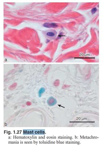
3. Mast cell
The mast cell is found in the dermis around capillaries and in the periphery of subcutaneous tissues. The shape is roundish or spindled, and the diameter is 10 mm (Fig. 1.27). Mast cells produce and maintain various vasodilatory and hyperlucent chemical mediators. Mast cell granules stain red-violet in toluidine blue and methylene blue, and present with metachromasia. Cutaneous mast cells resemble basophils in form and function; however, their characteristics differ slightly from those of other organs, because they differentiate in skin during intrauterine life.
A mast cell intracytoplasmic granule appears as a circular structure with a diameter of 0.3 mm to 0.5 mm under electron
microscopy. Multiple mast cell granules are evenly distributed in the cytoplasm. Chemotransmitters in the granules are released outside the cell under various stimuli, such as in type I allergic reactions (Fig. 1.28, Chapter 3).
The main components of the released substances are histamines and heparins, followed by various enzymes, including neutrophil chemotactic factors (NCF), eosinophil chemotactic factors of anaphylaxis (ECF-A), tryptase, chymase and tumor necrosis factor (TNF)-like substances. The mast cell may produce and release inflammatory substances such as prostaglandins, leukotorienes and plateletactivating factors.
4. Plasma cell
The plasma cell is a differentiated B cell that has been stimulated by an antigen. It produces antibodies and is involved in humoral immunity. The shape of the plasma cell varies from circular to pear-shaped, and the diameter ranges from 8 mm to 14 mm, which is twice as large as a leukocyte. It has a wheel-shaped nucleus with peripheral chromatin (Fig. 1.29).
5. Dermal dendrocyte
The dermal dendrocyte is found in the dermal upper layer (in and between the papillary layer and the reticular layer). It is thought to be an immunocompetent cell (Chapter 3), and it is characterized by containing clotting factor XIIIa.
d.Vascular channels and nerves
1. Blood vessels
Multiple branches of arteries distributed in skin (Figs. 1.30 and 1.31) are connected with each other in the dermal deep layer to form a horizontal network (subcutaneous plexus). With numerous branches ascending from the subcutaneous plexuses, the arteries form a second network in the papillary lower layer (subpapillary plexus). The arterioles ascend through the papillary layer, forming capillary loops in the dermal papillaries before moving to venules that connect to each other to form two kinds of plexuses, whereby the blood flows into the cutaneous veins (Fig. 1.30). There are also characteristic plexuses in the periphery of the cutaneous appendages. The peripheral regions of the eccrine glands are particularly rich in vascular networks, which control blood flow volume and body temperature by perspiration.
Moreover, hair follicles in the anagen (growth) stage are also richly supplied with blood vessels, present in the surrounding dermal tissue. There is another apparatus that circulates the blood directly from arteries to blood vessels: This is the arteriovenous anastomosis, which is controlled by sympathetic nerves. The arteriovenous anastomosis controls the peripheral blood flow and is involved in body temperature regulation. Glomus apparatuses, which have spherical anastomotic branches, are seen everywhere in the skin. They are particularly well developed in the fingers, at
apical ends of the toes, and below the nails. Many layers of smooth muscle cells (epithelial cells or glomus cells) cover the peripheral walls of the endothelial cells.
Under the electron microscope, endothelial cells, pericytes, and the multilayered basement membrane (lamina densa) in the peripheral interstitium may be observed in the capillaries. The stick-shaped Weibel-Palade granule, which has a diameter of 200 nm and a length of 1 mm or less, contains factor VIII associated with histamines and blood coagulation. It is found in the endothelial cell. Pericytes have a vasoconstrictive effect, and they are seen on the perimeter of the walls of the endothelial cells.
2. Lymphatic vessel
Lymph vessels are distributed around the subpapillary layer region and extend through the postcapillary lymph vessels to the dermal and subcutaneous lymph vessels. The endothelial cells of the lymph capillaries are thin, without pericytes or lamina densa. They are partly ruptured and are surrounded by loose collagen fibers and elastic fibers (Fig. 1.32). The closer the endothelium is to the dermal deep layer, the more continuous it becomes with the valva in the lumen. The structure of lymph vessels is not as regular as that of the blood vessels. Aggregated cutaneous lymphatic fluid passes through the regional lymph nodes and flows into the blood vessels.
3. Nervous system
The nerve fiber bundle is covered with a membrane in the dermal lower layers. The nerve fibers change from being myelinated to non-myeliated where the nerve bundle branches into many fibers in the dermis, and these branched fibers are distributed within the superficial dermis and peripheral appendages (Fig. 1.33). The sensory nerves transmit tactile, pressure, pain and temperature sensation. The autonomic nerves control the blood vessels, sweat glands and other appendages.
1) Sensory nerve
The sensory nerve structures include free nerve endings sensing pain, Merkel cells (described above) that perceive tactile sensation in the epidermal basal layer, and nerve end bulbs that accept tactile, pressure and vibration sensation.
Free nerve ending The free nerve endings are distributed in the dermal upper and papillary layers. Some of them adhere to Merkel cells in the dermal papillary layer, whereas others infiltrate into the dermis directly. Nonmyelinated nerves transmit pain sensations.
End corpuscle
The end corpuscle is a specific sensory nerve terminal covered with a membrane. Various end corpuscles are described below.
Meissner end corpuscle: The nerve fiber spirally ascends through the Schwann cell (inner bulb cell) in the dermal papillae
of the palms, soles, lips of the mouth, and external genitals, perceiving tactile and pressure sensations (Fig. 1.34).
Pacinian corpuscle: It is seen in the dermal deep layer and subcutaneous tissue of the palms, soles, and external genitals. The
central nerve fiber is multi-layered with concentric membranes. It is oval, with a major axis of 1 mm, and can be clearly seen by light microscopy (Fig. 1.35). It reacts to vibration.
2) Autonomic nerves
The autonomic nerves are principally distributed in the sweat glands, arrector pili muscles, blood vessels and glomus apparatuses, to control the functions of these organs. The cholinergic nonmyelinated sympathetic nerves are distributed in the eccrine
sweat glands. Mitochondria, and dense core and non-core vesicles containing chemical substances are observed. The adrenergic
sympathetic nerves are distributed in the arrector pili muscles and blood vessels.
- 근막유착에서 발생하는 통증을 이해할 수 있는 자료