드디어 발 촉진의 기준점, 반드시 기억해야할 landmark를 찾았다.
대박
panic bird....
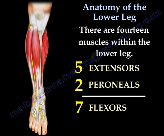
lower leg 14개의 근육
5 extensor
2 peroneals
7 flexor
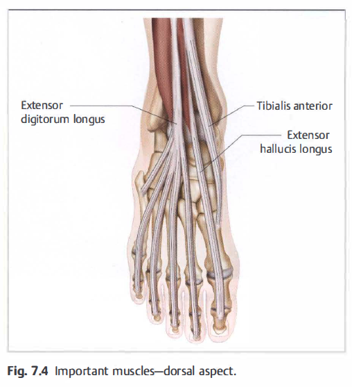
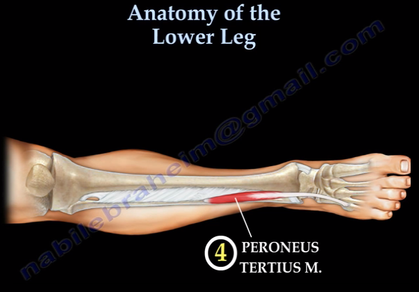
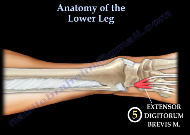
lower leg의 5 extensor
1) tibialis anterior
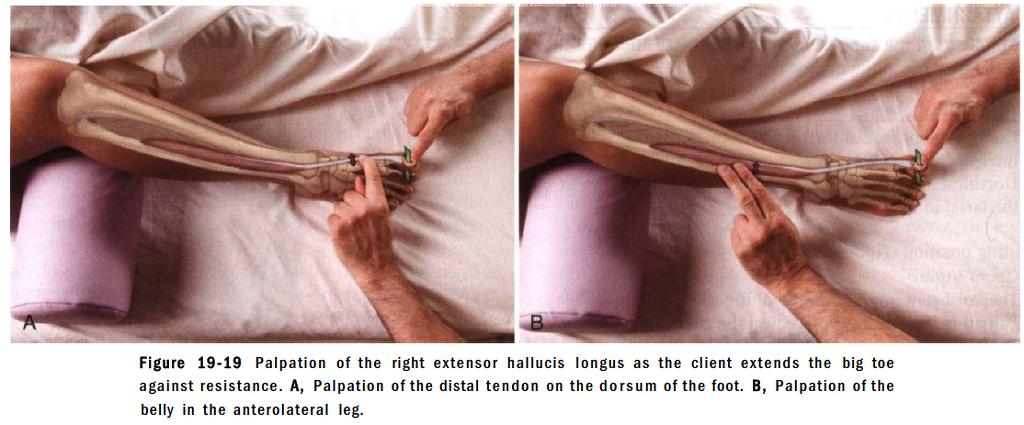
2) extensor hallucis longus
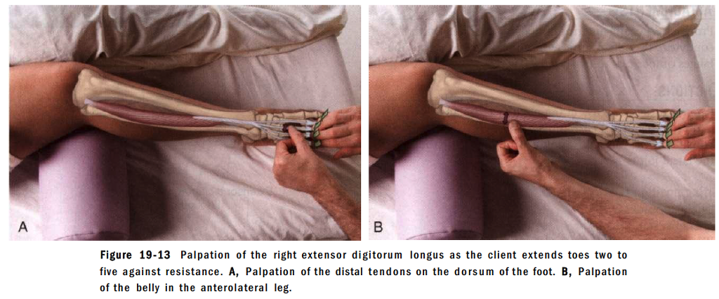
3) extensor digitorum longus
4) peroneus tertius msucle
5) extensor digitorum brevis
주요 촉진 가능한 부위
talus, navicualr tuberosity, calcaneous sustentaculum tali, 1st cuneiform, 1st metasal bone and cuneiform joint line
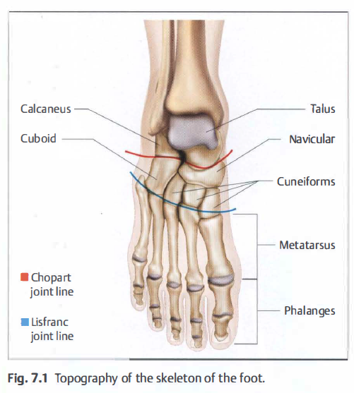
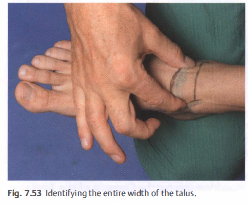
거골의 촉진
전경골근과 장무지신근
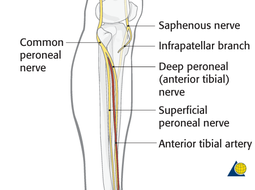
anterior tibial artery 촉진 가 능
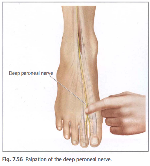
장무지신근과 장지신근 사이에 deep peroneal nerve 촉진
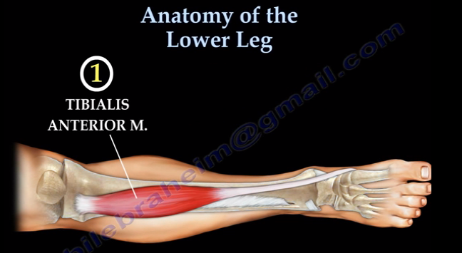
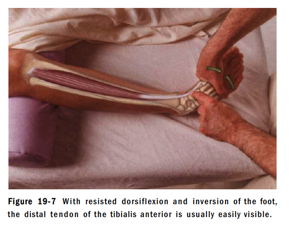
tibialis anterior 촉진법
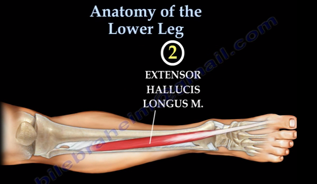
extensor hallucis longus촉진법
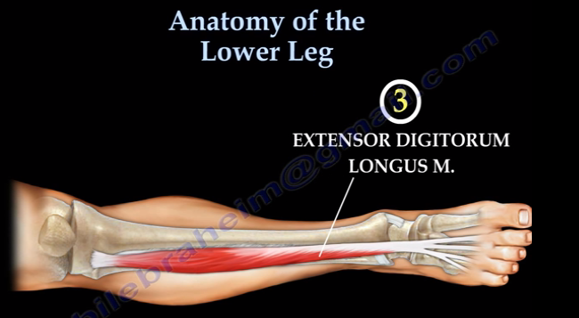
extensor digitorum longus 촉진법
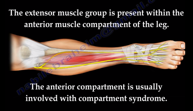
전방구획(anterior compartment)
deep peroneal nerve의 지배를 받는 구획을 가진 부위(근막)
전방구획증후군이 흔하게 발생하는 부위.
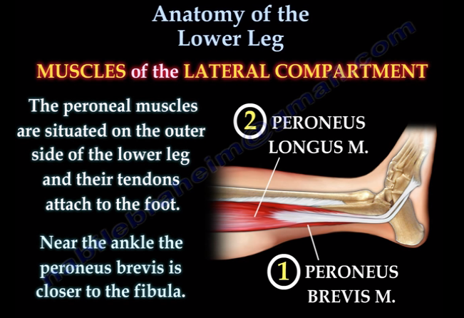
lower leg의 2 peroneal muscle
peroneal longus and brevis msucle
superficial peroneal nerve의 지배를 받는 구획
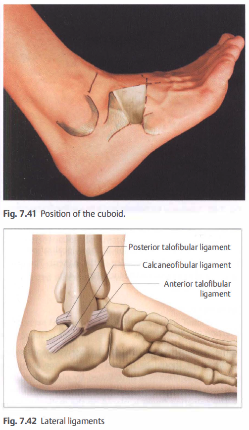
발목바깥쪽 촉진의 landmark
lateral mallelous
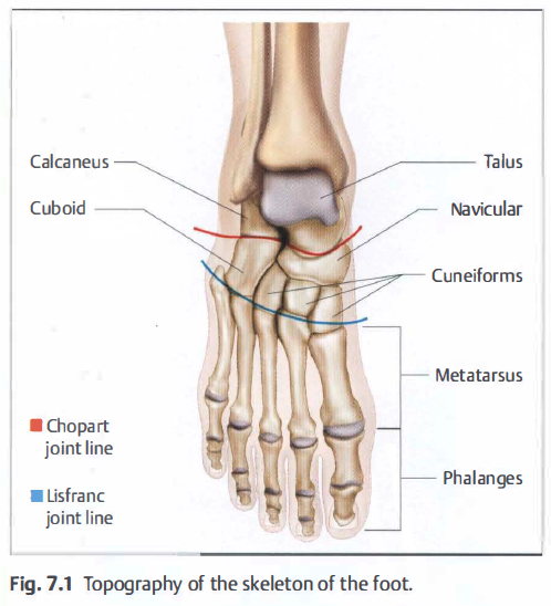
peroneal tubercle of calcaneous
5th metatarsal head
tarsal canal
- 족근동증후군의 위치
- 종골과 거골이 만나 이루는 발의 내외측으로 이어지는 canal
- 내측으로는 sustentaculum tali위쪽으로 이어짐.
- interosseous talocalcaneal ligament가 있어 신경이 많이 지배하는 부위
The subtalar or talocalcaneal joint consists of two separate parts, divided by the tarsal canal (Fig. 80.8), which is funnel shaped with the wide portion at its lateral end. The lateral end of the canal is easily palpated in front of the fibular malleolus between talus and calcaneus, especially when the foot is inverted. The canal runs posteromedially, to have its medial opening just behind and above the sustentaculum tali. In the canal a strong ligament, the interosseous talocalcaneal ligament, binds the two bones.
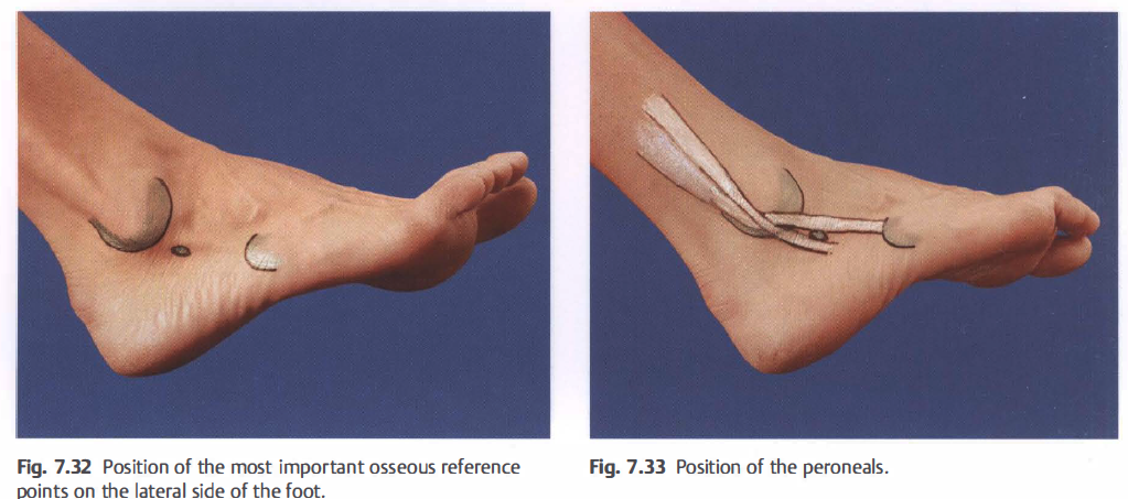
peroneal tublecle = trochlear process of calcareous 는 중요한 촉진 landmark.
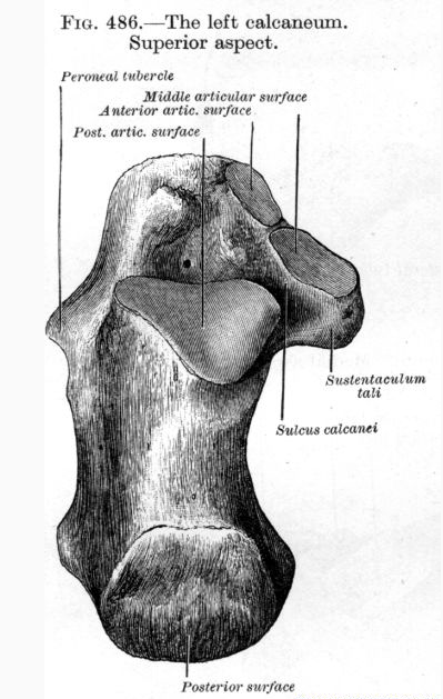
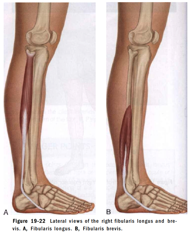
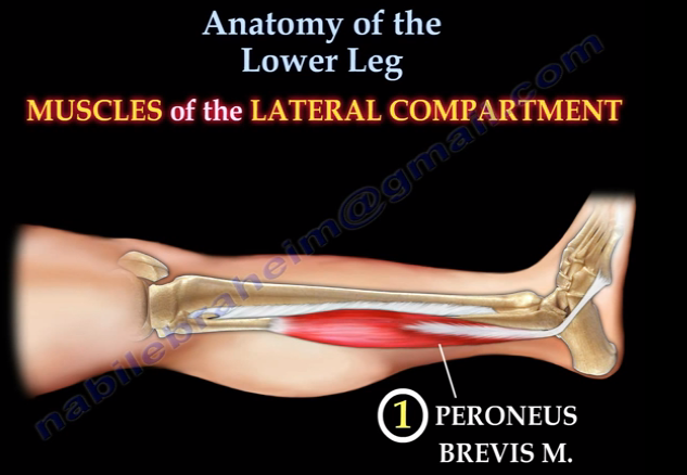
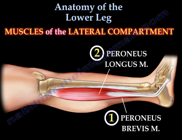
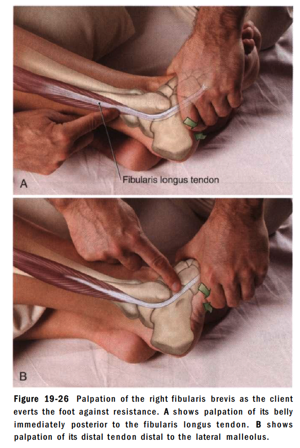
peroneus longus and brevis촉진법
lower leg의 7 flexor muscle
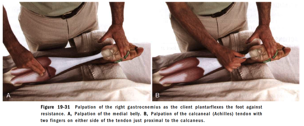
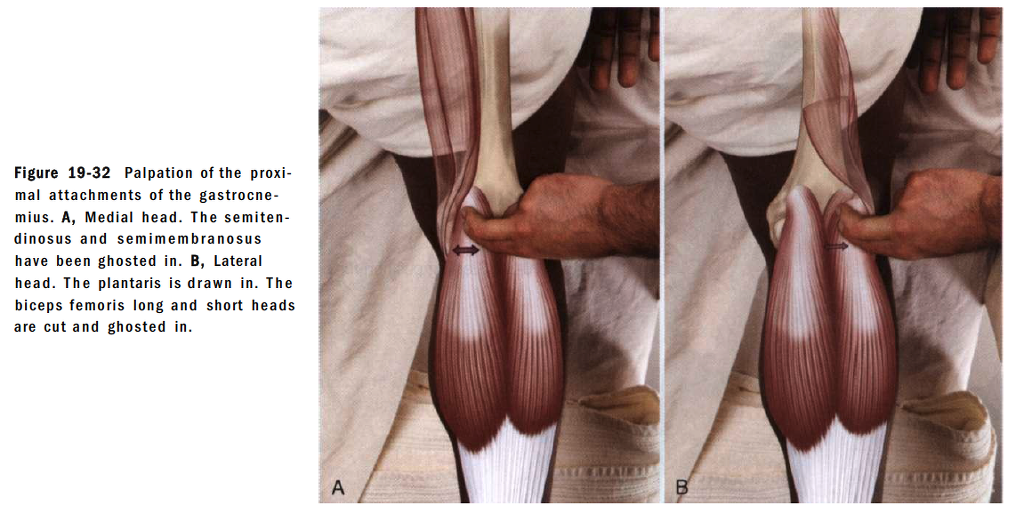
1) gastronemius
2) soleus
3) plantaris msucle
4) popliteus muslce
5) tibialis posterior
6) flexor hallucis longus
7) flexor digitorum longus
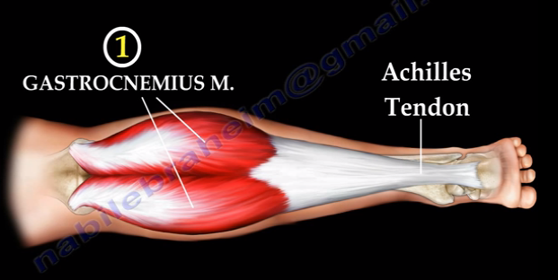
gastrocnemius 촉진법
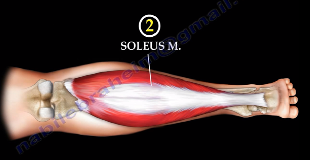
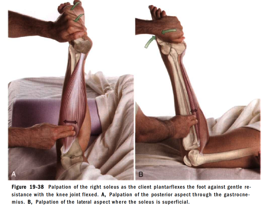
soleus촉진법
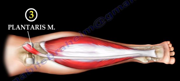
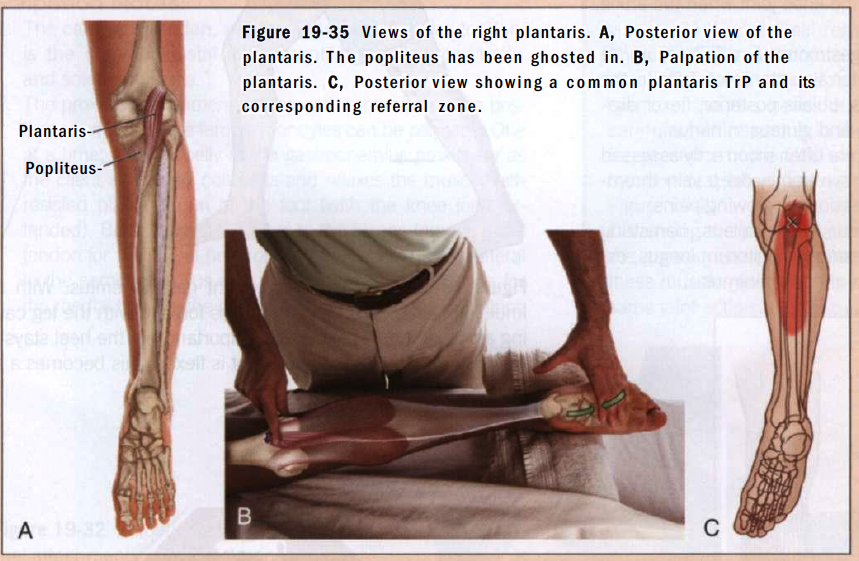
plantaris 촉진법
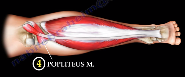
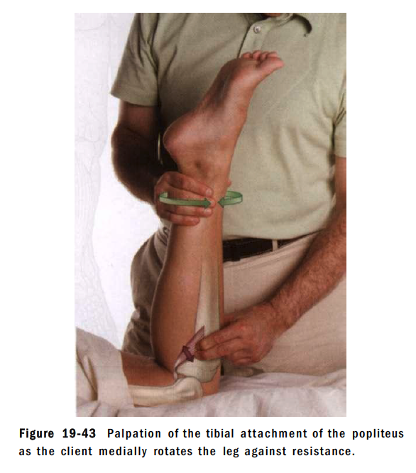
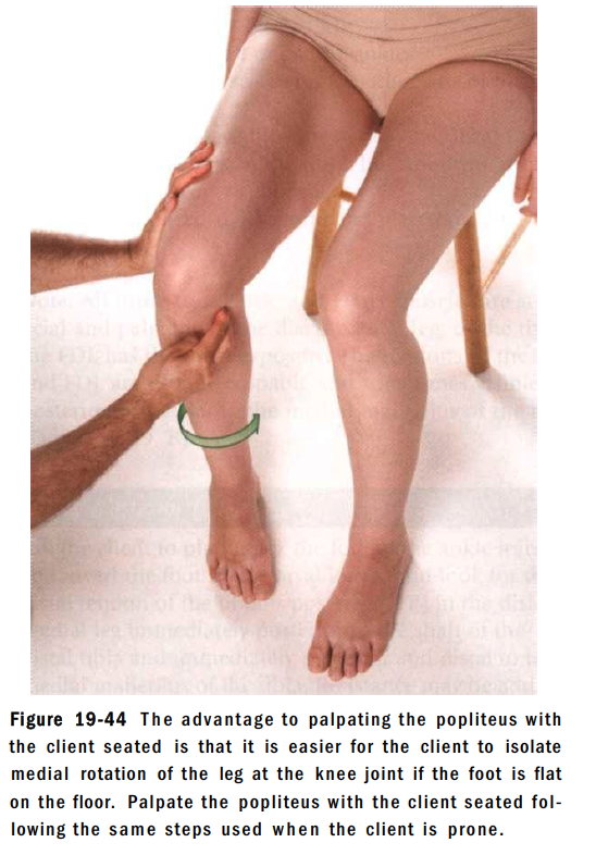
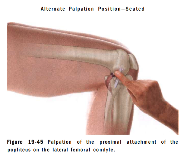
popliteus muscle 촉진법
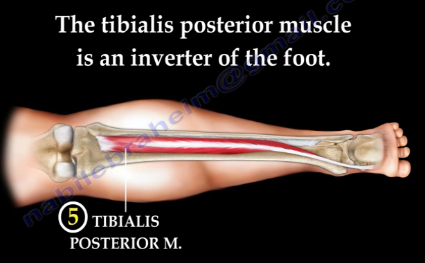
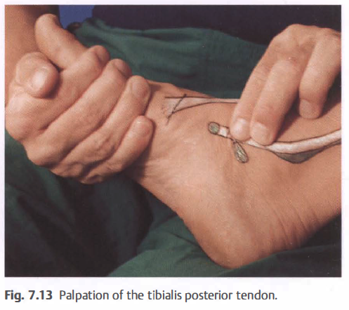
tibialis posterior 촉진법
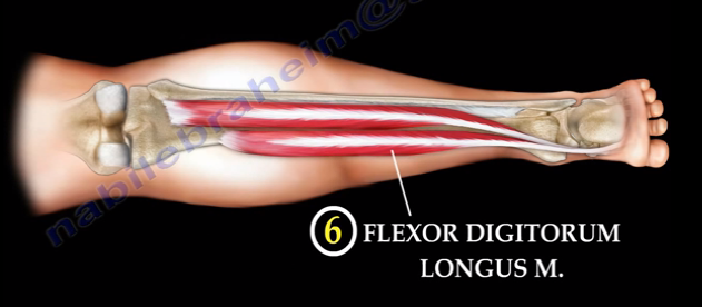
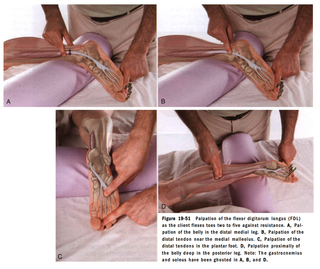
flexor digitorum longus촉진법
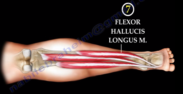
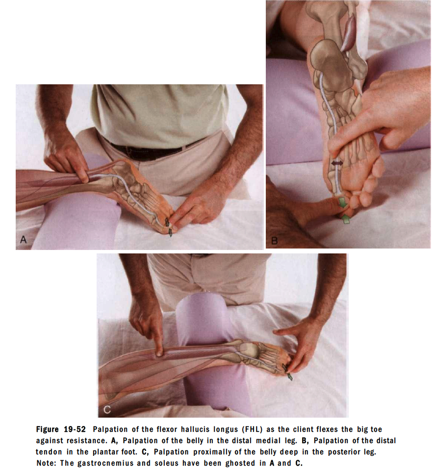
flexor hallucis longus 촉진법
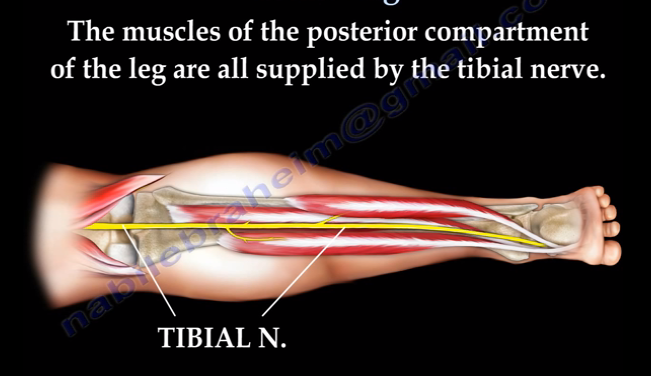
7개의 굴곡근은 후방구획으로 나뉘고 tibial nerve가 지배함.
후방구획은 superficial compartment와 deep compartment로 두개의 구획으로 나뉨.
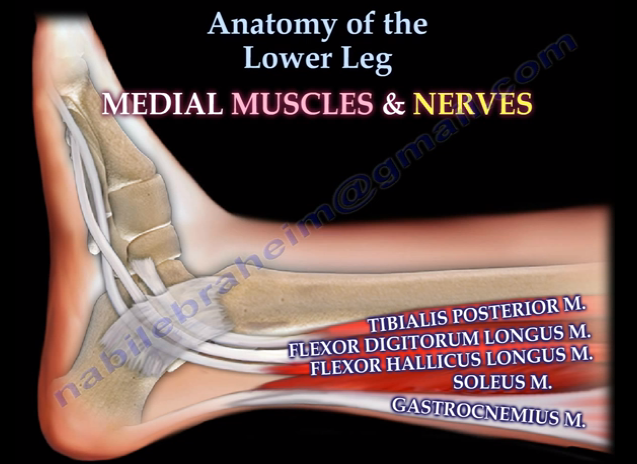
이 구조를 내측에서 다시 분석
1) tibialis posterior
2) flexor digitorum longus
3) flexor hallucis longus
4) soleus
5) gastronemius
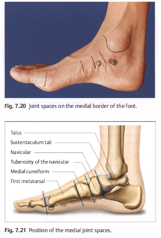

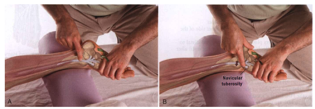
주상골 tuberoritiy와 종골의 sustentaculum tali를 기준으로
tibialis posterior tendon은 주상골 tuberosity에 부착함.
flexor digitorum longus는 사이로 지나감
flexor hallucis longus는 sustentaculum tali아래에서 촉진