viscerosomatic reflex, somatovisceral reflex의 관점에서
mind and body connection관점에서
psychomatic disease관점에서
설명하기 힘든 각종 통증을 이해하기 위한 관점에서
지금 이 자료를 잘 공부해야 한다.
이 간단하게 정리된 자료를 바탕으로 정확한 review논문을 찾아들어가기로 한다.
All dorsal and ventral roots at all spinal levels contain SOMATIC nerve fibers-
Dorsal roots at T1-L2 will also contain returning afferent sympathetic fibers
Ventral root at T1-L2 will also contain outgoing efferent sympathetic preganglionic fibers
Dorsal roots at S2, S3, S4 will also contain incoming afferent parasympathetic fibers
Ventral roots at S2, S3, S4 will also contain outgoing efferent preganglionic parasympathetic fibers
- 모든 dorsal and ventral 신경근은 척수레벨에서 체성신경섬유를 포함함.
- 흉추 1에서 요추 2번까지 dorsal root는 또한 되돌아오는 구심 교감신경섬유를 포함함.
- 흉추 1에서 요추 2번까지 ventral root는 또한 밖으로 나가는 원심성 교감신경 preganglionic 섬유를 포함함.
- 천추 2, 3, 4에서 dorsal root는 또한 들어오는 구심성 부교감신경 섬유를 포함함.
- 천추 2, 3, 4에서 ventral root는 또한 밖으로 나가는 원심성 preganlionic 부교감신경 섬유를 포함함.
panic bird...
1) Definitions
Ganglion-is a collection of cell bodies outside the central nervous system.
a- A dorsal root ganglion is a collection of afferent (sensory) cell bodies.
b-A sympathetic (paravertebral or prevertebral) ganglion is a collection of cell bodies of postganglionic sympathetic neurons
c- A parasympathetic (terminal / visceral OR head & neck) ganglion is a collection of cell bodies of postganglionic para-sympathetic neurons. b & c are also synapse sites but the term ganglion refers to a collection of cell bodies.
- 강글리온은 중추신경계 바깥에 존재하는 세포체의 집합
- 구심성 세포체의 집합은 dorsal root ganglion
- 교감신경 강글리온은 postganglionic 교감 신경 세포체의 집합.
- 부교감신경 강글리온은 postganglionic 부교감 신경 세포체의 집합
- 교감신경, 부교감신경 강글리온은 또한 synapse site 하지만 세포체의 집합으로 언급됨.
Nucleus-is a collection of cell bodies inside the central nervous system. These will be mentioned in Head & Neck section of Unit II-but will be studied in detail in neuroscience.
- 신경세포 핵은 중추신경계 내측에 있는 세포집합체(cell body)
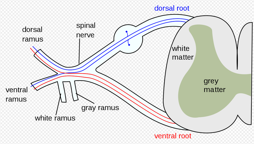
Roots- this term is used to describe:
The afferent pathway into spinal cord: dorsal root (dorsal root of the spinal nerve)
The efferent pathway out of the spinal cord: ventral root (ventral root of the spinal nerve)
The ventral primary ramus of a spinal nerve as it exits the vertebral canal & is named by the corresponding vertebral level is C5 root L4 root.
- 신경근
- 척수로 전달되는 구심신경로 : dorsal root
- 척수바깥으로 전달되는 원심신경로 ; ventral root
All dorsal and ventral roots at all spinal levels contain SOMATIC nerve fibers-
Dorsal roots at T1-L2 will also contain returning afferent sympathetic fibers
Ventral root at T1-L2 will also contain outgoing efferent sympathetic preganglionic fibers
Dorsal roots at S2, S3, S4 will also contain incoming afferent parasympathetic fibers
Ventral roots at S2, S3, S4 will also contain outgoing efferent preganglionic parasympathetic fibers
- 모든 dorsal and ventral 신경근은 척수레벨에서 체성신경섬유를 포함함.
- 흉추 1에서 요추 2번까지 dorsal root는 또한 되돌아오는 구심 교감신경섬유를 포함함.
- 흉추 1에서 요추 2번까지 ventral root는 또한 밖으로 나가는 원심성 교감신경 preganglionic 섬유를 포함함.
- 천추 2, 3, 4에서 dorsal root는 또한 들어오는 구심성 부교감신경 섬유를 포함함.
- 천추 2, 3, 4에서 ventral root는 또한 밖으로 나가는 원심성 preganlionic 부교감신경 섬유를 포함함.
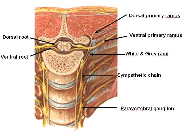
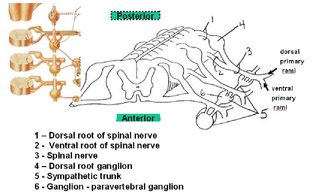
The dorsal and ventral roots join to form the spinal nerves. Spinal nerves then give off a dorsal primary rami and a ventral primary rami. The ventral rami may join to form a plexus as in cervical plexus, brachial plexus, lumbar plexus, sacral plexus; or form segmental spinal nerves like intercostals and segmental lumber nerves. The ventral rami are called roots based on the vertebral level at which they exit the spine.
- 배측과 복측 신경근은 합해져서 spinal verve를 만듬.
- 척수신경은 dorsal primary rami와 ventral primary rami를 give off
- ventral 가지는 신경총을 만듬(cervical plexus, brachial plexus, lumbar plexus, sacral plexus등)
Ramus Rami
In the neuroanatomy of the spinal cord there are 2 different types of rami
Dorsal & Ventral rami (divisions of a spinal nerve)
White and Grey rami (pathway in and out of the sympathetic chain)
DORSAL & VENTRAL RAMI
Dorsal ramus (dorsal primary ramus) is the dorsal branch of a spinal nerve which innervates posterior structures (e.g. back). It contains somatic sensory, somatic motor, sympathetic efferents (to sweat glands, blood vessels, erector pili, etc of the back) and sympathetic afferents.
- 배측 가지는 뒤쪽 구조를 신경지배하는 dorsal branch.
- 그것은 체성 감각, 운동, 교감 원심성 섬유와 교감 구심성 섬유를 포함함.
Ventral ramus (ventral primary rami) is the ventral or anterior branch of the spinal nerve. Like the dorsal ramus it contains somatic sensory, somatic motor, sympathetic efferents, and sympathetic afferents.
Spinal nerves do not carry para-sympathetic fibers. So there are no parasympathetic fibers passing through the dorsal or ventral rami.
- 복측가지는 척수신경의 전방가지로 배측가지와 마찬가지로 체성 감가, 운동, 교감신경의 구심, 원심섬유를 포함함.
- 척수신경은 부교감신경 섬유를 운반하지 않음. 그래서 부교감신경 섬유는 배측 또는 복측 가지를 통해 통과하지 않음.
WHITE & GREY RAMI
The white ramus (white rami communicans) connects the spinal cord with the sympathetic chain. All afferent and efferent sympathetic fibers returning to the spinal cord from the sympathetic chain or exiting from the spinal cord to the sympathetic chain travel through white rami. Since the sympathetic nervous system enters and exits the spinal cord at T1-L2 (thoraco-lumbar outflow) . there are white rami only at T1-L2.
- 흰색가지 연결체는 척수와 교감신경을 연결함.
- 모든 구심과 원심 교감신경섬유는 교감신경 사슬로부터 척수로 되돌아감.
- 교감신경계는 흉추 1-요추2번에서 척수로 연결되기 때문에 흰색연결체가 흉추 1-요추 2번에 존재함.
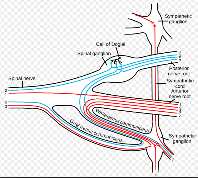
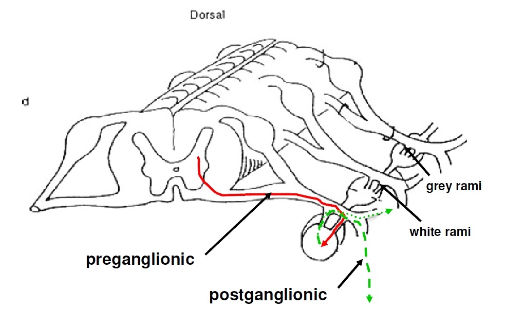
Efferent fibers: All preganglionic efferent sympathetic fibers exit the spinal cord (thoraco-lumbar outflow) via a ventral root and enter the sympathetic chain via a white rami. These are myelinated fibers hence the name white rami. Once in these preganglionic efferent fibers are enter the sympathetic chain there are then several options for the efferent signal to reach the desired target (see exit options below) .
- 원심섬유
- 모든 preganglionic 원심성 교감신경 섬유는 ...
Afferent fibers: Afferent sympathetic fibers returning from the target site enter the sympathetic chain. They then pass through the white rami and into the dorsal root and into the spinal cord... These afferent sympathetic fibers have their cell bodies in the dorsal root ganglion (like SOMATIC sensory fibers)
White rami therefore contain: efferent, preganglionic, myelinated, sympathetic fibers afferent sympathetic fibers.
- 흰색가지 : 원심성, preganglionic, 유수신경, 교감신경 구심섬유
The grey ramus (grey rami communicans) connects the sympathetic chain with the spinal nerves. All sympathetic fibers going to the skin and musculo-skeletal system leave the sympathetic chain via the grey ramus and enter a spinal nerve. While there are white rami only at T1-L2 there are grey rami at all levels of the sympathetic chain.
Efferent fibers follow Exit Option # 1 (below).
- 회색 가지는 척수와 함게 교감신경을 연결함
- 모든 교감신경 섬유는 피부와 근골격계로 이어져 회색가지를 거쳐 교감신경 사슬을 떠나 척수신경으로 들어감.
Afferent fibers: afferent sympathetic fibers returning from peripheral sites travel with the peripheral nerves back to the spinal nerves. They then exit the spinal nerve and enter the sympathetic chain via the grey ramus. They then exit the sympathetic chain via the white ramus and enter the spinal cord via the dorsal root.
Grey rami therefore contain: efferent, post ganglionic, unmyelinated, sympathetic fibers afferent sympathetic fibers.
- 회색가지 : 원심성, post ganglionic, 무수신경, 교감신경 구심섬유
In the cervical spine region there is usually a superior, middle, and inferior sympathetic ganglion (as the ganglia at some adjacent spinal levels may fuse together). These cervical ganglia would each therefore need to send fibers to spinal nerves at more than one level. As such, each cervical ganglia may have several grey rami. (e.g. middle cervical ganglion sends fibers to C4, C5, and C6 spinal nerves so it may have grey rami going to each of these spinal nerves).
Plexus-collection of nerve fibers.
Simplistically, a plexus is like a series of complex interstate interchanges. Nerve fibers are coming and going, changing lanes, entering and exiting the plexus to get where they need to go. In Unit I you have studied the brachial and lumbo-sacral plexus. A plexus can have several different types of fibers passing through it
e.g.--the brachial plexus contains somatic and sympathetic efferent and afferent fibers coming from and going to the upper extremity;
e.g. the cardiac plexus has both sympathetic and parasympathetic efferents and afferents coming to and going from the heart. The cardiac, pulmonary, esophageal and other plexi in the chest and abdomen are a jumble of all four type of autonomic nerve fibers (efferent and afferent sympathetic & efferent and afferent parasympathetic). You cannot look at these fibers at the gross anatomy level and tell which is which and it would be also extremely difficult to dissect these fibers out and trace them back to their source.
Cranial Nerves-- there are twelve cranial nerves
Cranial nerves III, VII, IX, X are part of the parasympathetic system (i.e. the cranial portion of the cranialsacral
outflow). The cranial nerves serve many functions. Some are purely sensory (e.g. II optic nerve) others carry multiple types of fibers (somatic, sympathetic, parasympathetic, special sensory). Some carry fibers which originate in other cranial nerves.
- 뇌신경은 12개
- 뇌신경 III, VII, IX, X 은 부교감신경 시스템
- 시신경은 순수한 감각신경, 다른 신경은 체신경, 교감신경, 부교감신경, 특수감각을 다루는 신경.
2) Autonomic afferent and efferent fibers.
Both sympathetic and parasympathetic systems have afferent and efferent fibers. Afferent autonomic sensory fibers: The afferent fibers for both sympathetic and parasympathetic have ONLY one fiber. The afferent autonomic fibers return sensory or feedback information on the autonomic nervous system to the spinal cord or directly to the brain (for some parasympathetic fibers). Afferent autonomic fibers returning to the spinal cord have their cell bodies in the dorsal root ganglion. Afferent autonomic fibers returning to the brain have their cell bodies in the brain (nucleus).
- 교감과 부교감신경 시스템은 둘다 구심과 원심섬유를 가짐.
- 구심 자율 감각신경섬유 : 교감과 부교감 신경을 위한 구심섬유는 오직 하나의 섬유를 가짐
- ...
Afferent autonomic sensory fibers returning to the spinal cord or brain will follow the same general path back from the target site as the efferent fibers followed to the target site. They do follow separate paths as they exit or enter the spinal cord (efferent fibers exit the spinal cord in the ventral roots & afferent fibers enter the spinal cord in the dorsal roots). This anatomy is similar for the brain (afferent fibers return via sensory nerves to sensory nuclei in the central nervous system while the efferent motor fibers exit from motor nuclei via motor nerves)
Efferent autonomic fibers: The efferent system for both sympathetic and parasympathetic has TWO nerve fibers (pre-ganglionic fiber and post ganglionic fiber).
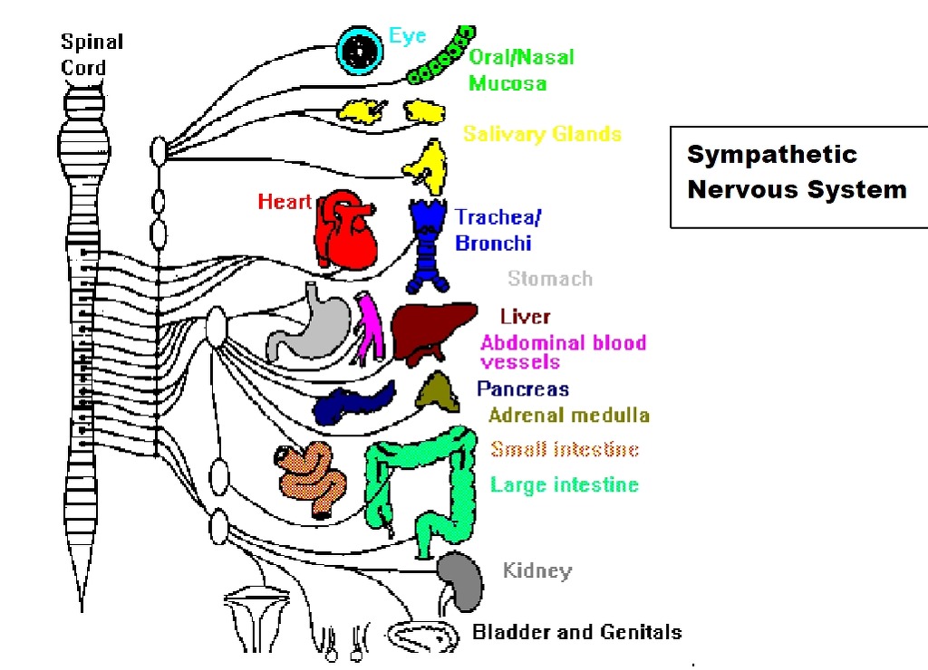
Sympathetic: Sympathetic efferent fibers exit the spinal cord at the T1-L2 levels (thoraco-lumbar outflow) via the ventral root. They then enter the sympathetic chain via the WHITE rami. From this point there are four main distribution options which will be discussed below.
Parasympathetic: Parasympathetic efferent fibers exit: the central nervous system via cranial nerves III, VII, IX, or X.
the sacral cord at the S2-S4 level
3) Sympathetic Pathways
All efferent sympathetic fibers exit the spinal cord at T1-L2 via the ventral roots and then enter the sympathetic chain via the white rami. Once in the sympathetic chain these preganglionic efferent fibers have four potential options for "exiting" the sympathetic chain. Before exiting, these preganglionic fibers can travel up or down the chain or stay at the same vertebral level as they entered. Remember these preganglionic fibers can synapse with and therefore activate many postganglionic fibers and commonly do so at multiple levels.
Exit Option # 1: The pre-ganglionic fiber enters the sympathetic chain via the white rami. The preganglionic fiber may ascend or descend the sympathetic chain or stay at the same vertebral level it entered. This preganglionic fiber synapses in the paravertebral ganglion which is part of the sympathetic chain. The post ganglionic sympathetic fiber then exits the sympathetic chain via a grey rami and joins the spinal nerve. These fibers may then travel in either the dorsal primary rami (to reach structures of the back) or in a ventral primary rami (to reach ventral structures) This is the pathway for innervation of the skin and musculo-skeletal system (UNIT I & as shown in the example drawing above).
Example of this pathwayBSympathetic innervation of the radial artery at the wrist.
Efferent preganglionic fiber leaves ventral spinal cord at the upper thoracic level (thoraco-lumbar
outflow) via the ventral root.
Enters sympathetic chain via the WHITE rami
Passes up the sympathetic chain to the C6 vertebral level (or possibly C5 or C7)
Synapses in the paravertebral ganglion at one of these levels
Post ganglionic sympathetic efferent fiber leaves sympathetic chain via the GREY rami and enters
spinal nerve
The post ganglionic fiber then travels in spinal nerve, into ventral primary rami, into the brachial
plexus, into median nerve, down arm to level of the radial artery in the forearm, travels along the
radial artery to its target destination.
Afferent fibers returning from this target site would return via the same pathway except they would use the
dorsal root to enter the spinal cord.
Target site on radial artery ÷median nerve ÷ brachial plexus ÷ C 6 cervical root ÷ ventral primary ramus
÷ spinal nerve ÷ grey ramus ÷ sympathetic chain ÷ white ramus ÷ dorsal root (with cell body in dorsal
root ganglion) ÷ spinal cord
(no synapse-remember afferents are single fiber with cell body in the dorsal root ganglion)
Exit option # 2: The preganglionic fiber enters the sympathetic chain via the white rami. It then
synapses in the paravertebral ganglion (above or below or at the level of entry).
The post ganglionic fiber then leaves the ganglion "directly" via sympathetic fibers (NOT by the grey rami) .
For sympathetic innervation of the thorax, these postganglionic fibers form groups of postganglionic fibers
traveling to specific organs (e.g. cardiac nerves).
Example of this pathwayBSympathetic innervation of the heart.
Efferent preganglionic fiber leaves ventral spinal cord at the upper thoracic level (thoraco-lumbar
outflow) via the ventral root.
Enters sympathetic chain via the WHITE rami
Passes up the sympathetic chain to one of the cervical ganglia
Synapses in the paravertebral ganglion at one of these levels
Post ganglionic sympathetic efferent fiber leaves sympathetic chain directly (NOT via grey rami)
and joins with other similar post ganglionic fibers. These fibers travel to the heart as cardiac
nerves (or cardiac splanchnic nerves) .
Afferent fibers returning from this target site would return via the same pathway except they have
no synapse and would use the dorsal root to enter the spinal cord.
For sympathetic innervation of the head and neck these postganglionic fibers travel from the cervical
ganglion (usually the superior cervical ganglion) along major blood vessels supplying the head and neck.
A large number of fibers travel along the carotid artery-hence the term carotid plexus. These post
ganglionic sympathetic fibers traveling via the carotid plexus can then reach their target destination in
several ways
a- on the surface of branches of the carotid (internal and external) artery
b- leave the carotid plexus & travel with other nerves including somatic and parasympathetic fibers
c- leave the carotid plexus and go directly to their destination
d- leave the carotid plexus as an identifiable sympathetic nerve (deep petrosal nerve) which then
merges with other nerves to reach its destination (e.g. sympathetic innervation of the lacrimal
gland)
Example of this pathwayBSympathetic innervation of the eye (disruption of this pathway results in Horner=s
syndrome)
Efferent preganglionic fiber leaves ventral spinal cord at the upper thoracic level (thoraco-lumbar
outflow) via the ventral root.
Enters sympathetic chain via the WHITE rami
Passes up the sympathetic chain the superior cervical ganglion
Synapses in the superior cervical ganglion
Post ganglionic sympathetic efferent fiber leaves sympathetic chain directly (NOT via grey rami)
and joins with other similar fibers and travels on the surface of the carotid artery(carotid plexus) to
join with ciliary branches of the opthalmic nerve (branch of CN V) to reach the eye.
Afferent fibers returning from this target site would return via the same pathway except they have
no synapse and would use the dorsal root to enter the spinal cord
Exit option # 3: Pathway for sympathetic innervation of the abdomen.
The preganglionic fiber enters the sympathetic chain via the white rami. This preganglionic fiber may
ascend or descend before exiting or may exit at the same level of entry.
This preganglionic fiber then exits the sympathetic chain (without synapse) and joins with other
preganglionic fibers doing the same thing.
These preganglionic fibers form the splanchnic nerves (e.g. greater, lesser, and least splanchnic nerves)
which travel to the pre-vertebral ganglia in the abdomen (e.g. celiac, superior mesenteric, inferior
mesenteric and aorto-renal ganglia.)
These preganglionic sympathetic fibers then synapse in these ganglia and the postganglionic sympathetic
fibers travel to various organs.
These post ganglionic fibers may travel in groups of postganglionic fibers and/or along the surface of blood
vessels to reach their target. This is the way the sympathetic system innervates the abdominal and pelvic
organs (Unit III)
Example of this pathwayBSympathetic innervation of the stomach.
Efferent preganglionic fiber leaves ventral spinal cord at the mid to lower thoracic level (thoracolumbar
outflow) via the ventral root.
Enters sympathetic chain via the WHITE rami
Exits the sympathetic chain without synapse
Travels with other similar fibers as part of the greater splanchnic nerve
Enters prevertebral ganglion (celiac ganglion)
Synapse in the celiac ganglion
Post-ganglionic fibers travel along branches of the celiac trunk to the stomach
Afferent fibers returning from this target site would return via the same pathway except they have
no synapse and would use the dorsal root to enter the spinal cord
Exit option # 4: This is the pathway for sympathetic innervation of the adrenal gland. This pathway is
the exception to the autonomic efferent two nerve system as there is only one nerve fiber.
This nerve fiber exits the sympathetic chain the same as exit option # 3. These fibers then travel in
splanchnic nerves but pass through prevertebral ganglia without synapse and go directly to the adrenal
gland. The adrenal gland is directly activated (ie no synapse and therefore no ganglion).
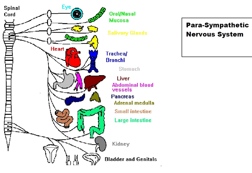
4) Para - Sympathetic Pathways
exit via the cranial outflow Cranial Nerves III, VII, IX, X
exit via the sacral outflow S2-S4
NO parasympathetics to the musculo-skeletal system
Parasympathetic pathways to the chest and abdomen
Vagus nerve (CN X)
Preganglionic fibers exit the brain stem (cell bodies are in the bran stem) and travel in the
vagus nerve
Preganglionic parasympathetic fibers to target organ
Synapse on or near target organ in terminal ganglion
Postganglionic parasympathetic fibers to target
Afferent vagal fibers returning from these target sites in the chest and abdomen would
return via the vagus nerve (they would have no synapse and the cell body of the sensory
fiber would be in the brain stem)
Vagus nerve (CN X) provides parasympathetic supply to chest/thorax and abdomen (to the level of
the descending colon
Sacral outflow
Preganglionic fibers from S2, S3, S4 travel as pelvic splanchnic nerves to lower abdominal,
pelvic, and perineal target organs
Synapse on or near target organ in terminal ganglion
Postganglionic parasympathetic fibers to target
NOTE: the term pelvic splanchnic nerves refers specifically to preganglionic parasympathetic fibers from
the sacral outflow. Splanchnic nerves elsewhere carry sympathetic fibers (e.g. greater splanchnic, lesser
splanchnic, etc)
Parasympathetic pathways in the head and neck
Parasympathetic pre-ganglionic fibers exit the brain stem as part of cranial nerves III, VII, IX, and X
(NOTE: These cranial nerves carry other types of fibers in addition to parasympathetic fibers)
These preganglionic parasympathetic fibers from CN III, VII, and IX travel to parasympathetic
ganglion found in the head. (parasympathetic fibers carried by CN X go to the chest and
abdomen). These ganglion are:
1) ciliary ganglion
2) pterygopalatine ganglion
3) otic ganglion
4) submandibular ganglion
The pre-ganglionic parasympathetic fibers synapse in these ganglia and the postganglionic
parasympathetic fibers continue on to the target site.
Usually these post ganglionic parasympathetic fibers will travel along with other nerve fibers going
to the same target location.