수근관 증후군, 가이온터널 증후군, 협착성 건초염, TFCC, 손가락 퇴행성 관절염(Herberden's node, bouchard's node), 류마티스 관절염, 손가락 염좌, 듀프이렌트(Dupuytren's contracture) 구축, 콜래스 골절 등
손은 미세한 움직임, 강한 움직임이 동시에 필요하고
움직임의 역학적 이해가 필수인데, 가장 탐구되지 않고, 치료에서 소외되고 있는 관절이다.
손과 손목의 움직임은 팔꿈치에서부터 시작하는 근육이 담당하고
어깨의 움직임까지 영향을 미치는 중요한 관절임을 인식하자.
panic bird...
흔한 손의 질환
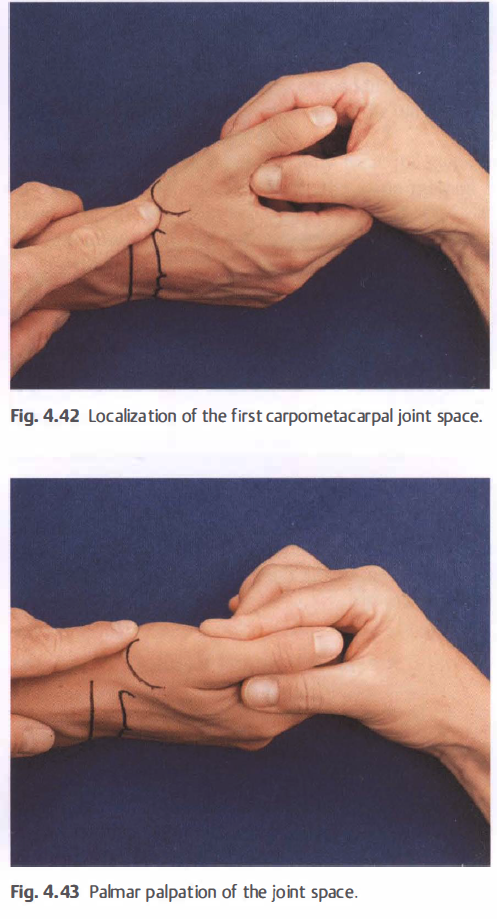
• Arthritis, mainly caused by rheumatic disease or trauma. Joint inf lammation is not only seen in the wrist area; it can also be observed in the distal radioulnar joint (DRUJ) and the first carpometacarpal joint. This does not make differentiation during assessment easier.
• Restrictions in mobility, mostly as a result of immobilization, for example, following fractures near the joint (Colles fracture). When presented with a hand with restricted mobil ity, the crucial issue for therapists is to localize the point of restriction. Should the therapist start treating the radiocarpal joint, or should the local carpal bones be assessed first?
• Instability in the hand region is a common cause for symptoms. It involves circumscribed hypermobility and either presents as capsular and ligamental strain or as an abnormal position (subluxation) of joints under loading.
A variety of areas can be found to be instable: in the carpus (lunate and along the ulnar column), in the distal radioulnar joint, and in the first carpometacarpal joint. The challenge for therapists is to use their understanding of local biomechanics and the precise localization of appropriate structures to find the causative hypermobility and therefore the source of symptoms. Local in vivo anatomy is immensely helpful here.
o Soft-tissue conditions. The passage of tendons and the fixed points of the long muscles in the wrist provide sufficient opportunity for the development of overuse problems. The entire spectrum of possible pathological conditions can be observed here: tenosynovitis, myotenosynovitis, and insertion tendinopathy. On the dorsal side, the tendons travel in tendon compartments. On the palmar aspect, nine tendons and one nerve are bundled together in the carpal tunnel. Symptoms
in this region can often be treated with precisely applied interventions.
o Nerve compression. Similar to the situation at the elbow, three large peripheral nerves can also be compressed
in the hand. The median nerve can be compromised in the carpal tunnel. A variety of provocative tests for the carpal tunnel
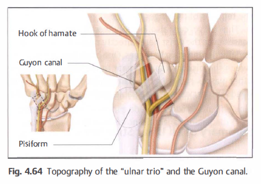
syndrome are based on the careful localization of the point of constriction. Cyclists occasionally compress the ulnar nerve in the Guyon canal. The radial nerve can be compressed as it passes through the fascia of the forearm into the superficial tissue.
질병을 상상하기 전에 먼저 구조와 기능을 상상해 보자.
손목과 손가락의 생체역학적 지식을 위한 구분
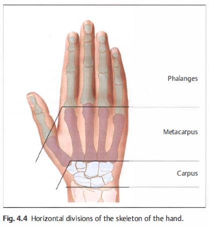
1. 수평분류
phalanges, metacapus, capus, rulnar, radius
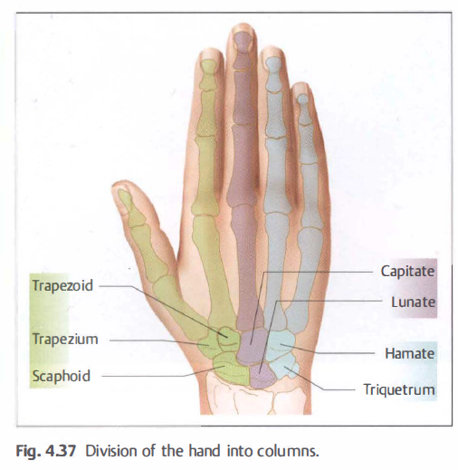
2. 축성 분류
radial column, central column, ulnar column
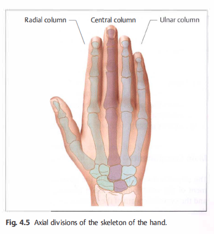
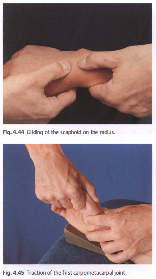
Radial Column: This column comprises the first and second rays as well as the trapezium and scaphoid. Experience
shows that arthrotic changes are most frequently detected here. The scaphoid articulates with four neighboring carpal bones and the radius so that it controls the biomechanics of both capitate and lunate (Fig. 4.6). Hypomobility
is mainly observed in the radial column.

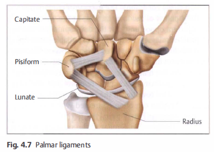
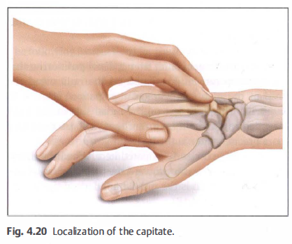
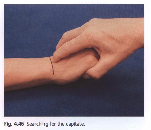
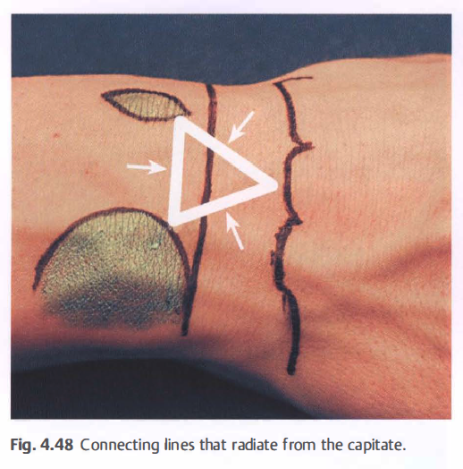
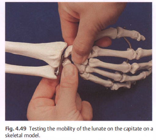
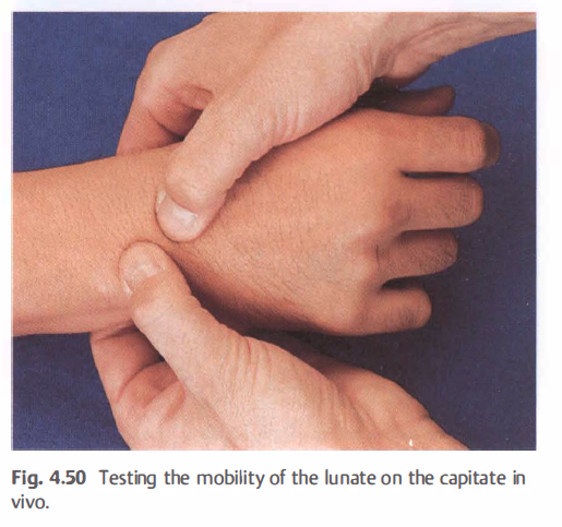
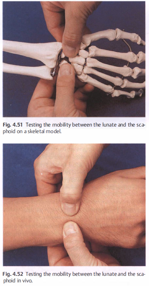
Central Column: The central column is formed by the longest ray of the hand (third metacarpal and the middle finger), capitate, and lunate. The column's special clinical feature is the frequent presence of local instability. This refers to dislocation and the consequential permanent fixation in a non physiological position when loaded. It is primarily the lunate that dislocates, usually in a palmar direction, aided by weakness in the ligaments on the palmar aspect (Fig. 4.7). This is the space of Poirier, which is located in the joint space between capitate and lunate. Providing the therapist is able to identify the carpal bones, the presence of instability can be proven by testing the local mobility of the articulations of the lunate.
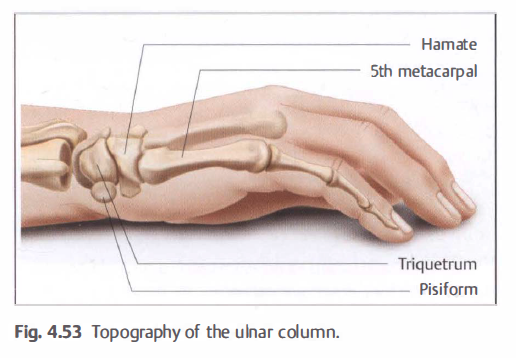
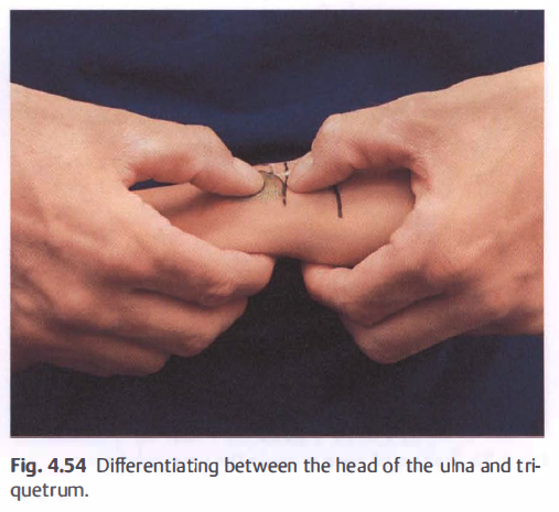
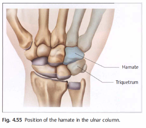
Ulnar Column: The connections between the fourth and fifth rays, hamate, and triquetrum opposite the articular
disk of the DRUJ are known to be hypermobile and can cause symptoms.
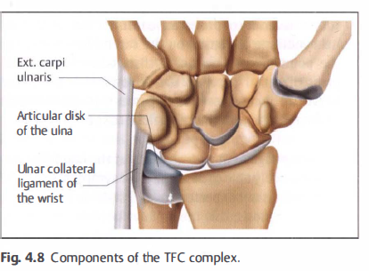
Triangular and Fibrocartilage Complex(TFCC)
삼각섬유연골 복합체
The triangular and fibrocarti lage (TFC) complex controls stability in this region and the region surrounding the distal
radioulnar joint. TFC stands for triangular and fibrocartilage (Fig. 4.8).
Functions
• Stabilizes the carpus on the ulna and radius.
• Load bearing.
• Stabilizes the DRUj.
Main Components
The articular disk of the DRUJ, the ulnar collateral ligament of the wrist joint, the deep ulnocarpal ligaments, and the synovial sheath of the extensor carpi ulnaris tendon. The margins of the TFC complex have a vascular and nociceptive supply and can therefore also be a direct source of pain.
수근관의 구조와 기능
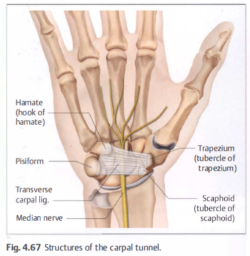
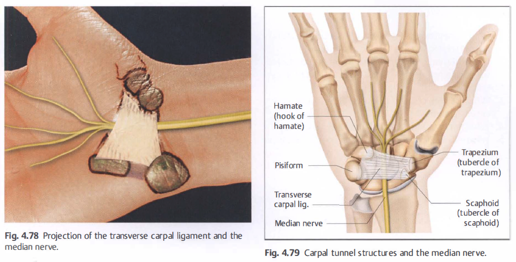
Construction of the Carpal Tunnel
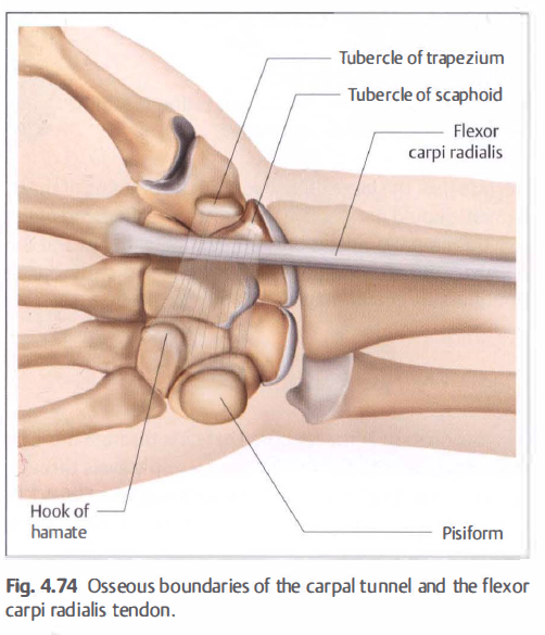
The two rows of carpal bones form a transverse arch. The term "row" is actually quite confusing. The construction of
the carpal arch becomes clear when the therapist examines the parts of bone that protrude pal marly (Fig. 4.9):
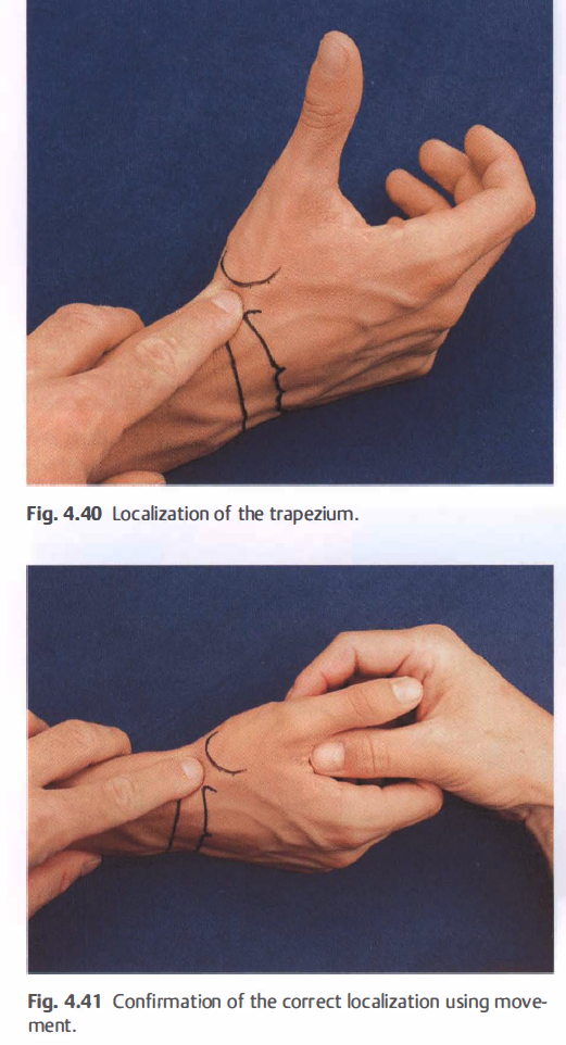
• Radial: scaphoid tubercle and trapezium.
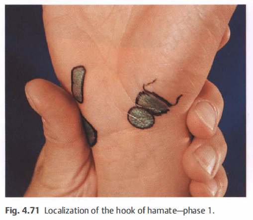
• Ulnar: pisiform and the hook of hamate. The transverse carpal ligament encloses this carpal arch, forming the carpal tunnel. The following structures pass through the carpal tunnel:
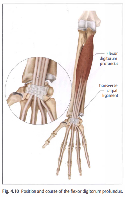
• The four tendons of the f lexor digitorum profundus (Fig. 4.1 0).
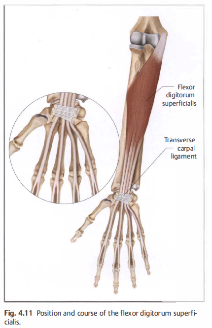
• The four tendons of the flexor digitorum superficial is (Fig. 4.11 ).
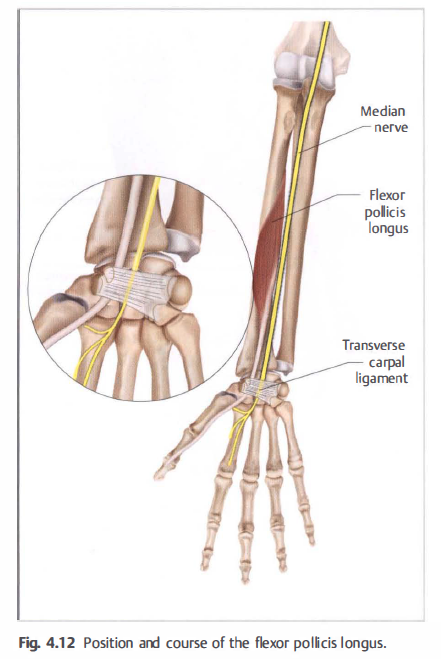
• The tendon of the flexor pollicis longus ( Fig. 4.1 2).
• Median nerve (Fig. 4.1 2).
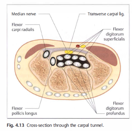
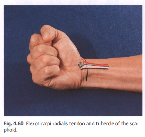
In the past, the flexor carpi radialis tendon was considered part of the carpal tunnel. Its course underneath the ligament
is listed as a separate passage in topographical anatomy ( Fig. 4.1 3).
손목 신전근 힘줄

Radiocarpal joint line 촉진
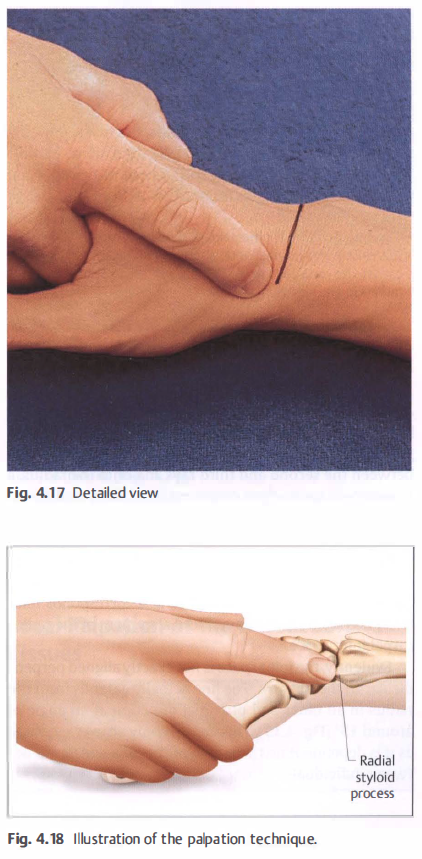
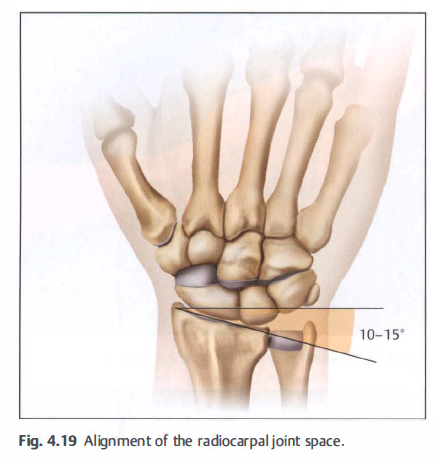
손목관절의 정렬
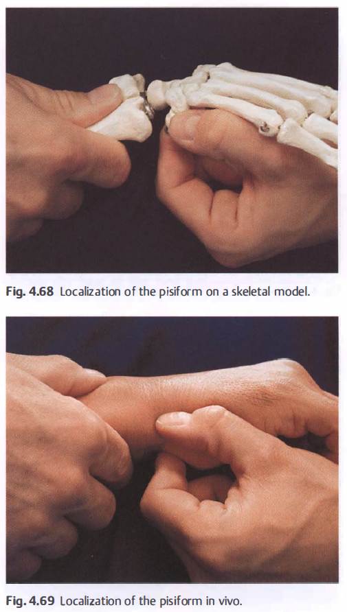
유두골의 촉진
수근골의 촉진
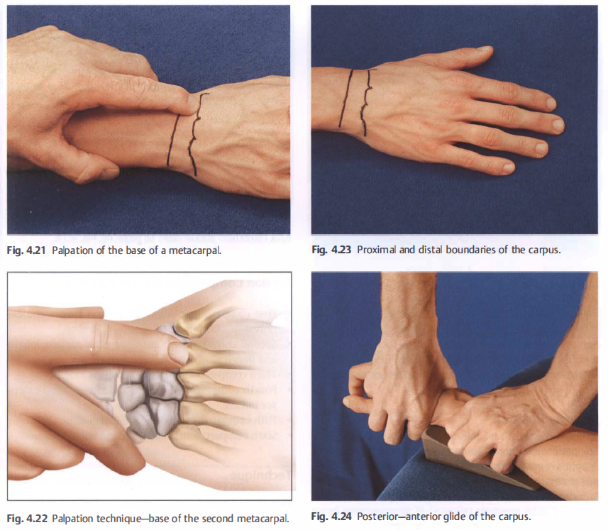
손등의 연부조직 촉진
1) Radial fossa(anatotomical sfuffbox)
2) extensor tendons and their compartment
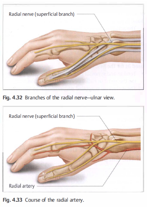
3) radial nerve, cephalic vein, and radial artery
4) distal radioulnar joint(DRUJ) space
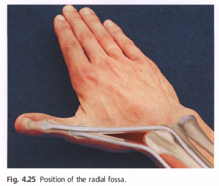
• Proximal = radius.
Dorsal = extensor pollicis longus tendon.
Palmar = extensor pollicis brevis tendon.
손목 신전근 건과 구조물
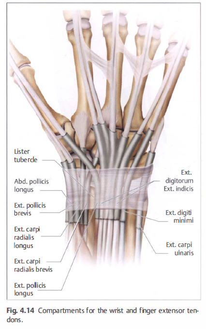
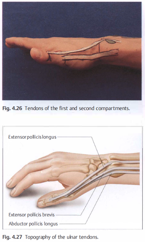
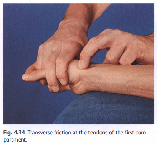
• First compartment: abductor pollicis longus and extensor pollicis brevis.
• Second compartment: extensor carpi radialis longus and brevis.
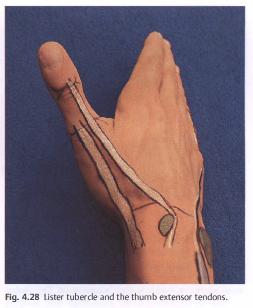
• Third compartment: extensor pollicis longus.
• Fourth compartment: extensor digitorum and extensor indicis.
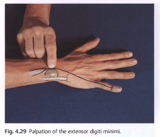
• Fifth compartment: extensor digiti minimi.
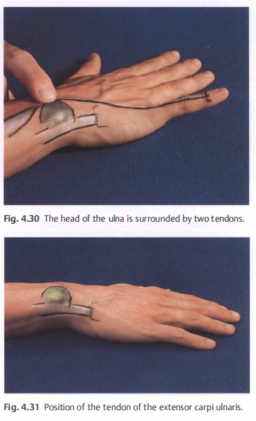
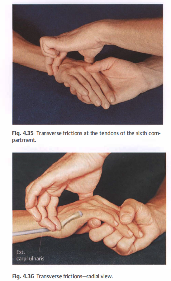
• Sixth compartment: extensor carpi ulnaris.
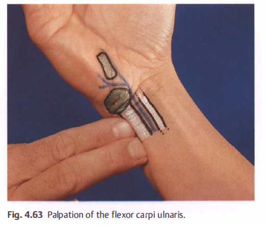
척골측 건의 촉진
요골신경, 요골동맥
교차마찰 마사지
수근골의 촉진
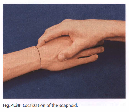
주상골 활주
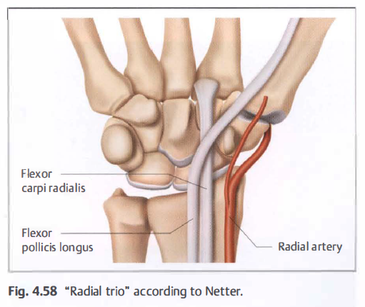
요골 트리오
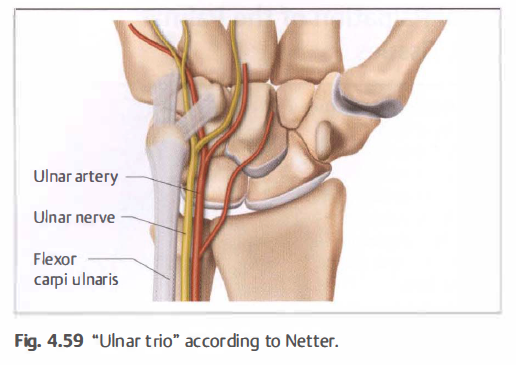
척골 트리오
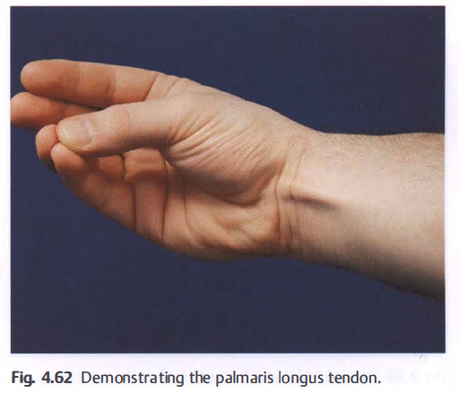
palmaris longus
flexor carpi ulnaris
척골 트리오와 가이온 터널
수근관의 구조