인체에서 가장 큰 기관, 전신 조절자 --> 근막

https://pmc.ncbi.nlm.nih.gov/articles/PMC11346343/
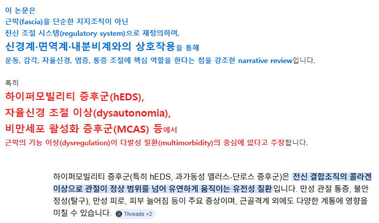
이 논문은
근막(fascia)을 단순한 지지조직이 아닌
전신 조절 시스템(regulatory system)으로 재정의하며,
신경계·면역계·내분비계와의 상호작용을 통해
운동, 감각, 자율신경, 염증, 통증 조절에 핵심 역할을 한다는 점을 강조한 narrative review입니다.
특히
하이퍼모빌리티 증후군(hEDS),
자율신경 조절 이상(dysautonomia),
비만세포 활성화 증후군(MCAS) 등에서
근막의 기능 이상(dysregulation)이 다발성 질환(multimorbidity)의 중심에 있다고 주장합니다.
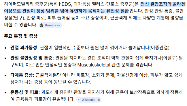

| 하이퍼모바일 에러스-단로스 증후군(hEDS)과 하이퍼모빌리티 스펙트럼 장애(HSD)는 관절 과이동성, 만성 통증, 자율신경 기능 이상, 면역 dysregulation, 조직 취약성을 특징으로 하는 다계통 결합조직 장애입니다. 유전적 마커가 없어 진단이 늦어지는 경우가 많습니다. 이 리뷰는 근막(fascia) 중심의 병태생리를 제안하며, 분자·조직학적·생역학적 증거를 종합하여 hEDS/HSD의 발병 기전을 설명합니다. 특히 섬유아세포(fibroblast) dysregulation, 세포외기질(ECM) 재형성, 그리고 전신 증상을 중점적으로 다룹니다. 주요 발견 (근막 병태생리 중심)
지속적인 근막 재형성과 근섬유아세포 활성화, ECM dysregulation, 면역-기계전달(mechanotransduction) 이상으로 설명할 수 있습니다. 근막은 단순한 지지조직이 아니라 전신적인 신호 전달 네트워크로 작용 |
https://pmc.ncbi.nlm.nih.gov/articles/PMC10871156/

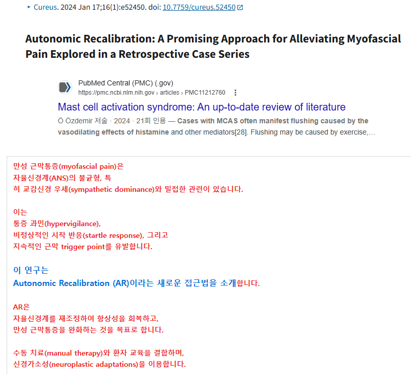
| 만성 근막통증(myofascial pain)은 자율신경계(ANS)의 불균형, 특 히 교감신경 우세(sympathetic dominance)와 밀접한 관련이 있습니다. 이는 통증 과민(hypervigilance), 비정상적인 시작 반응(startle response), 그리고 지속적인 근막 trigger point를 유발합니다. 이 연구는 Autonomic Recalibration (AR)이라는 새로운 접근법을 소개합니다. AR은 자율신경계를 재조정하여 항상성을 회복하고, 만성 근막통증을 완화하는 것을 목표로 합니다. 수동 치료(manual therapy)와 환자 교육을 결합하며, 신경가소성(neuroplastic adaptations)을 이용합니다. 방법 및 기법
Head-to-toe palpation startle assessment 설명 이 평가는 Autonomic Recalibration (AR) 치료와 Primal Reflex Release Technique (PRRT)의 핵심적인 초기 평가 도구. John Iams가 개발한 PRRT에서 유래한 방법으로, nociceptive startle exam (NSE) 또는 one-minute nociceptive exam이라고도 불립니다. 목적
교감신경 과활성화 패턴을 확인한 후, PRRT를 이용해 전신적인 자율신경 재설정(ANS reset)을 시도합니다. 수행 방법
임상적 의미 (이전 논문 맥락에서)
주요 사례 및 결과 (Retrospective Case Series) 3명의 다양한 환자 사례를 제시:
2주간의 지속적인 셀프케어로 통증 점수가 평균 67% 개선되었다고 보고되었습니다. AR(autonomic recalibration) 적용 후, nociceptive trigger point가 치료 시점에 모두 소실되었으며, 교감신경 우세가 감소하고 자율신경 균형이 회복된 것으로 관찰되었습니다. 결론 및 함의 AR은 만성 근막통증 관리에 있어 유망한 대안적 접근법으로, 기존 진통제(특히 오피오이드) 의존을 줄일 수 있는 비약물적 옵션입니다. 자율신경 불균형을 직접 타겟으로 하여 통증의 근본 원인을 다루는 trauma-informed manual therapy입니다. 임상적 의미:
|

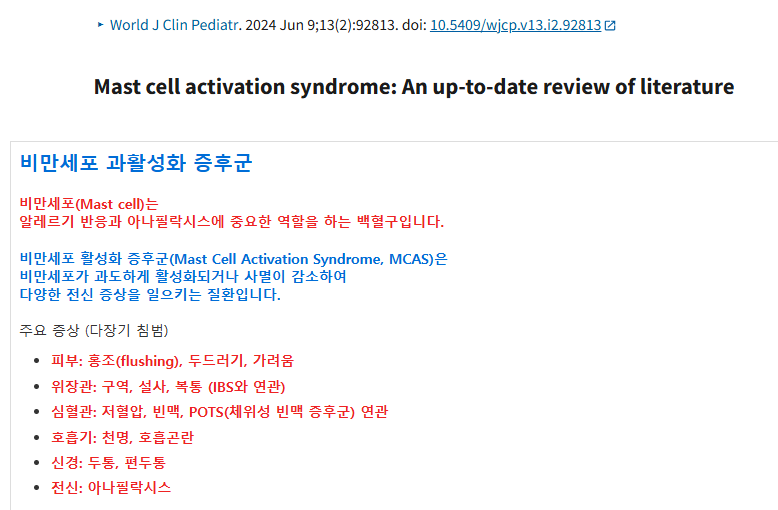
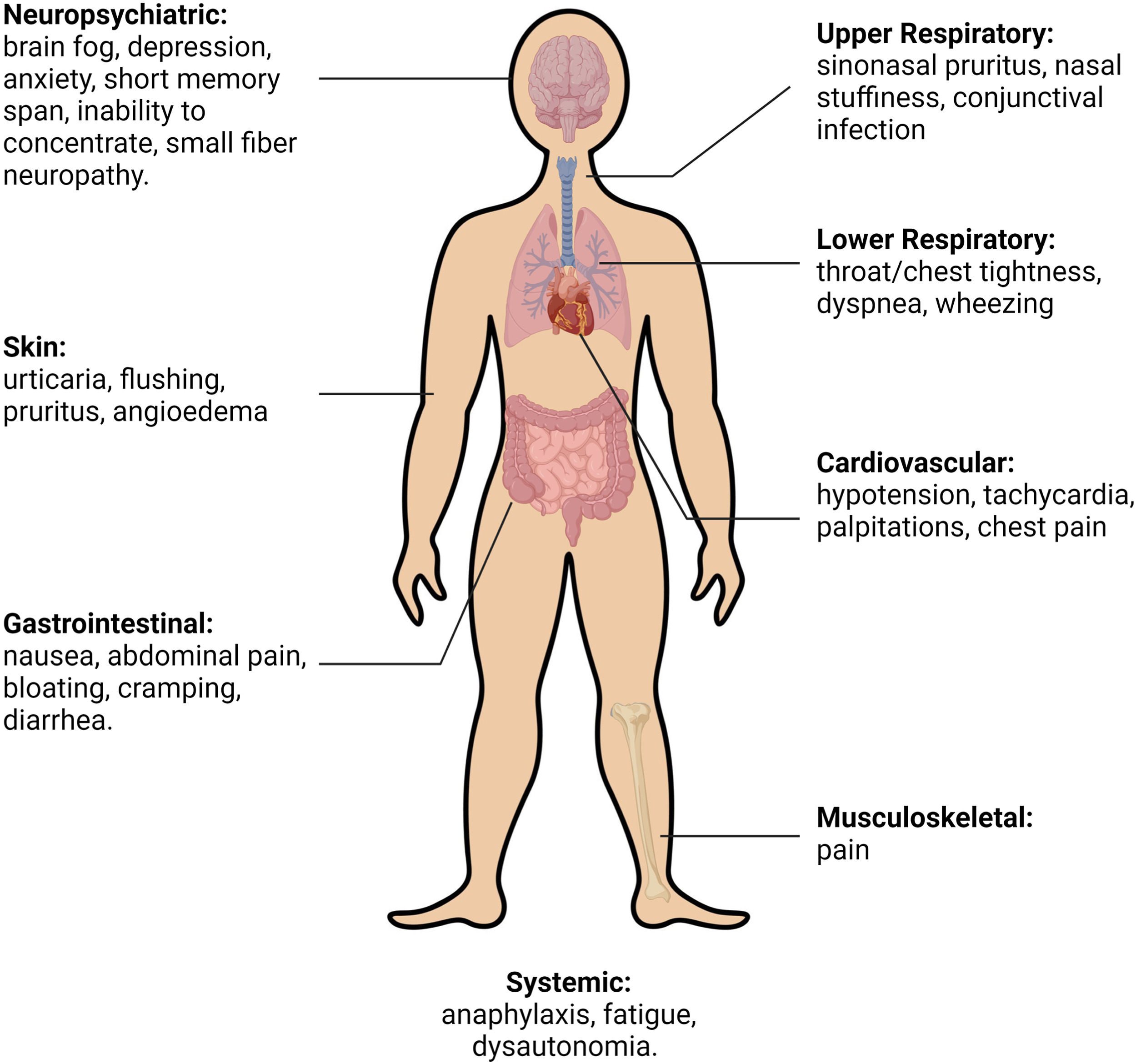
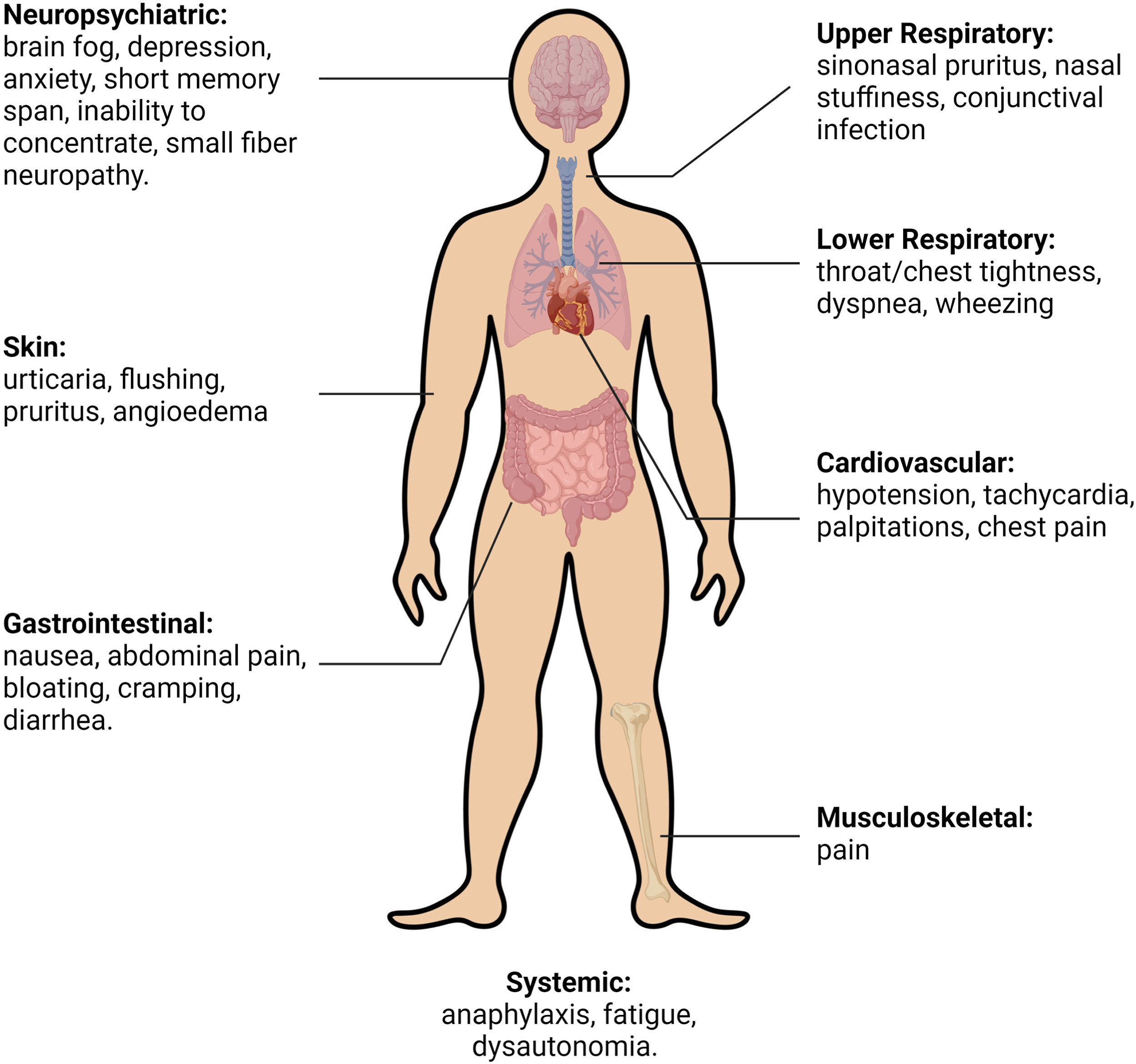
| 비만세포 과활성화 증후군 비만세포(Mast cell)는 알레르기 반응과 아나필락시스에 중요한 역할을 하는 백혈구입니다. 비만세포 활성화 증후군(Mast Cell Activation Syndrome, MCAS)은 비만세포가 과도하게 활성화되거나 사멸이 감소하여 다양한 전신 증상을 일으키는 질환입니다. 주요 증상 (다장기 침범)
이 논문은 MCAS의 역학, 병인, 진단, 증상, 치료에 대한 최신 문헌을 종합적으로 검토한 서술적 리뷰입니다. 진단 기준 (주요 내용) MCAS 진단은 다음 조건을 모두 충족해야 합니다:
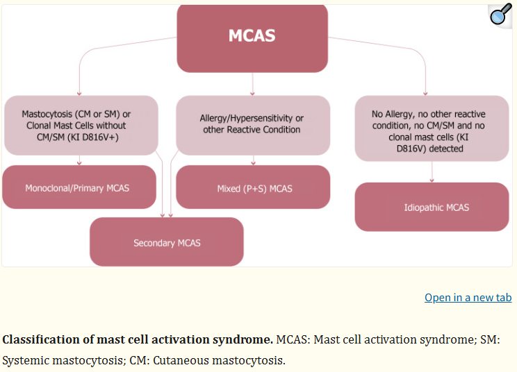 분류
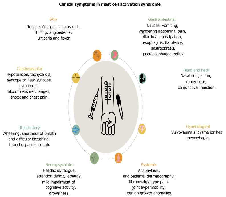 주요 증상 (다장기 침범)
|
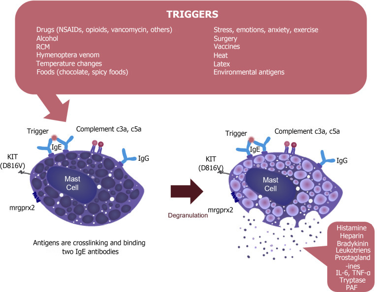
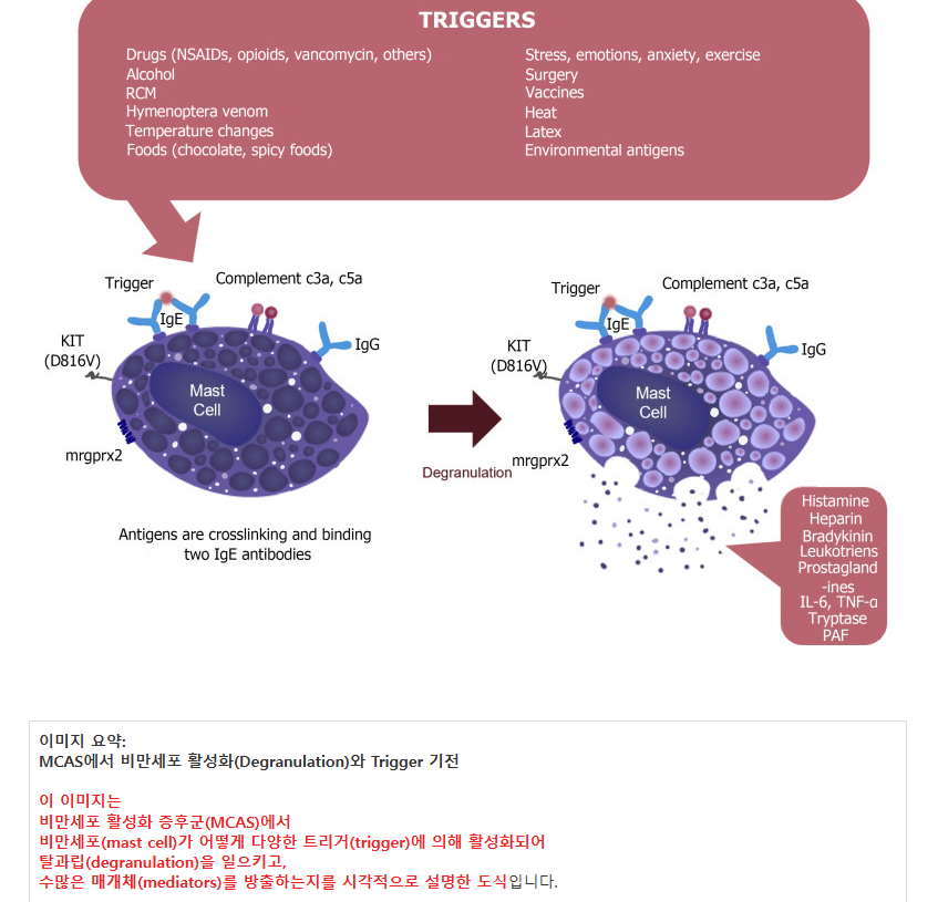
| 이미지 요약: MCAS에서 비만세포 활성화(Degranulation)와 Trigger 기전 이 이미지는 비만세포 활성화 증후군(MCAS)에서 비만세포(mast cell)가 어떻게 다양한 트리거(trigger)에 의해 활성화되어 탈과립(degranulation)을 일으키고, 수많은 매개체(mediators)를 방출하는지를 시각적으로 설명한 도식입니다. 1. 상단: 주요 트리거 목록 (Triggers) 비만세포는 알레르기 외에도 매우 다양한 자극에 과민하게 반응합니다. 주요 트리거는 다음과 같습니다: 약물 및 물질 관련:
IgE 매개뿐만 아니라 비면역성(non-immunologic) 경로로도 비만세포를 직접 자극할 수 있습니다. 2. 중앙: 비만세포 활성화 과정 (Mechanism)
피부, 소화기, 심혈관, 호흡기, 신경계 등 다계통(multi-system) 증상을 일으킵니다. https://cafe.daum.net/panicbird/QrbQ/70 https://cafe.daum.net/panicbird/QrbQ/68 임상적 의미 (이전 대화 맥락)
MCAS의 핵심 병태생리를 한눈에 보여주며, “왜 MCAS 환자가 특정 약물·음식·스트레스에 민감한가”를 잘 설명 |
| MCAS와 hEDS의 연관성은 환자 커뮤니티와 일부 임상가 사이에서 자주 논의되는 주제이지만, 과학적 증거는 아직 논쟁적이며 명확한 인과관계는 입증되지 않았습니다. 1. 관찰되는 임상적 중복 (Comorbidity) hEDS (hypermobile Ehlers-Danlos syndrome) 또는 HSD (Hypermobility Spectrum Disorder) 환자들에서 MCAS-like 증상 (홍조, 두드러기, 가려움, 복통·설사, 저혈압, 피로, 두통, 아나필락시스 유사 반응 등)이 흔히 동반됩니다.
다계통 증상(소화기, 피부, 심혈관, 신경계)의 겹침 때문에 진단이 복잡해지며, 일부 환자는 “unconfirmed MCAS” (확인되지 않은 MCAS)로 과진단되는 경향도 있습니다. 2. 가능한 병태생리적 기전 (이론적 설명) hEDS와 MCAS가 함께 나타날 수 있는 이유로 여러 가설이 제시됩니다:
|
https://www.jacionline.org/article/S0091-6749(24)00569-4/fulltext

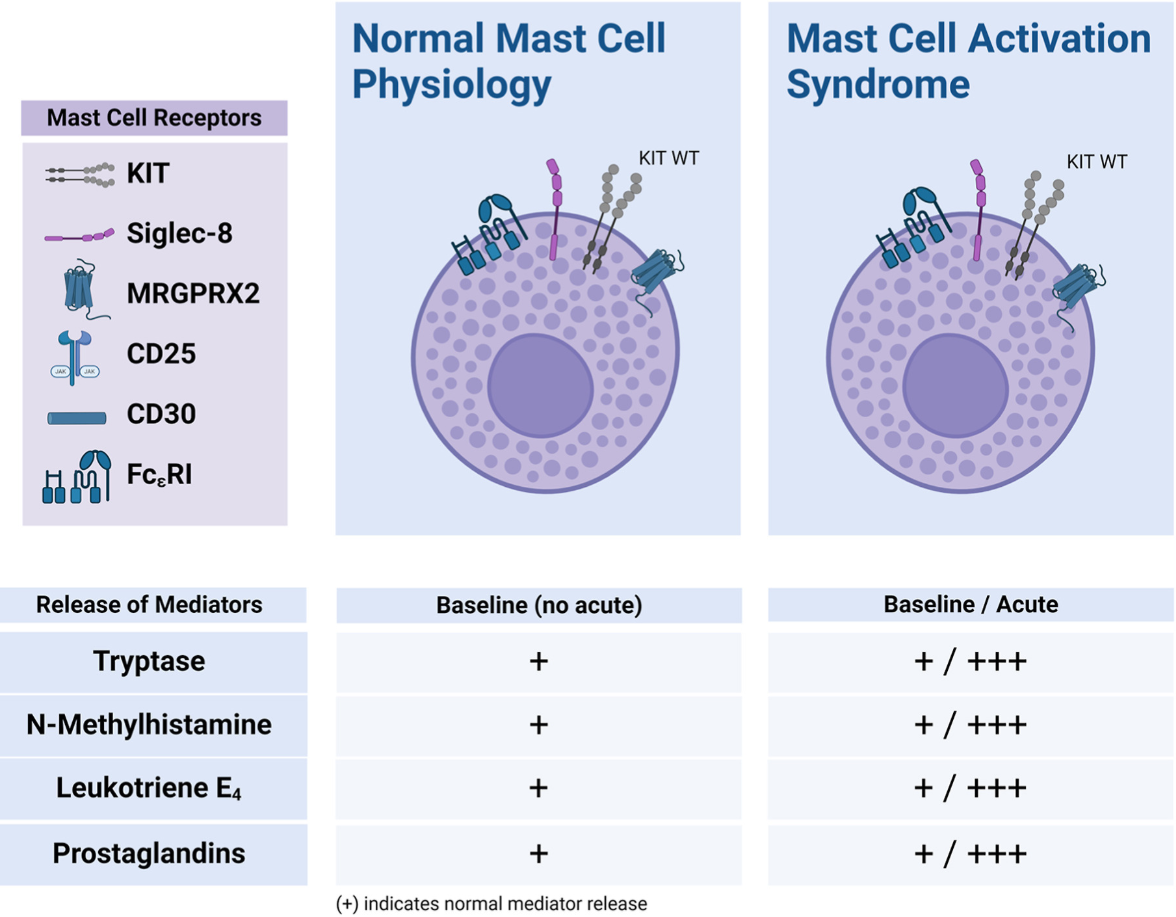
| 비만세포 활성화 증후군(MCAS)은 비만세포(Mast Cell)에서 매개체(히스타민, 트립타아제, 프로스타글란딘 등)가 과도하게 방출되어 반복적인 전신 증상을 일으키는 상태. 환자와 의료진 사이에서 관심이 급증했으나, MCAS가 독립된 원발성 증후군인지, 아니면 알레르기·염증·클론성 질환의 일부인지 명확하지 않습니다. 이 논문은 기존 문헌을 종합하고, 진단·기전·치료의 지식 격차를 파악하며, 향후 연구 방향을 제시하는 것을 목적으로 합니다. MCAS 진단 기준 (2022 Consensus 기준) 진단을 위해 다음 3가지 조건을 모두 충족해야 합니다:
주요 증상 및 동반 질환 다장기 증상 + 흔한 동반증:
|
Abstract
근막은
신경이 풍부하게 분포하고 혈관·림프관이 많으며,
호르몬·신경전달물질 수용체를 가지고 있어
전신 건강의 “감시자(watchman)” 역할을 합니다.
기존 의학에서
근막을 “무생물적 충전재(inert filler)”로 간과한 결과,
만성 근골격계 통증과 복합 증상을 제대로 치료하지 못하고 있습니다.
본 리뷰는
근막의 해부학적 구성, 기능, 그리고 질환에서의 dysregulation을 종합적으로 검토하며,
임상 진료와 연구 방향의 패러다임 전환을 촉구합니다.
주요 내용 상세 설명
1. 서론 및 배경
- 만성 근골격계 통증은 전 세계적으로 큰 부담이지만 연구·진단·치료가 부족합니다.
- 전통적 관점: 근막 = passive connective tissue (단순 지지).
- 새로운 관점: 근막 = dynamic regulatory organ으로, 신경·면역·내분비 시스템과 긴밀히 상호작용.
- 문제 제기: hEDS, MCAS, dysautonomia, Long COVID, plantar fasciitis 등에서 근막 dysregulation이 핵심 병태생리임에도 의료진이 이를 인지하지 못함.
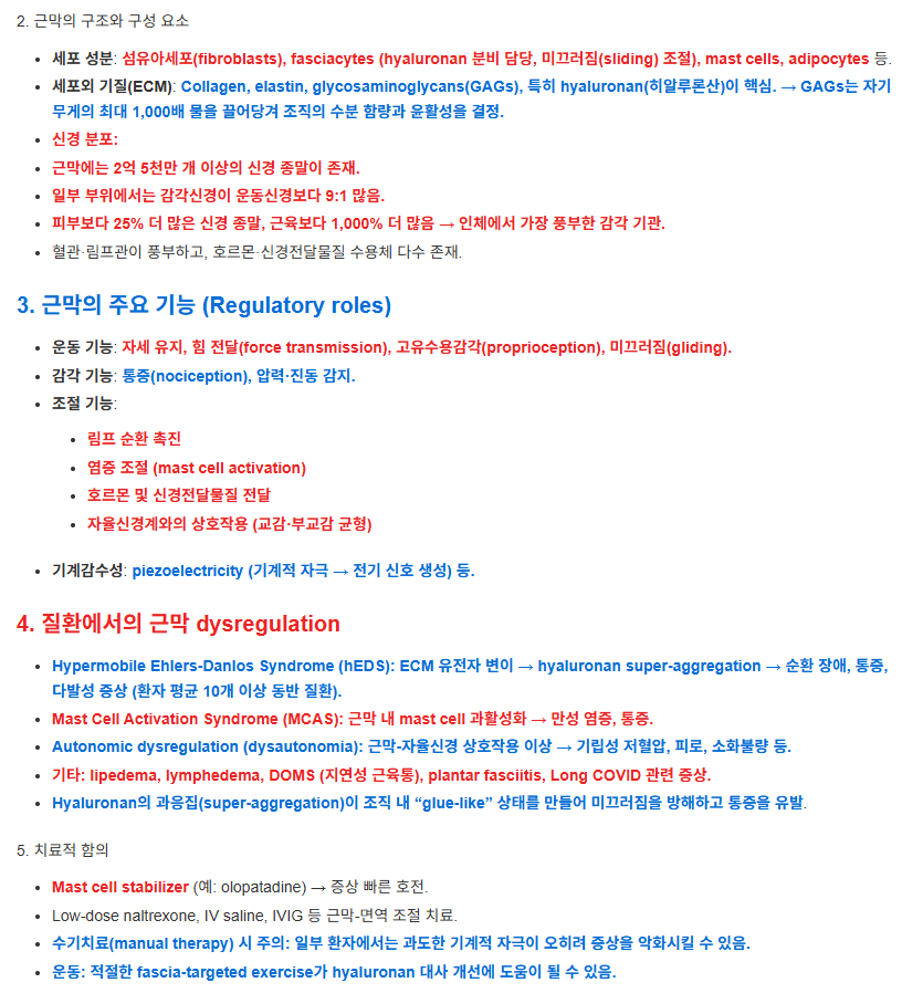
2. 근막의 구조와 구성 요소
- 세포 성분: 섬유아세포(fibroblasts), fasciacytes (hyaluronan 분비 담당, 미끄러짐(sliding) 조절), mast cells, adipocytes 등.
- 세포외 기질(ECM): Collagen, elastin, glycosaminoglycans(GAGs), 특히 hyaluronan(히알루론산)이 핵심. → GAGs는 자기 무게의 최대 1,000배 물을 끌어당겨 조직의 수분 함량과 윤활성을 결정.
- 신경 분포:
- 근막에는 2억 5천만 개 이상의 신경 종말이 존재.
- 일부 부위에서는 감각신경이 운동신경보다 9:1 많음.
- 피부보다 25% 더 많은 신경 종말, 근육보다 1,000% 더 많음 → 인체에서 가장 풍부한 감각 기관.
- 혈관·림프관이 풍부하고, 호르몬·신경전달물질 수용체 다수 존재.
3. 근막의 주요 기능 (Regulatory roles)
- 운동 기능: 자세 유지, 힘 전달(force transmission), 고유수용감각(proprioception), 미끄러짐(gliding).
- 감각 기능: 통증(nociception), 압력·진동 감지.
- 조절 기능:
- 림프 순환 촉진
- 염증 조절 (mast cell activation)
- 호르몬 및 신경전달물질 전달
- 자율신경계와의 상호작용 (교감·부교감 균형)
- 기계감수성: piezoelectricity (기계적 자극 → 전기 신호 생성) 등.
4. 질환에서의 근막 dysregulation
- Hypermobile Ehlers-Danlos Syndrome (hEDS): ECM 유전자 변이 → hyaluronan super-aggregation → 순환 장애, 통증, 다발성 증상 (환자 평균 10개 이상 동반 질환).
- Mast Cell Activation Syndrome (MCAS): 근막 내 mast cell 과활성화 → 만성 염증, 통증.
- Autonomic dysregulation (dysautonomia): 근막-자율신경 상호작용 이상 → 기립성 저혈압, 피로, 소화불량 등.
- 기타: lipedema, lymphedema, DOMS (지연성 근육통), plantar fasciitis, Long COVID 관련 증상.
- Hyaluronan의 과응집(super-aggregation)이 조직 내 “glue-like” 상태를 만들어 미끄러짐을 방해하고 통증을 유발.
5. 치료적 함의
- Mast cell stabilizer (예: olopatadine) → 증상 빠른 호전.
- Low-dose naltrexone, IV saline, IVIG 등 근막-면역 조절 치료.
- 수기치료(manual therapy) 시 주의: 일부 환자에서는 과도한 기계적 자극이 오히려 증상을 악화시킬 수 있음.
- 운동: 적절한 fascia-targeted exercise가 hyaluronan 대사 개선에 도움이 될 수 있음.
https://cafe.daum.net/panicbird/QrbQ/69

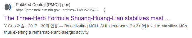
| 금은화(Lonicerae Japonicae Flos), 황금(Scutellariae Radix), 연교(Fructus Forsythiae)로 구성된 전통 중국 의학 3초본 처방입니다. 기존에는 호흡기 감염 치료제로 사용되었으나, 알레르기 질환(천식, 두드러기, 습진 등)에서도 항알레르기 효과가 관찰되었습니다. 이 연구는 SHL의 항알레르기 작용 기전을 밝히기 위해, 특히 비만세포(Mast Cell, MC) 안정화와 칼슘 신호에 초점을 맞췄습니다. 주요 발견 (기전) SHL은 미토콘드리아 칼슘 uniporter (MCU)를 활성화하여 작용합니다.
SHL의 칼슘 저하 효과와 탈과립 억제 효과가 거의 사라짐 → MCU 활성화가 핵심 기전임을 증명. |
https://www.mdpi.com/2073-4409/12/8/1187

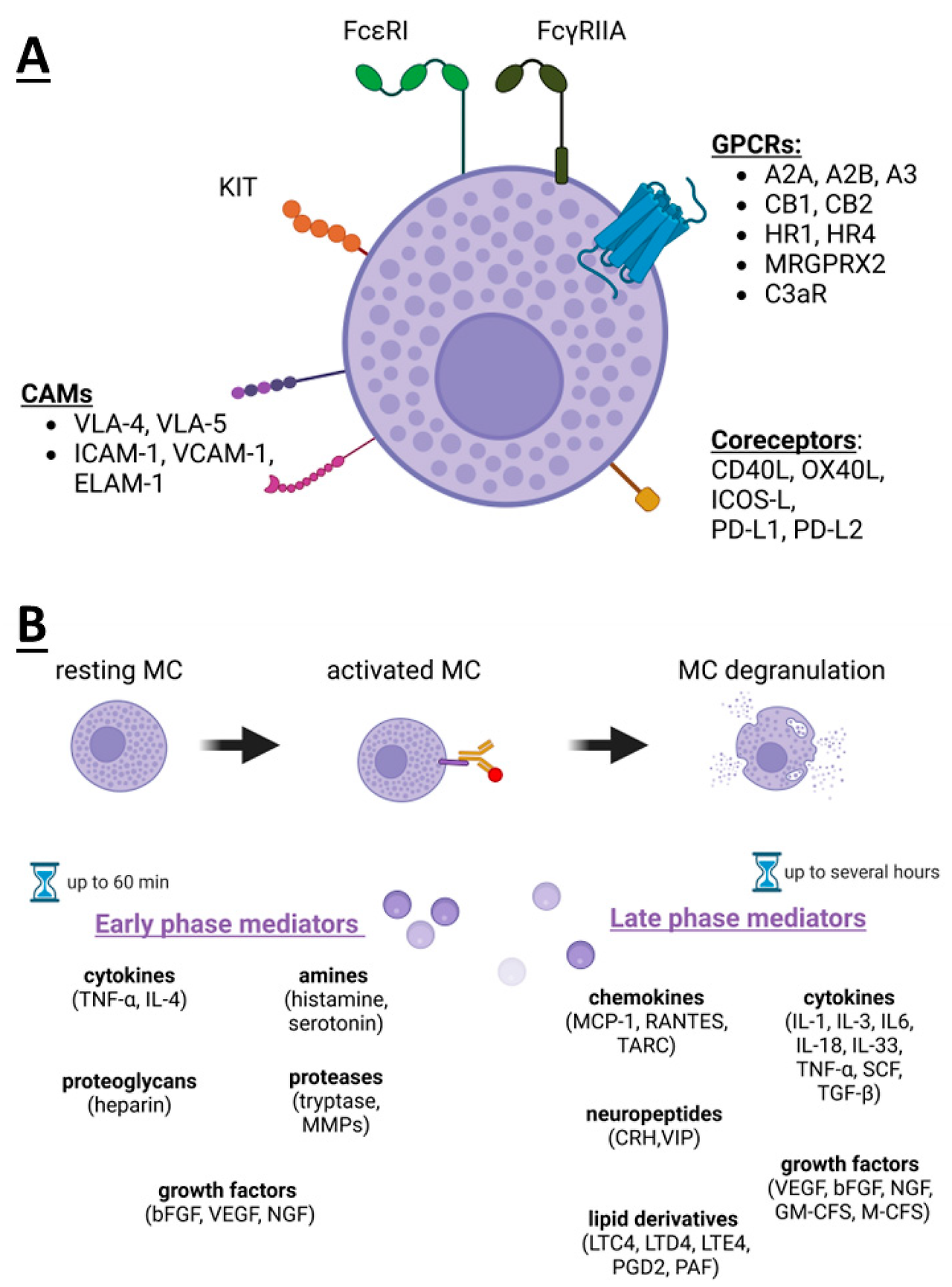
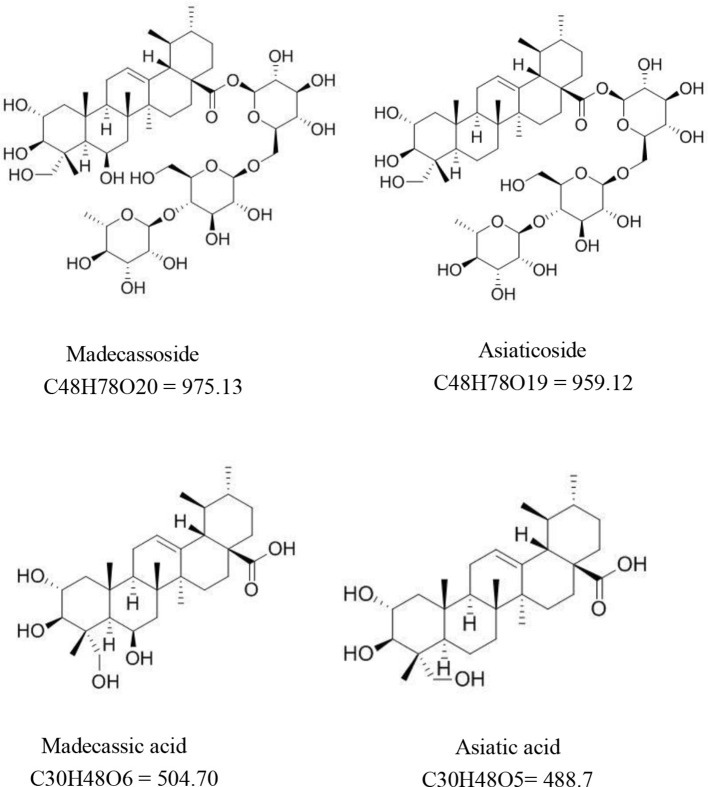
마데카솔의 주성분 .. 병풀(적설초)
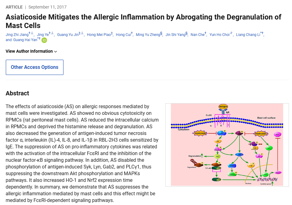
논문의 모식도에서 보여주듯:
비만세포 탈과립을 억제하고 FcεRI-의존적 신호 전달 경로를 차단함으로써 알레르기 염증을 효과적으로 완화합니다. 이 결과는 병풀이 아토피 피부염, 알레르기 비염, 두드러기, 그리고 비만세포 활성화 증후군(MCAS) 같은 질환의 보조 치료제로 활용될 가능성을 강력히 시사 |

| Centella asiatica (병풀, Gotu Kola)와 그 트리터펜 성분들이 피부 질환(여드름, 화상, 아토피 피부염, 창상 등)에 미치는 약리 효과와 분자 기전을 종합적으로 검토. 2011년 이후 연구를 중심으로 in vitro, in vivo, 임상 증거를 분석. 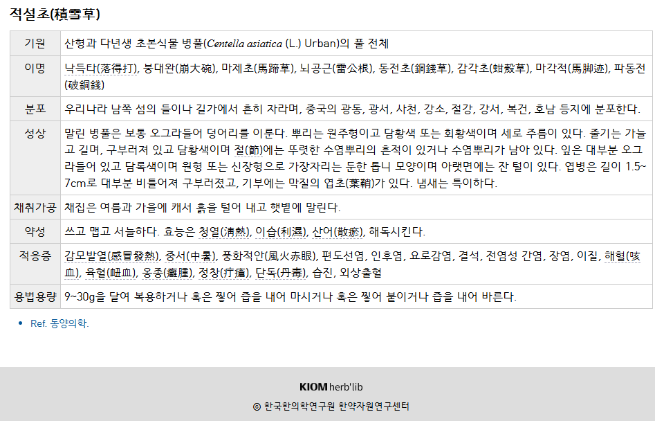 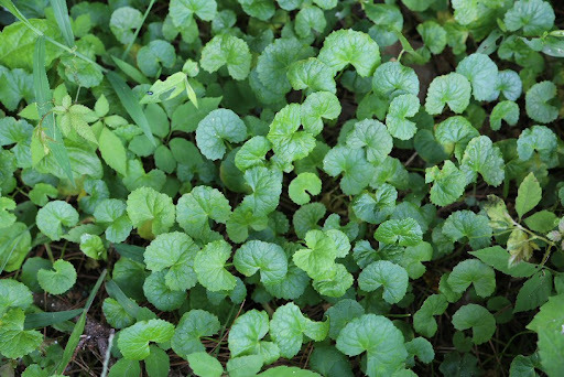 주요 활성 성분
병풀과 트리터펜 성분들은 항염증·창상 치유 효과가 강력하며, 특히 아토피 피부염에서 mast cell 침윤 감소가 유의미한 기여를 할 수 있음. 유럽에서는 Madecassol® 등 제제가 이미 사용 중. 더 많은 고품질 임상 연구가 필요 |
https://pmc.ncbi.nlm.nih.gov/articles/PMC7498642/

| 이 리뷰는 병풀(Centella asiatica)과 그 주요 트리터펜 성분(asiaticoside, madecassoside, asiatic acid, madecassic acid)의 다양한 질환에 대한 치료 잠재력을 종합적으로 검토합니다. 신경계, 내분비, 피부, 심혈관, 위장관, 면역, 호흡기, 부인과 질환 등을 다루며, 특히 항염증, 항산화, 항세포사멸, 미토콘드리아 기능 개선 기전에 초점을 맞춥니다. 전통 의학 용도와 2015~2019년 PubMed 연구를 중심으로 in vitro, in vivo, 임상 증거를 분석했습니다. 주요 활성 성분 및 약동학
|
결론
근막은
신경·면역·내분비 시스템을 연결하는 통합적 조절 시스템입니다.
근막 dysregulation을 제대로 이해하고 관리하면
복합 만성 질환 환자의 진단·치료가 크게 개선될 수 있음
미래에는 fascia-specific 연구와 교육이 필요
https://pmc.ncbi.nlm.nih.gov/articles/PMC12244286/
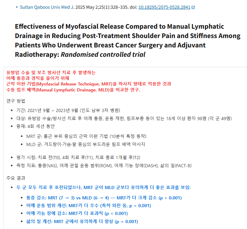
| 유방암 수술 및 보조 방사선 치료 후 발생하는 어깨 통증과 경직을 줄이기 위해 근막 이완 기법(Myofascial Release Technique, MRT)을 마사지 형태로 적용한 것과 수동 림프 배액(Manual Lymphatic Drainage, MLD)을 비교한 연구. 연구 방법
유방암 치료 후 어깨 통증·경직·기능 저하에 근막 이완 기법(MRT)이 수동 림프 배액(MLD)보다 더 효과적이며, 통증 감소, 운동 범위 회복, 기능 개선, 삶의 질 향상에 도움 |

| 근막 이완 요법(MFR)은 임상에서 다양한 근골격계 및 통증 관련 질환 치료에 널리 사용되지만, 여러 질환에 대한 체계적인 효과 평가가 부족해 적용이 일관되지 않는 문제가 있습니다. 이 리뷰는 MFR의 다양한 질환 치료 적용을 종합적으로 검토하고, 그 효능을 평가하며, 추가 연구가 필요한 영역을 제시합니다. 특히 만성 통증, 섬유근육통, 수술 후 회복, 신경계 질환 등에서의 적용을 중점적으로 다룹니다. 주요 내용 및 적용 질환 리뷰는 기존 연구들을 바탕으로 다음과 같은 질환에서의 MFR 효과를 정리했습니다:
결론 및 시사점 MFR은 다양한 질환에서 통증 완화, 기능 개선, 삶의 질 향상에 효과적인 비침습적 치료법으로, 신경 재활과 수술 후 회복 분야에서 특히 유망 |
문치연 탐구
근막 질환의 생화학적 신호만 중요한가?
mechanotransduction이 필요조건인 이유 탐구
https://cafe.daum.net/panicbird/QrbQ/71

서로 다른 근막 유형(특히 deep fascia의 aponeurotic vs epimysial)의
신경 분포에 대한 지식이 부족한 상황에서,
본 연구는 마우스
thoracolumbar fascia (TLF, aponeurotic)와
gluteal fascia (GF, epimysial)를
Transmission Electron Microscopy(TEM)과
floating immunohistochemistry(S100, PGP9.5, Tyrosine Hydroxylase 항체)로
분석했습니다.
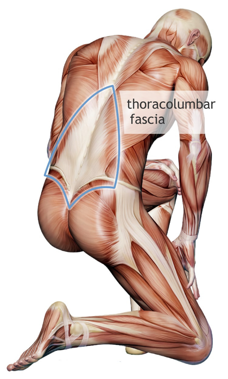
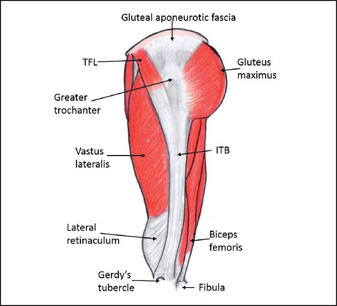
결과,
근막 전체에 rhomboid하고 dense한 신경 네트워크가 존재하며,
TLF가 GF보다 신경 밀도가 유의하게 높았습니다.
자율신경 섬유 밀도는 비슷했으며,
TLF에서는 신경 소체(corpuscles)가 발견되지 않았습니다.
이는
두 근막이 proprioception(고유수용감각)과 motor unit coordination(운동 단위 조절)에서
서로 다른 역할을 한다는 가설을 뒷받침합니다.
연구 배경 및 목적
- 기존 연구: 근막은 오랫동안 “무생물적 지지조직”으로 여겨졌으나, 2007년 Fascia Congress 이후 proprioception과 nociception(통증) 역할이 부각됨.
주요 방법
- 동물: 7마리 성체 C57-BL 마우스 (수컷, 8주령)
핵심 결과
- 신경 네트워크 패턴: 근막 전체에 rhomboid (마름모꼴)이고 dense한 신경망이 퍼져 있음 (3차원 floating 기법으로 처음 명확히 시각화).
- 밀도 차이 (통계적으로 유의미):
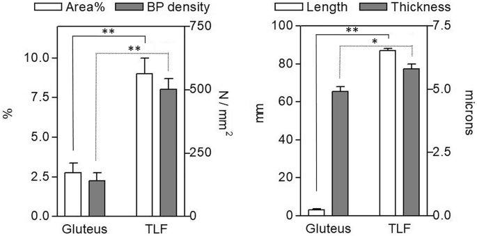
- TLF (aponeurotic): innervated area 9.01 ± 0.98%, branching points 500.9 ± 43.1/mm², 신경 길이 87.1 ± 1.0 mm, 두께 5.8 ± 0.2 µm
- GF (epimysial): innervated area 2.78 ± 0.6%, branching points 140.3 ± 31.6/mm², 신경 길이 3.2 ± 0.6 mm, 두께 4.9 ± 0.2 µm
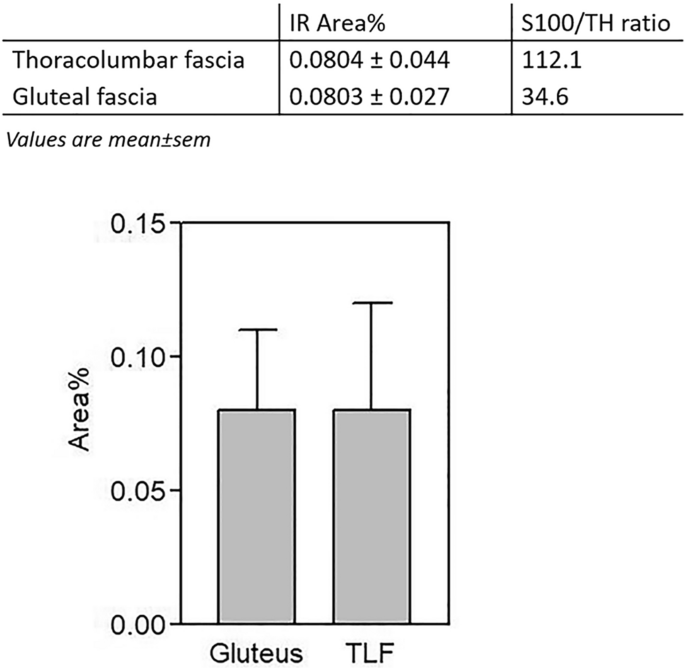
- 자율신경: 두 근막 모두 동일한 낮은 밀도 (0.08%)
- 신경 소체: TLF에서는 발견되지 않음 (free nerve endings 중심)
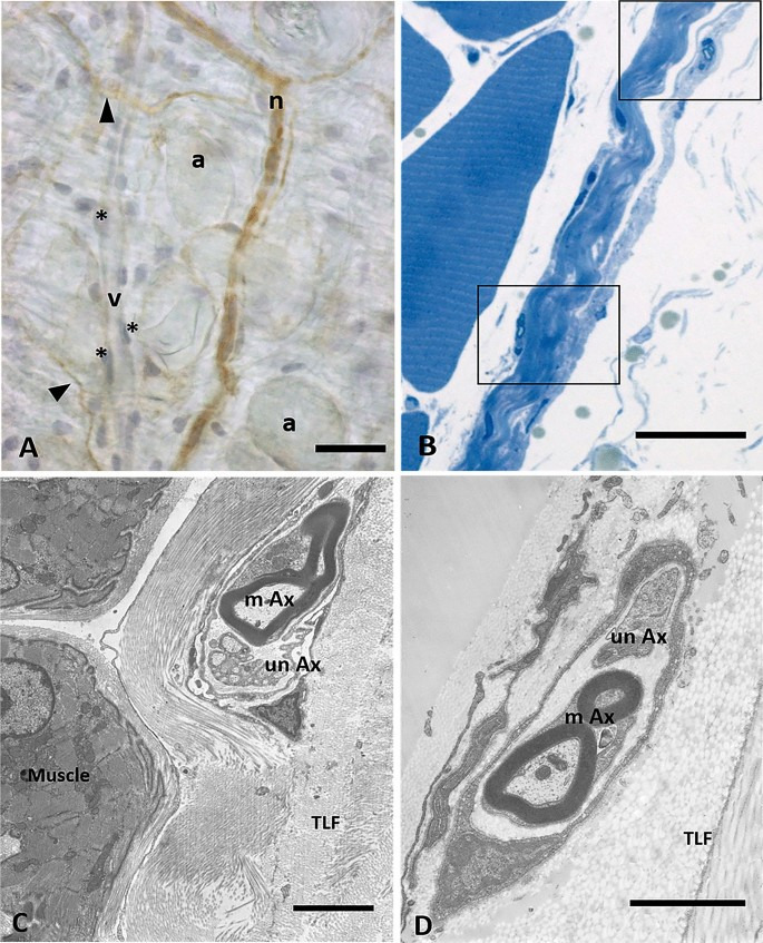
- TEM: 신경 섬유의 초미세 구조 확인 (myelinated/unmyelinated fibers)
결론 및 임상적 함의
- TLF (aponeurotic): 풍부한 free nerve endings → proprioceptor 역할. 근육 긴장 조절, 비특이적 요통(non-specific low back pain) 병태생리에 관여.
- GF (epimysial): 상대적으로 적은 신경망 → 개별 근육의 motor units 조정 역할.
- 근막은 더 이상 “수동적”이 아니라, 감춰진 신경 네트워크(hidden neural network)를 가진 감각 기관으로 재평가되어야 함.
- 임상적 의미: myofascial pain syndrome, 요통, 수기치료(manual therapy), fascia manipulation의 과학적 근거 제공. 만성 통증 치료에서 근막 신경망을 표적으로 할 수 있음.
- nature
- scientific reports
- articles
- article
Evidence of a new hidden neural network into deep fasciae
- Article
- Open access
- Published: 16 June 2021
Evidence of a new hidden neural network into deep fasciae
- Caterina Fede,
- Lucia Petrelli,
- Diego Guidolin,
- Andrea Porzionato,
- Carmelo Pirri,
- Chenglei Fan,
- Raffaele De Caro &
- Carla Stecco
Scientific Reports volume 11, Article number: 12623 (2021) Cite this article
Abstract
It is recognized that different fasciae have different type of innervation, but actually nothing is known about the specific innervation of the two types of deep fascia, aponeurotic and epymisial fascia. In this work the aponeurotic thoracolumbar fascia and the epymisial gluteal fascia of seven adult C57-BL mice were analysed by Transmission Electron Microscopy and floating immunohistochemistry with the aim to study the organization of nerve fibers, the presence of nerve corpuscles and the amount of autonomic innervation. The antibodies used were Anti-S100, Anti-Tyrosine Hydroxylase and Anti-PGP, specific for the Schwann cells forming myelin, the sympathetic nerve fibers, and the peripheral nerve fibers, respectively. The results showed that the fascial tissue is pervaded by a rhomboid and dense network of nerves. The innervation was statistically significantly lower in the gluteal fascia (2.78 ± 0.6% of positive area, 140.3 ± 31.6/mm2 branching points, nerves with 3.2 ± 0.6 mm length and 4.9 ± 0.2 µm thickness) with respect to the thoracolumbar fascia (9.01 ± 0.98% of innervated area, 500.9 ± 43.1 branching points/mm2, length of 87.1 ± 1.0 mm, thickness of 5.8 ± 0.2 µm). Both fasciae revealed the same density of autonomic nerve fibers (0.08%). Lastly, corpuscles were not found in thoracolumbar fascia. Based on these results, it is suggested that the two fasciae have different roles in proprioception and pain perception: the free nerve endings inside thoracolumbar fascia may function as proprioceptors, regulating the tensions coming from associated muscles and having a role in nonspecific low back pain, whereas the epymisial fasciae works to coordinate the actions of the various motor units of the underlying muscle.
Article Open access10 March 2026
Article Open access18 April 2023
Article Open access10 July 2023
Introduction
For many years fascia was considered as an inert tissue, which wraps and gives mechanical support to muscles and other organs. The first demonstrations that fascia is innervated date back to 1957: Stilwell reported some histological findings about sensory nerves in deep fasciae1. In 1974, Sakada and co-authors studied the masticatory area but without giving a specific meaning to the presence of mechanoreceptors in the fascia2. The first event in which many Authors described a huge presence of sensory nerves in the fasciae with a role in proprioception and nociception was the First International Fascia Congress (2007, Harvard Medical School, Boston)3. It is now demonstrated that the different fasciae have different type of innervation: the visceral fascia is rich in autonomic innervation4, the superficial fascia shares with the skin mechano- and thermic-receptors, and the deep fascia has a role in proprioception5. Furthermore, also different areas show different density and type of innervation. The superficial fascia is the second most highly innervated soft tissue after the skin, with a density of nerve structures of 33.0 ± 2.5/cm2 and 64.0 ± 5.2/cm2, respectively, and a mean size of 19.1 ± 7.2 μm; the deep fascia has a nerve density of 19 ± 5.0/cm2 and presents a thin but huge network of small nerve fibers (mean diameter 15.5 ± 9.4 μm)6. Tesarz demonstrated that the different sublayers of thoracolumbar fascia (TLF) have different amounts of nerve fibers, more numerous in the superficial and deep sublayers rather than the middle one7.
The two types of deep fascia, aponeurotic and epimysial, have totally different functions and mechanical properties: the aponeurotic fascia envelops various muscles and keep in place and connect them, whereas the epymisial fascia is specific for each muscle and strongly connected with them, defining their form and volume5. But despite that, the two type of fasciae are usually considered together and nothing is known about their specific innervation. One of the most studied aponeurotic fasciae is the TLF, being related to non-specific low back pain8,9,10,11. After chronic inflammation, the density of nociceptive fibers in TLF increased, from 4 to 15%12. Schilder13 demonstrated that the free nerve endings of TLF are more sensitive to chemical stimulation by injections of hypertonic saline, compared to the underlying muscles and subcutis, maintaining a long lasting hypersensitivity and a longer pain duration. Yahia found corpuscular receptors in human TLF8, unlike Mense, who demonstrated in rat TLF the only presence of free nerve endings: they can have both nociceptive and proprioceptive functions, due to a low mechanical threshold14. The increase of calcitonin gene related peptides (CGRP) and substance P (SP) positive nociceptive fibers in the inner and outer layers of the inflamed TLF can explain the mechanism of low back pain10.
On the contrary, up to now, no studies exist about the innervation of the epimysial fasciae.
The aim of this work was to deeply study the organization of nerve fibers in the deep fasciae: in TLF as aponeurotic fascia, and in gluteal fascia, as epimysial fascia. The use of mouse tissues permitted an exclusive analysis of innervation in a tridimensional way, to better understand the differences in various areas and the presence of autonomic innervation and nerve corpuscles. First of all this work aims to demonstrate that the fasciae are richly innervated tissues, then it allows to evidence for the first time differences in the density of innervation and the presence of corpuscles according to the type of fascia, aponeurotic or epymisial, and the role it plays in proprioception or in coordination of the actions of the motor units.
Results
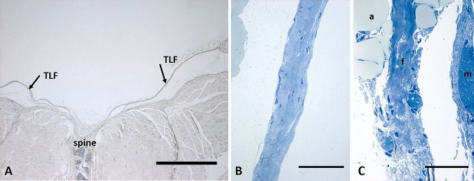
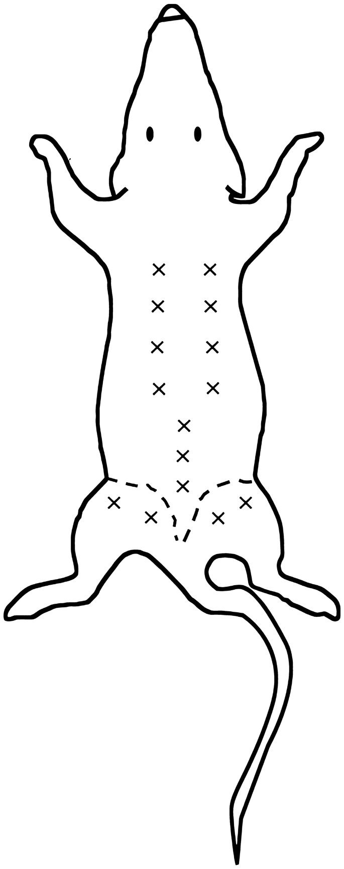
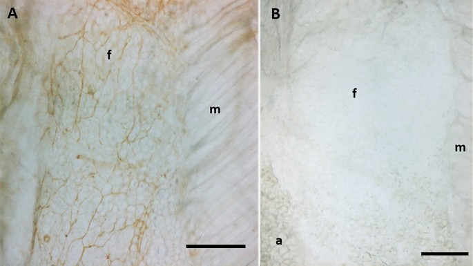
The mean thickness of the posterior layer of TLF was 34.3 µm ± 12.3 µm, whereas the mean thickness of the gluteal region was 21.3 µm ± 10.4 µm (Fig. 1, analysis performed in seven semithin sections stained with toluidine blue, 5 < areas < 10 for each section, using Image J software). The analysis of density innervation per area or per side (right/left) of TLF and gluteal fascia didn’t highlight any significant difference comparing random areas stained with the same antibody (data not shown): all the areas of TLF, from thoraco to sacral region (identified in Fig. 2), exhibited the same distribution of innervation and the same density; on the other side also the gluteal fascia didn’t show any difference according to the mapped region. Figure 3A shows that the fascial tissue is pervaded entirely by a dense network of nerves individualized by S100 antibody, ending on the border with the muscle, which is not at all equally innervated. The specificity of the staining was demonstrated by the absence of reaction in the negative control (Fig. 3B).
Figure 1
The alternative text for this image may have been generated using AI.
Thickness of thoracolumbar and gluteal fascia: (A) 5 µm section of fascia, spine and muscle, stained with ematoxylin and eosin; arrows indicate the thoracolumbar fascia (TLF); scale bar: 1000 µm; (B) 0.5 µm semithin section of thoracolumbar fascia stained with Toluidine Blue; scale bar: 50 µm; (C) 0.5 µm semithin section of gluteal fascia stained with Toluidine Blue; a: adipocytes; f: gluteal fascia; m: muscle; scale bar: 50 µm.
Figure 2
The alternative text for this image may have been generated using AI.
Analysis of density innervation per area and side (right and left): “ × ” indicate each area analyzed after immunohistochemical stains: from thoraco to sacral region of thoracolumbar fascia (both right and left side), and gluteal fascia (right and left side).
Figure 3
The alternative text for this image may have been generated using AI.
Different density of innervation of fascia and muscle: A: S100 immunohistochemistry reaction of thoracolumbar fascia (f) and latissimus dorsi muscle (m). B: negative control with the omission of the primary antibody. (a): adypocytes; (f): fascia; (m): muscle. Scale bar: 300 µm.
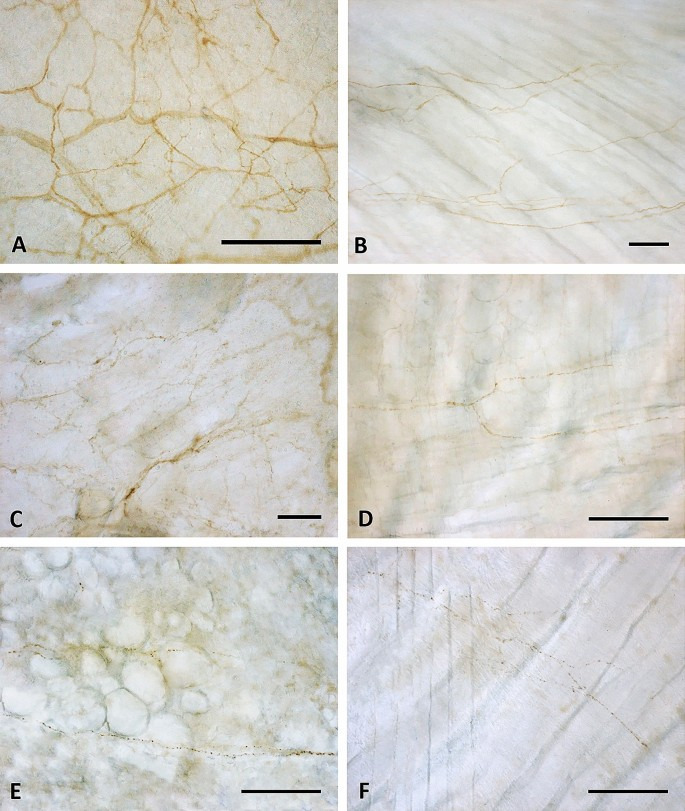
Tyrosine Hydroxylase showed less positivity (Fig. 4E–F), with only some positive single nerve filaments. On the contrary, the distribution of PGP 9.5 and S100 reaction were qualitatively similar (Fig. 4C–D,A–B, respectively), with a rhomboid and dense thin network of nerves homogeneously distributed in all the samples area analyzed. The reaction with PGP 9.5 antibody showed, however, thinner positive filaments less contrasted: for this reason the quantitative and morphometric analysis to compare TLF and gluteal fascia were performed on S100-stained images. The results are reported on Figs. 4 and 5. Both by qualitative (Fig. 4B,D,F) and quantitative (Fig. 5) analysis emerged in a clear and evident way that the gluteal fascia was less innervated with respect to the TLF. All the analyzed parameters (percentage of innervated area, density of branching points, length and thickness of the nerve structures) were statistically significantly lower in the epymysial gluteal fascia with respect to the aponeurotic thoracolumbar fascia. In particular, the latter showed a percentage of innervated area equal to 9.01 ± 0.98%, with 500.9 ± 43.1 branching points per mm2. Instead in the gluteal fascia the positive area was equal to 2.78 ± 0.6%, with a number of branching points of 140.3 ± 31.6/mm2 (p < 0.01, t-test gluteal fascia vs. TLF). Moreover, the length of the nerves in the gluteal fascia was 3.2 ± 0.6 mm, showing a very statistically significant difference (p < 0.01) with respect to the TLF (mean length 87.1 ± 1.0 mm). In the latter one, also the thickness of the nerve structures was higher (5.8 ± 0.2 µm in TLF vs 4.9 ± 0.2 µm in gluteal fascia, p-value < 0.05).
Figure 4
The alternative text for this image may have been generated using AI.
Innervation of thoracolumbar and gluteal fascia: Thoracolumbar fascia (A,C,E) and gluteal fascia (B,D,F) samples stained with S100 (A,B), PGP 9.5 (C,D) and Tyrosine Hydroxylase (E,F) antibodies. Scale bars: 100 µm.
Figure 5
The alternative text for this image may have been generated using AI.
Morphometric analysis of the pattern of S-100 positive fibers in thoracolumbar and gluteal fascia: positive area (%), branching points (BP) density (number/mm2), length of nerve structures (mm), thickness of nerve structures (µm) in thoracolumbar fascia (TLF) and gluteal fascia. *p < 0.05; **p < 0.01, t test.
The TH immunohistochemistry showed that in both the fasciae the positive area is around 0.08%, thus leading to a ratio S100/TH positivity of 112.1 in the thoracolumbar fascia and 34.6 in the gluteal fascia (Fig. 6).
Figure 6
The alternative text for this image may have been generated using AI.
Autonomic innervation in thoracolumbar and gluteal fascia: fraction of area (IR area %) positive to Tyrosine Hydroxylase (TH) and values of S100/TH ratio, in thoracolumbar fascia (TLF) and gluteal fascia, expressed as mean ± standard error mean.
A deeper analysis by TEM of the nerves that cross the fascia permitted us to highlight that the majority of the nerve structures (with both myelinic and amyelinic axons) are in the midst of collagen bundles, and not in the muscle (Fig. 7B–C–D) or in the adipose tissue (Fig. 7A).
Figure 7
The alternative text for this image may have been generated using AI.
Analysis of nerves inside the fascial tissue: (A) Floating thoracolumbar fascia stained with anti-S100 antibody and ematoxylin: the nervous structures are S100 positive (n: small nerve, arrows indicate single nerve fibers), whereas blood vessels are not stained (v: vessel; *: endothelial cells; a: adipocytes). (B) Semithin section of thoracolumbar fascia, whose boxes show nerve structures in the midst of collagen bundles of the fascial layers. (C) and (D): TEM images of a small nerve fiber in the inner layer (C) or in the outer layer (D) of the TLF, with both myelinc and unmyelinic axons. m: muscle; TLF: thoracolumbar fascia; mAx: myelinic axon; unAx: unmyelinic axon. Scale bars: (A) and (B) 30 µm; (C) 3 µm; (D) 2 µm.
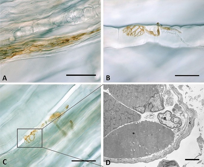
Lastly, the analysis found no presence of any corpuscle in all of the thoracolumbar samples analyzed. In the gluteal fascia, and more specifically in the perimysium and endomysium closely connected to this fascia, Golgi tendon organs (Fig. 8A), neuromuscular junctions (Fig. 8B) and muscle spindles (Fig. 8C–D) have been identified.
Figure 8
The alternative text for this image may have been generated using AI.
S100-positive corpuscles in gluteal fascia: Golgi tendon organ (A), neuromuscular junction (B) and muscle spindles (C) in gluteal fascia. In (D) is shown the TEM analysis of the muscle spindle: a connective tissue capsule (c) wraps a small group of muscle fibers (*) and nerve fibers (#). Scale bars: 50 µm (A,B,C), 5 µm (D).
Discussion
This work highlights for the first time the concept that we cannot consider all muscular fasciae as a similar structure. Indeed the TLF and the gluteal fasciae present different densities of innervation with nerve structures of different thicknesses. However, according to our results, both the gluteal and TLF have the same density of autonomic nerve fibers (0.08%). Among all the possible mediators of the autonomic nervous system, we have checked tyrosine hydroxylase that catalyzes the rate limiting step in this synthesis of catecholamines. The positive finding of these fasciae to this enzyme suggests a possible role of the autonomic nervous system in the opening and closing of the vessels inside fasciae, and consequently, it can play a role in the ischemia of fascial tissue. As the amount of sympathetic nerve elements are similar in the two fasciae, we can hypothesize that they are sensitive to ischemia in a similar way. Furthermore, the finding of sympathetic nerve fibers in TLF and gluteal fascia allows to consider that people subjected to a state of chronic stress may have increased levels of pain in those areas, as sympathetic activity is greater under psychological stress15. Indeed, it is well recognized that chronic stress can alter the physiological cross-talk between brain and biological systems, leading to compromised functions on the nervous, immune, endocrine, and metabolic systems16. This result can help to explain the increased levels of low back pain in patients with chronic stressfull state, a known effect but the mechanisms of which are not yet fully understood17.
Although the gluteal and TL fascia present the same density of autonomic innervation, they show a totally different amount of free nerve endings, leading to hypothesize that the major amount is due to the presence of sensitive free nerve endings and suggesting that they can have a different role in proprioception and pain perception. This study also confirms the absence of mechanoreceptors, like Pacini and Ruffini corpuscles, in the thoracolumbar fascia and gluteal fascia, as already demonstrated by Tesarz et al.7. It is probable that these receptors are usually localized in the superficial adipose tissue and superficial fascia, with the function of perceiving mechanical stimuli6, and where the superficial fascia joints the deep fascia, as in the retinacula surrounding joints, in the palmar and plantar fascia18,19,20,21, to increase the proprioception of these areas.
The fascial tissue showed a greater and homogeneous distribution of the nerve network with respect to the adjacent muscular tissue (Fig. 3-A). These results are in line with an our previous work about the innervation of the human hip joint, which showed that muscles (vasto-lateral and gluteus medius) were crossed by large nerves bundles (mean number 12 ± 6.1/cm2, mean diameter 36.4 ± 13.4 μm), presumably motor nerves, whereas the fasciae were invaded by networks of small nerve fibers (33 ± 2.5/cm2, mean size 19.1 ± 7.2 μm in the superficial fascia, and 19.0 ± 5.0/cm2, mean diameter 15.5 ± 9.4 μm, in the deep fascia)22. These findings lead to the revealing that the pain perception is higher in the fascia with respect to the muscle, highlighting the importance to preserve the fascial structures during surgery and to target to the fascial structures during a manual treatment. It is well know that the TLF could be a source of pain, as demonstrated by Langevin et al.9, Tesarz et al.7, Schilder et al.23, e. g., probably because it can feel the tensions coming from different muscles. Indeed the TLF is not related to a specific muscle, but gives insertions to both the latissimus dorsi, gluteus maximus and external oblique muscles24, and also on the inner side it adheres to the serrati posteriors fascia and to the erector spinae aponeurosis5. All these muscles stretch the TLF and can create a deformation of the network of the free nerve endings that we have just described. They form a thin and delicate net, strongly connected with the extracellular matrix of the fascial tissue, and consequently are particularly responsive to stretch, shear loading and mechanical stimuli. According to Hoheisel and coauthors25 the free nerve endings inside TLF may function as proprioceptors, in the absence of Pacini and Ruffini receptors. Based on this information, we can consider the thoracolumbar fascia as a large proprioceptive element that merges all the tensions coming from its associated muscles. Due to trauma, overuse, poor posture etc., these tensions can become unbalanced, resulting in an anomalous deformation of the free nerve endings, creating deficient motor patterns and eventual pain perceived by the CNS. Bednar et al.26 found an alteration in both the histological structure and the degree of innervation of TLF in patients with chronic low back pain. This possibility has surely to be further studied to better explain the possible role of TLF in nonspecific low back pain.
There are minimal studies about the possible role of the gluteal fascia in pain, but the density and type of innervation found in this work suggest that this gluteal fascia is probably less sensitive and consequently plays a minor role as pain generator. The gluteal fascia is totally adherent to the underlying muscle and is connected with some muscle spindles and Golgi corpuscles. These nerve corpuscles are specialized to feel the state of contraction of the various muscular bundles and to regulate them. It is well known that their capsules are totally in continuity with the perimysium and endomysium of the muscle27, and consequently any alteration of the intramuscular connective tissue can alter the sensitivity of these receptors, as demonstrated for dystrophy28. We also know that the intrafusal spindle muscular fibers can stretch the surrounding connective tissue, and consequently through fascial continuity among endomysium, perimysium and epimysium affect the tension of the gluteal fascia. Therefore, we can hypothesize that the nerve network in the gluteal fascia can unify all the tensions coming from the muscle spindles of the gluteus maximus muscle and transmit by way of the Central Nervous System a decoded input about the state of contraction of the gluteus maximus. So, we can suggest that the epymisial fasciae works more to coordinate the actions of the various motor units present in the underlying muscle, rather than just regulating the tensions originating from many directions as the TLF does.
Methods
Samples collection
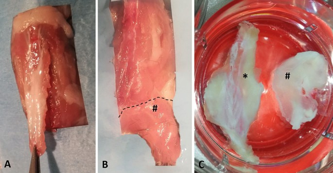
The experiments were performed on seven adult C57-BL mice (all males, mean age 8 weeks). All animal procedures were approved by the ethical committee of the University of Padova, in agreement with the guidelines of the Italian Department of Health, and in compliance with the ARRIVE (Animal Research: Reporting of In Vivo Experiments) guidelines. The specimens were collected from Thoracolumbar Fascia (TLF), as an example of the aponeurotic fascia, and gluteal fascia (GF), as an example of epimysial fascia. The posterior layer of the TLF, from the sacral to the thoracic region was separated from the spine (Fig. 9A) and maintained as an unique specimen with its orientation during all the steps. The gluteal fascia layer was removed from the sacral column to the gluteal region (Fig. 9B).
The fascia collected from one mice was fixed for 24 h in 10% formalin solution and for two weeks in 10% EDTA, then embedded in paraffin and cut in four-μm-thick sections for classical histological analysis and ematoxylin stains (Fig. 1-A). The fascia tissues from the others six mices were divided in three parts: one small piece of tissue (2 × 5 mm) was collected, subdivided in 15 small fragments and processed for semithin sections analysis and TEM analysis (see Transmission Electron Microscopy paragraph), and two were processed for Immunohistochemistry floating reaction (Fig. 9C), using randomly one of these for S100 staining (total of six samples), and one for TH or PGP stainings (three samples for each antibody), following the protocol described in the paragraph below.
Figure 9
The alternative text for this image may have been generated using AI.
Samples collection and macroscopic images of thoracolumbar fascia and gluteal fascia: (A) collection of thoracolumbar fascia; (B) collection of gluteal fascia (#); (C) floating samples of thoracolumbar fascia (*) and gluteal fascia (#).
Immunohistochemical method
The floating fascia samples of thoracolumbar and gluteal region were fixed in formalin 10% in Phosphate Buffer Saline (PBS). After repeated washings in PBS, endogen peroxidases were blocked with 1% H2O2 in PBS for 20 min at room temperature. The floating tissues were then pre-incubated with a blocking buffer (BSA 0.1% in PBS) for 60 min at room temperature and then incubated in rabbit polyclonal Anti S100 (Dako, dilution 1:4000), Rabbit Anti Tyrosine Hydroxylase (TH, GeneTex, dilution diluited 1:700), Rabbit Anti PGP (GeneTex, dilution 1:500) in the same pre-incubation buffer and maintained overnight at 4 °C. We selected these antibodies because S100 is specific for Schwann cells forming myelin, TH stains the sympathetic nerve fibres, the anti-PGP is widely used as a marker for all peripheral nerve fibers. After repeated PBS washing, the floating thoracolumbar and gluteal fasciae were incubated for 1 h in goat anti rabbit HRP (Jackson ImmunoResearch—Laboratories, Inc., Cambridge -UK) diluted 1:300 in the same pre-incubation buffer and washed in PBS. Negative controls underwent the same protocol steps, with the omission of the primary antibodies. The reaction was then developed with 3,3′-diaminobenzidine (Liquid DAB + substrate Chromogen System kit Dako Corp, Carpinteria, CA, USA) and stopped with distilled water.
Image acquisition
The images were acquired by using Leica DMR microscope (Leica Microsystems, Wetzlar, Germany).
For the image processing at least 10 pictures for each sample were acquired at 40X enlargement, to analyze the pattern of nerve fibers. Each sample was also entirely analyzed under light microscope to visualize any corpuscles.
Image processing and analysis
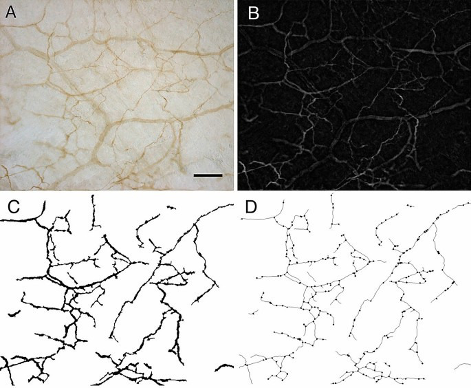
To morphometrically characterize the pattern of nerve fibers, the images of S100-stained samples of thoracolumbar and gluteal fascia underwent steps of image processing and analysis performed with ImageJ software as illustrated in Fig. 10.
Figure 10
The alternative text for this image may have been generated using AI.
Images processing and morphometric analysis of the pattern of nerve fibers: (A) original picture of one S100 reaction; (B) top-hat filter applied in picture A to enhance the contrast; (C) binary image of nerve network; (D) final binary skeleton derived from picture C and identification of the branching points. Scale bar: 100 µm.
Briefly, after shading correction, removal of background signals and manual adjustment of each image to eliminate non-nervous structures, a top-hat filter29 was applied (Fig. 10B) to enhance the contrast between the pattern of nerve fibers and the background. An adaptive discrimination procedure30 was then applied to select nerve profiles. This method operated with a local threshold: the mean grey value of a neighboring region was calculated for every pixel (by a 15 × 15 pixel low-pass filter) and this value plus an offset threshold constant defined the local threshold for that pixel. After interactive editing of the remaining artifacts, a binary image of the nerve network was then obtained (see Fig. 10C). By using binary thinning procedures31 the binary skeleton of this image was finally derived (Fig. 10D) and the branching points identified32. Nerve density was estimated from the binary image by evaluating the area fraction covered by the fibers, while from the binary skeleton image the total length of the nerve network and the density of branching points were estimated. From these primary morphometric parameters, the mean thickness of the nerve fibers was also derived as the ratio between the area of the network and its length.
Furthermore, nerve density was estimated also from images of TH-stained samples to estimate the fraction of area (%) positive to Tyrosine Hydoxylase and to calculate the S100/TH ratio.
Statistical analysis
Student’s t test was used to verify significant differences when comparing data of area of positivity to S100 (%), branching points density (number/mm2), length (mm) and thickness (µm) of the nerve structures, and relative percentage of positivity of tyrosine hydroxylase of the thoracolumbar fascia and the gluteal fascia.
Transmission electron microscopy (TEM)
The specimens of thoracolumbar and gluteal fasciae were fixed in 2.5% glutaraldehyde (Serva Electrophoresis, Heidelberg, Germany) in 0.1 M phosphate buffer, pH 7.3, and post-fixed in 1% osmium tetroxide (Agar Scientific Elektron Technology, Stansted, UK) in 0.1 M phosphate buffer, dehydrated in a graded alcohol series and then embedded in Epoxy Embedding Medium Kit (45349, Sigma-Aldrich, St. Gallen, Switzerland). Semithin (0.5 µm) and ultrathin (60 nm) sections were cut with the ultramicrotome RMC-PTX PowerTome (Boeckeler Instruments, Arizona –USA). Semithin sections were stained with 1% Toluidine blue solution. Ultrathin sections were collected on 300-mesh copper grids, counterstained with 1% uranyl acetate and then with Sato’s lead. Specimens were observed by a Hitachi H-300 Transmission Electron Microscope (Japan).
Data availability
All data generated or analysed during this study are included in this published article.
References