CAFE

생리학, 생화학

Mucosal Immunology of Food Allergy

작성자문형철|작성시간20.02.21|조회수727 목록 댓글 0

BEYOND REASON


원인은 모르지만 음식알레르기가 증가하고 있음. 


Figure 1Mechanism of oral tolerance induction.

Antigens are captured in the lamina propria and Peyer’s patch and carried to the mesenteric lymph node (MLN) by CD103+ dendritic cells, which induce gut-homing (α4β7+) induced regulatory T cells (iTregs) by a mechanism dependent on TGF-β, retinoic acid (RA) and indoleamine-2,3-dioxygenase (IDO). Dendritic cells induce gut-homing IgA-secreting plasma cells also through RA-dependent mechanisms. Gut-homing iTregs are expanded in the lamina propria by IL-10-expressing CX3CR1+macrophages. These iTregs can then suppress systemic immune responses, including allergic sensitization, in an antigen-specific manner. The immune mechanism of Th3 induction has not been established as for iTregs, but Th3 cells induced by feeding also suppress systemic immune responses.


Figure 2. Impact of environmental factors on allergic sensitization.

Green arrows and text indicate factors either known or suggested to promote allergic sensitization; red arrows and text indicate factors suppressive to allergic sensitization (known or suggested). Experimental adjuvant can induce allergic sensitization through IL-33 release from epithelial cells, driving Th2 responses via OX40L on dendritic cells. In skin, damage and decreased barrier function can function as physiological adjuvants driving this process, through the cytokine TSLP acting on dendritic cells, and through activating intraepithelial lymphocytes (IELs) to promote a Th2 response. Dietary factors including vitamin D, vitamin A, aryl hydrocarbon receptor (AHR) ligands, and folate are thought to promote regulatory responses or suppress inflammatory responses, while a high-fat diet (HFD) promotes inflammatory responses. The gut microbiota or its constituents can suppress aspects of the allergic immune response, directly as shown on the left or through the induction of regulatory T cells as shown on the right. Effector mechanisms of food allergy involve IgE antibodies and cells such as basophils and mast cells. Microbiota can suppress basophils, or through regulatory T cells (iTregs) suppress Th2 cells that are central to generating IgE and allergic effector cells. Factors that promote generation of Th2 cells (adjuvants) promote food allergy.


Figure 3. Mechanisms of systemic and local manifestations of food allergy.

Mast cells are central to both local and systemic manifestations of food allergy. Antigen disseminated systemically can trigger distal reactions (urticaria, bronchospasm) through mechanisms dependent on histamine and platelet activating factor (PAF). Gastrointestinal manifestations of food allergy in mice are dependent on repeated exposure to the food allergen that drives an allergic inflammation (dependent on Th2-derived cytokines, including IL-4, IL-13, and IL-9) and mastocytosis that is necessary for the local symptoms. PAF and serotonin mediate the local acute gastrointestinal response (diarrhea) to allergen exposure.


Review
Mucosal Immunology of Food Allergy

Under an Elsevier user license
open archive

Food allergies are increasing in preval‎ence at a higher rate than can be explained by genetic factors, suggesting a role for as yet unidentified environmental factors. In this review, we summarize the state of knowledge about the healthy immune response to antigens in the diet and the basis of immune deviation that results in immunoglobulin E (IgE) sensitization and allergic reactivity to foods. The intestinal epithelium forms the interface between the external environment and the mucosal immune system, and emerging data suggest that the interaction between intestinal epithelial cells and mucosal dendritic cells is of particular importance in determining the outcome of immune responses to dietary antigens. Exposure to food allergens through non-oral routes, in particular through the skin, is increasingly recognized as a potentially important factor in the increasing rate of food allergy. There are many open questions on the role of environmental factors, such as dietary factors and microbiota, in the development of food allergy, but data suggest that both have an important modulatory effect on the mucosal immune system. Finally, we discuss recent developments in our understanding of immune mechanisms of clinical manifestations of food allergy. New experimental tools, particularly in the field of genomics and the microbiome, are likely to shed light on factors responsible for the growing clinical problem of food allergy.

Main Text

Introduction

Food allergy is an immune-mediated adverse reaction to food and is a growing clinical problem. It is not currently understood why some individuals develop allergic sensitization to allergenic foods while the majority of individuals are immunologically tolerant, but evidence suggests that environmental factors are important. In this review, we will outline what is known about the healthy immune response to foods and what is currently understood about the immune mechanisms leading to allergic sensitization. Although the field is young and there is a lack of a comprehensive understanding of risk factors associated with development of food allergy, we will review emerging literature on the role of diet, gut microbiota, and exposure to food allergens through non-oral routes in the development of food allergy. Finally, immune mechanisms responsible for the different clinical manifestations of food allergy will be discussed.

Food Allergy

Food allergy is defined as “an adverse health effect arising from a specific immune response that occurs reproducibly on exposure to a given food” [1]and encompasses a range of disorders from IgE-mediated anaphylaxis to delayed cell-mediated reactions affecting the gastrointestinal tract, respiratory tract or skin. For the purpose of this review, we will focus primarily on the food allergies mediated by immunoglobulin E (IgE); readers are referred to recent reviews for information on cell-mediated disorders, including food-protein-induced enterocolitis syndrome [2] or eosinophilic esophagitis [3]. IgE-mediated reactions typically occur within two hours of ingestion of the food, and involve the skin, gastrointestinal tract, respiratory tract and, less frequently, the cardiovascular system. In the most severe case of anaphylaxis, multiple organ systems are involved and can include cardiovascular collapse.

Although true preval‎ence rates of IgE-mediated food allergy have been difficult to accurately estimate, as reviewed by Sicherer [4], a systematic review of the literature concluded that food allergy affects greater than 2% and less than 10% of the general population [5]. A population-based study from Australia demonstrated a greater than 10% rate of IgE-mediated food allergy in response to oral challenge in a cohort of infants at one year of age [6]. The majority of children allergic to milk or egg will outgrow their food allergies, but in contrast peanut, tree nut, fish and shellfish allergies are most commonly lifelong. The preval‎ence of food allergy is increasing [7]. In repeated studies of the preval‎ence of peanut allergy in a US population based on random telephone survey performed in 1997, 2002, and 2008, it was found that the preval‎ence of peanut allergy increased from 0.4% to 0.8% to 1.4% over the three time points 8910. Rates of peanut allergy over 1% are consistent with reports from Canada [11], the UK [12], and Australia [13], including studies that employed physician eval‎uation and food challenges. Although there is a significant genetic component to food allergy, the rapid rise in the preval‎ence of food allergy suggests an important contribution from environmental factors. We will review emerging research on the role of factors that may contribute to the rising rate of allergic sensitization to foods through modulatory effects on the mucosal immune system.

Introduction to the Mucosal Immune System

The gastrointestinal tract is the largest reservoir of immune cells in the body, and the function of the mucosal immune system is to protect the large surface area of the gastrointestinal tract from invading pathogens and to keep the commensal microbiota compartmentalized. The mucosal immune system is divided from the gut lumen by a single layer of columnar epithelial cells, which secrete a number of factors that contribute to barrier function, including mucins, antimicrobial peptides, and trefoil factors. The epithelial cells also transport antibodies, particularly IgA, into the intestinal lumen where these antibodies can contribute to barrier function by excluding the uptake of antigens or microbes. (Readers are referred elsewhere [14] for a comprehensive review of regulation of the epithelial barrier function.) Below the epithelium lies the mucosa, which is densely populated by resident immune cells, including CD4+ and CD8+ T effector and regulatory T cells, antibody-secreting B cells, and mononuclear phagocytes (macrophages and dendritic cells): see Box 1 for a glossary of immunological terms used in this review. Eosinophils are also resident cells in the normal intestinal mucosa. These scattered immune cells make up the effector sites of the mucosal immune system, and function to recognize and clear pathogenic challenges from the environment. Inductive sites — locations where antigen-specific cellular and humoral immune responses are first generated — include Peyer’s patches and isolated lymphoid follicles that sit directly within the gut mucosa, and the mesenteric lymph nodes that drain the gastrointestinal tract, including Peyer’s patches, via the lymph. A specialized subset of epithelial cells termed M cells overlies the Peyer’s patches and contributes to the selective uptake of particulate antigens at this site. In contrast, soluble antigens are primarily taken up across the epithelium lining the villi and are carried into the mesenteric lymph nodes.

Box 1

Glossary of immunology terminology.

Innate immunityAn immediate response to infection or damage that is mediated through recognition of conserved patterns. Recognition occurs via pattern recognition receptors, such as Toll-like receptors, NOD-like receptor proteins, and endocytic/phagocytic pattern recognition receptors, such as mannose receptor or DC-SIGN. Effector mechanisms of innate immunity are diverse and include the release of anti-microbial peptides, killing through release of cytotoxic molecules, phagocytosis and intracellular killing of microbes, complement activation, release of innate cytokines (IL-1β, IL-6, and tumor necrosis factor-α, TNF-α) and release of acute-phase proteins from the liver that assist in pathogen clearance.
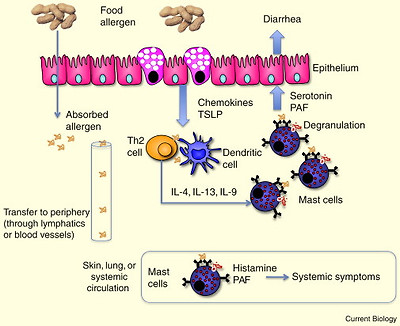
Adaptive immunityAdaptive immunity is mediated by T and B lymphocytes, which recognize specific antigens through their T-cell receptor (TCR) and B-cell receptor (BCR), respectively. The adaptive immune responses are slower than the innate immune response, but have the property of memory, such that subsequent responses to the same antigen are more rapid. T cells can be divided into cytotoxic (CD8+) and helper (CD4+) T cells. CD4+ T cells can be further subdivided based on the cytokines they secrete. In general, Th1 cells express the transcription factor T-bet, secrete IFN-γ and assist in clearing intracellular pathogens by activating macrophages; Th2 cells express the transcription factor GATA-3, secrete IL-4, IL-5 and IL-13, and provide help to B cells for antibody production and promote allergic responses; Th17 cells express the transcription factor RORγt, secrete IL-17, and play an important role in microbial clearance and a destructive role in autoimmunity. Regulatory T cells suppress these effector T-cell responses, and regulatory T cells come in a variety of different phenotypes. B cells contribute to adaptive immunity by secreting antibodies that can neutralize pathogens, activate complement, and activate antibody-dependent cell-mediated cytotoxicity pathways. Antibodies of the IgE isotype trigger allergic reactions.
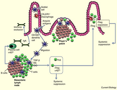
Dendritic cellsDendritic cells are resident cells of tissues, acquire antigen and process it for presentation on major histomcompatibility complex (MHC) molecules, and migrate to lymph nodes for presentation of antigen to T lymphocytes. Dendritic cells are the most potent antigen-presenting cells capable of priming naïve T cells.
CytokinesA cytokine is a small protein made by a cell that affects the behavior of other cells. Cytokines can be grouped into categories based on their function. Innate cytokines (IL-1β, IL-6, TNFα) are released by a variety of stromal cells and macrophages early in infection and play a role in pathogen clearance. Th1, Th2, and Th17 cytokines are outlined under ‘adaptive immunity’. Cytokines released from innate cells and antigen-presenting cells can act on naïve T cells to influence their developmental fate. IL-12 promotes the development of Th1 cells, IL-4 promotes the development of Th2 cells, and IL-6 and TGF-β promote the development of Th17 cells.
Chemokines and homing moleculesChemokines are chemotactic cytokines that act on G-protein-coupled surface receptors. Cells respond to chemokines through migration along a chemokine gradient. Tissue-specific homing is mediated through chemokine–chemokine receptor interaction as well as integrin–ligand interactions. Gut homing is mediated by the chemokine receptor CCR9 and its ligand CCL25 (expressed in the small intestine), as well as the integrin α4β7 and its ligand MAdCAM, which is also differentially expressed in the intestine. Skin homing is mediated by the chemokine receptors CCR4 and CCR10 in response to the ligands CCL17, CCL22, and CCL27, which are expressed in the skin. In addition, the integrin cutaneous lymphocyte antigen (CLA) on lymphocytes binds to E-selectin.
Regulatory T cellsRegulatory T cells can be CD4+ or CD8+, although the most commonly recognized regulatory T cell is the CD4+ Foxp3+ form. Foxp3+ regulatory T cells can be generated in the thymus (natural regulatory T cells ) or generated from naïve T cells in the periphery (induced regulatory T cells). Regulatory T cells use a variety of suppressive mechanisms, including cytokines (TGF-β and IL-10), cell–cell interaction (for example, involving the surface molecule CTLA-4), and can also suppress by cytolytic activity.
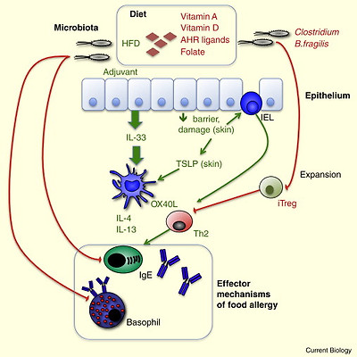
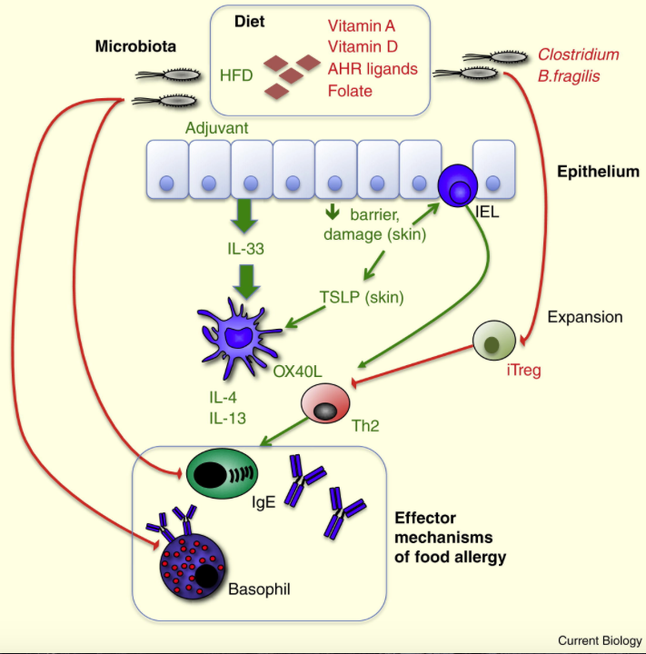
Co-stimulatory moleculesThese molecules are expressed on the surface of antigen-presenting cells and provide additional signals to shape the nature of the T-cell response. Co-stimulatory molecules can provide an activating signal, such as when B7.1 and B7.2 bind to CD28 on the T-cell surface. Alternatively, inhibitory signals can be provided by co-stimulatory molecules, such as when B7.1 and B7.2 bind to CTLA-4 instead of CD28 and provide an ‘off’ switch to T-cell activation. Expression‎ of other co-stimulatory molecules, such as OX40L, or Notch ligands on antigen-presenting cells can influence the differentiation of naïve T cells through binding to surface receptors on T cells.
Innate lymphoid cellsInnate lymphoid cells are innate cells that share characteristics with CD4+T cells, but do not recognize specific antigen within the context of MHC molecules. These cells represent a distinct category of different innate immune cells, but features in common include localization at mucosal sites and production of cytokines that play a role in mucosal homeostasis and regulation of the intestinal microbiota as well as in pathogenesis of intestinal and allergic disease.

A challenge faced by the mucosal immune system is to discriminate between harmful pathogens and harmless or beneficial commensal organisms. The lack of reactivity to the commensal flora is in part achieved by a specialized regulatory milieu that may also shape the immune response to antigens derived from the diet. Antigen-presenting cells and macrophages of the intestinal mucosa are hypo-responsive to many microbial ligands [15] and secrete high levels of immunoregulatory cytokines, such as interleukin-10 (IL-10) [16]. However, the mechanisms responsible for suppression of inappropriate immune reactivity to microbes or food antigens may be quite different. For example, targeted deletion of a number of immune regulatory components (such as IL-10) will result in a microbiota-dependent colitis [17], but spontaneous allergy to dietary components or allergic inflammation is rarely described in these models.

Normal Response to Foods: Mechanisms of Oral Tolerance

As was first described by Osborne and Wells in 1911 [18], antigens that are present in the diet induce a systemic non-responsiveness that has been termed ‘oral tolerance’. Antigens present in the diet could not elicit an immune response after parenteral immunization. This has also been demonstrated in human subjects after feeding and immunization with the neoantigen keyhole limpet hemocyanin 1920. The finding that this was an active regulatory response was demonstrated by transferring tolerance to naïve mice through the transfer of T cells. However, T-cell anergy and deletion — mechanisms of tolerance that cannot be transferred to naïve mice — have also been shown to contribute to the development of oral tolerance in some experimental systems.

T cells with a suppressive phenotype were described in the Peyer’s patches and later in the spleen after antigen feeding 212223, but several studies have shown that the Peyer’s patches are dispensable for the development of tolerance 2425. In contrast, the mesenteric lymph nodes have been shown to be required for the development of tolerance. Surgical or immunological ablation of the mesenteric lymph nodes prevents the development of oral tolerance 2627. The migration of immune cells to the intestine and from the intestine to the draining lymph nodes is controlled by the regulated expression‎ of chemotactic cytokines (chemokines) and chemokine receptors. The chemokine receptor CCR7, which is necessary for the migration of dendritic cells from the lamina propria to the mesenteric lymph nodes where CCR7 ligands are expressed, is also necessary for the development of oral tolerance [27]. Global expansion of dendritic cells with the growth factor Flt3L decreases the amount of antigen required to induce oral tolerance [28], and tolerance can be transferred to naïve animals by transfer of dendritic cells derived from the intestinal lamina propria [29]. There have been no reports about the effect of dendritic cell ablation on the development of oral tolerance.

Role of Gastrointestinal Antigen-Presenting Cells

The biology of the mononuclear phagocyte system in the gastrointestinal tract is currently the focus of a great deal of research and some debate. The initial description that dendritic cells could extend dendrites across the intestinal epithelium 3031 and capture bacteria from the intestinal lumen generated much interest in this cell subset. These CD11c+ dendritic cells express the chemokine receptor CX3CR1, which is necessary for the extension of the transepithelial dendrites into the lumen [32]. These cells are derived from monocytes in an M-CSF receptor-dependent manner, express the surface marker F4/80, do not constitutively express CCR7, and do not migrate in lymph 3334. By transcriptional analysis they are more closely related to tissue macrophages than tissue dendritic cells [35]. In contrast, CX3CR1 CD103+dendritic cells in the lamina propria constitutively express CCR7 and migrate to the mesenteric lymph nodes. These cells arise from common dendritic cell progenitors and pre-dendritic cells in a GM-CSF receptor-dependent manner 3334. Although CD103+ dendritic cells do not extend dendrites between epithelial cells, they sample antigens from goblet cells, which function as a conduit between the lumen and the mucosa [36]. In addition, they may sample antigens that are transported across the intestinal epithelium by a number of different mechanisms, including transcellular or paracellular pathways or sampling by M cells. CD103+ dendritic cells isolated from the mesenteric lymph nodes of mice and humans preferentially induce the generation of gut-homing CD4+ Foxp3+ regulatory T cells from naïve T cells. Although CD103 is a marker of these dendritic cells, it is not itself involved in the generation of gut-homing regulatory T cells. These CD103+ dendritic cells express high levels of the enzyme retinal dehydrogenase 2 (RALDH2), which converts retinal to retinoic acid. Both gut homing activity (via expression‎ of chemokine receptors and integrins) as well as the regulatory activity of the responder T cells are dependent on retinoic acid derived from CD103+ dendritic cells. An important source of the precursor for retinal comes from the diet in the form of vitamin A (readers are referred to a recent review [37] on the role of retinoic acid in mucosal immunity).

In addition to generating regulatory T cells through expression‎ of RALDH2, CD103+ dendritic cells also use a variety of other mechanisms that promote the development of regulatory T cells, involving other enzymes, such as indoleamine 2,3-dioxygenase, and secretion of immunosuppressive cytokines, such as transforming growth factor β (TGF- β), which promote the development of regulatory T cells from naïve T cells 383940. This subset of intestinal dendritic cells also promotes the development of IgA-secreting, gut-homing plasma cells through retinoic acid and IL-6 4142. Although CD103+dendritic cells are migratory, whereas CX3CR1 macrophages are not, a population of CX3CR1intermediate F4/80 CD103 CD11c+ dendritic cells has been shown to be present in intestine-draining lymph collected under steady-state conditions, and these cells are preferentially able to induce the secretion of cytokines interferon-γ (IFN-γ) and IL-17 from naïve T cells [43]. IFN-γ and IL-17 are cytokines produced by effector T cells and are involved in protection against pathogens. Their role in normal gut homeostasis or response to dietary antigens is poorly understood, but it is possible that different subsets of intestinal dendritic cells have different roles. For example, the CD103+dendritic cells may be regulatory under steady-state conditions, while CD103dendritic cells prime the immune system to respond to pathogens. The balance of these dendritic cells may play an important role in maintaining gut homeostasis. Furthermore, although CX3CR1high intestinal macrophages do not migrate to initiate T-cell responses, they express high levels of IL-10 in response to interaction with the intestinal microbiota and are thought to be important for the expansion of regulatory T cells that have homed back to the gut after initial priming in the mesenteric lymph nodes [44]. In addition to cues provided by the dendritic cells themselves to naïve T cells, stromal cells of the mesenteric lymph nodes express high levels of retinoic acid-generating enzymes and are important for the generation of a gut-homing phenotype in lymphocytes [45]. These stromal cells may also contribute to the immunoregulatory function of the gastrointestinal tissue.

Role of Regulatory T Cells

As mentioned previously, oral tolerance can be transferred to a naïve mouse through the transfer of T cells. This was initially shown using either CD4+ or CD8+ T cells. CD8+ T cells can mediate tolerance, as shown by the induction of tolerance by feeding mice the CD8 peptide SIINFEKL, but are not necessary for the development of oral tolerance and do not provide tolerance against allergic inflammation driven by CD4+ T helper 2 (Th2) cells [46]. Feeding of antigen to either mice or humans induces a regulatory CD4+ T cell termed the Th3 cell, characterized by surface expression‎ of latency associated peptide (LAP), a propeptide non-covalently associated with TGF-β that keeps the complex in an inactive form 4748. These cells do not express CD25 or Foxp3, and their mechanism of suppression is dependent on TGF-β. Th3 cells are induced by antigen feeding, and their transfer provides protection against experimental autoimmunity. Administration of antigen by the oral route also induces a population of CD4+ Foxp3+ CD25+ regulatory T cells, termed induced regulatory T cells, from Foxp3 precursors [49]. Th3 cells can also promote the development of induced regulatory T cells through their secretion of TGF-β [50]. Foxp3+ induced regulatory T cells are required for oral tolerance, as shown by studies in which they are specifically depleted 4451.

Although it has been clearly demonstrated that the default immune response to an experimentally fed antigen is one of tolerance mediated by regulatory T cells, it is not understood whether the lack of clinical reactivity to normal dietary antigens is an active regulatory response mediated by these T cells. In mice and humans, a lack of Foxp3+ T cells leads to enteropathy, eczema, and elevated IgE. Severe food allergy can also occur as one manifestation of Foxp3mutations [52]. Mice having a more selective defect in induced Foxp3+regulatory T cells, with normal levels of thymically derived ‘natural’ regulatory T cells, exhibit a Th2-skewed mucosal inflammation and the generation of an antibody response (of undetermined isotype) to antigens in the chow [53]. These data show that regulatory T cells may have a role in the constitutive suppression of responses to mucosally derived antigens. Children who have outgrown their milk allergy have an increased frequency of circulating CD4+CD25+ regulatory T cells after an oral milk challenge and reduced proliferation of milk-specific T cells; depletion of CD4+ CD25+ regulatory T cells restores the in vitro proliferative response in milk-tolerant subjects [54]. These data suggest that regulatory T cells may be involved in the development of clinical tolerance to food allergens. The presence of food-antigen-specific regulatory T cells has not yet been demonstrated in healthy human subjects. Figure 1summarizes known mechanisms of tolerance to dietary antigens, and readers are referred to a recent article [55] for a comprehensive review of the mechanisms of oral tolerance.

Figure 1. Mechanism of oral tolerance induction.

Antigens are captured in the lamina propria and Peyer’s patch and carried to the mesenteric lymph node (MLN) by CD103+ dendritic cells, which induce gut-homing (α4β7+) induced regulatory T cells (iTregs) by a mechanism dependent on TGF-β, retinoic acid (RA) and indoleamine-2,3-dioxygenase (IDO). Dendritic cells induce gut-homing IgA-secreting plasma cells also through RA-dependent mechanisms. Gut-homing iTregs are expanded in the lamina propria by IL-10-expressing CX3CR1+macrophages. These iTregs can then suppress systemic immune responses, including allergic sensitization, in an antigen-specific manner. The immune mechanism of Th3 induction has not been established as for iTregs, but Th3 cells induced by feeding also suppress systemic immune responses.

Factors Promoting Sensitization to Foods

Experimental Adjuvants

Mice, like most humans, do not normally develop allergic sensitization to potent food allergens, such as peanut. In the absence of an adjuvant, the default response is oral tolerance. The two adjuvants that have been used most widely to induce hypersensitivity to food are bacterial toxins: cholera toxin (CT) and staphylococcal enterotoxin B (SEB). When these are co-administered orally with a variety of food antigens, they induce an antigen-specific Th2 response and antigen-specific IgE production 5657. When mice are re-challenged with the food antigen in the absence of adjuvant, sensitized mice will undergo systemic anaphylaxis that can be measured by a drop in body temperature and is associated with other symptoms affecting the skin and respiratory tract. By examining how these adjuvants alter the mucosal immune system, we hope to gain a better understanding of immune pathways that can lead to allergic sensitization. Oral administration of CT leads to a depletion of dendritic cells from the lamina propria and migration to the mesenteric lymph nodes 5859. Dendritic cells from CT-fed mice have an enhanced capacity to induce Th2 responses from naïve T cells, as well as enhanced production of IFN-γ and IL-17, which are not pro-allergic but instead are important in host defense. The normally tolerogenic CD103+ dendritic cells upregulate the surface co-stimulatory molecules OX40L and Jagged2, and neutralization of OX40L effectively suppresses the Th2 response while having no effect on the IFN-γ or IL-17 response [59]. Therefore, generation of the Th2 response by the adjuvant CT was dependent on surface expression‎ of OX40L by the gastrointestinal dendritic cells. The central role of OX40L in the induction of allergic sensitization to peanut was then confirmed and extended by Chu et al. [60], who found that repeated feeding of peanut plus CT led to upregulation of OX40L on mesenteric lymph node dendritic cells, and that neutralization of OX40L during sensitization suppressed the generation of peanut-specific IgE and clinical signs of anaphylaxis upon allergen challenge. Furthermore, they found that the induction of OX40L on gut dendritic cells was downstream of the upregulation of the cytokine IL-33 by the intestinal epithelium, but was independent of thymic stromal lymphopoietin (TSLP) or IL-25: IL-33, IL-25, and TSLP are epithelial-derived cytokines that have been shown to play an important role in the induction of Th2 immunity at mucosal surfaces [61]. CT is unlikely to play a role in human food allergy, but these data suggest that comparable factors that can promote the upregulation of IL-33 by intestinal epithelium may be important factors in promoting the development of food allergy. Environmental factors derived from the diet or the microbiota have direct access to intestinal epithelial cells and may regulate epithelial gene expression‎ to promote allergic sensitization.

It is not clear whether CT suppresses the generation of regulatory T cells in response to antigen feeding in addition to promoting the development of Th2 effector cells. Ganeshan et al. [57] reported that feeding of SEB was associated with a decrease in TGF-β and Foxp3 expression‎ in splenocytes, but did not specifically look at the induction of regulatory T cells. It is likely that both a suppression of regulatory responses and an induction of effector Th2 responses are necessary for the generation of allergic sensitization to antigens in the diet. Neutralization of CTLA-4, a surface molecule that mediates the regulatory activity of regulatory T cells, leads to enhanced sensitization when adjuvant is present, but does not in itself break tolerance [62].

Dietary Factors

Environmental influences affecting the mucosal immune system are most likely to come from the diet, the commensal microbiota, or interactions between the two. Data from the US Department of Agriculture comparing dietary intake between 1970 and 2000 show significant increases in caloric intake as well as the contribution of different food groups to that energy intake (http://www.ers.usda.gov/Data/FoodConsumption). For example, energy from lipids/fats/oils and legumes/nuts/soy increased while energy from meat/dairy/eggs decreased. Studies utilizing the National Health and Nutrition Examination Survey (NHANES) database or other cohorts have reported associations between food-specific IgE levels and vitamin D [63], folate [64] and obesity [65]; however, it should be pointed out that concern has been raised about the accuracy of utilizing food-specific IgE levels reported in this general population as a proxy measure of clinical food allergy [66]. Some of these dietary associations with food allergy are supported by studies that potentially explain the immune basis of this association. Alternatively, there are dietary components that are well known to influence mucosal immunity, but whose role in food allergy remains unstudied. An example of the latter is vitamin A. As outlined above, the vitamin A metabolite retinoic acid is associated with the generation of regulatory T cells, IgA-secreting B cells, and upregulation of gut homing receptors on T and B cells 394167. Multiple steps are required for the transformation of dietary vitamin A to retinoic acid, but the last step is mediated by RALDH enzymes. Different isoforms of RALDH are highly expressed in the gastrointestinal tissues, and cell sources of retinoic acid include intestinal epithelial cells, CD103+ dendritic cells, and stromal cells of the mesenteric lymph node [68]. Vitamin A-depleted mice lack T cells and IgA-secreting B cells in the intestinal mucosa, but not elsewhere in the body [69]. Although retinoic acid promotes the development of regulatory T cells and suppresses the development of Th17 effector T cells in vitro, findings in vivosuggest that retinoic acid is important for induction of both regulatory and effector T cells in the gastrointestinal mucosa [70]. For example, in the context of high levels of the cytokine IL-15, which is a feature of celiac disease, retinoic acid can promote the development of pathogenic Th1 and Th17 effector cells rather than regulatory T cells and promote celiac-like disease in mice [71]. Thus, although much of the in vitro evidence suggests that vitamin A should be regulatory, it has the potential to contribute to the pathogenesis of intestinal disease. Additional mechanistic studies are needed, utilizing both animal models of food allergy as well as human subjects to determine how dietary vitamin A levels may specifically influence the development of food allergy.

Vitamin D is another nutrient of significant interest in the development of tolerance or allergic sensitization to food antigens. Serum vitamin D levels are generated both by dietary intake and synthesis of vitamin D in the skin in response to exposure to sunlight. Addition of vitamin D to immunizations given by the subcutaneous route induces protective mucosal immunity [72]. Like retinoic acid, vitamin D can influence T- and B-cell homing, and has been shown to suppress the development of Th17 cells in vivo [73]. Vitamin D deficiency also results in a depletion of a subset of intraepithelial CD8αα-expressing T lymphocytes from the intestinal mucosa 7475. CD8αα serves as a T-cell receptor repressor and has been hypothesized to give these intraepithelial lymphocytes a regulatory function in vivo. As with vitamin A, there is a lack of mechanistic studies in mouse models of food allergy to test the role of vitamin D in disease. However, there are some data from human subjects that hint at a potential protective role of vitamin D in food allergy. Incidence of food allergy varies with latitude in Australia [76] and, in the US, rates of epinephrine auto-injector prescriptions (as a proxy measurement of food allergy) and emergency room visits for acute allergic reactions vary according to latitude, with highest rates in the northeast [77]. This has been hypothesized to be due to associated variations in vitamin D levels resulting from differences in sun exposure. The association of serum vitamin D levels with allergic sensitization to common allergens was analyzed using data available in the NHANES database. It was found that low vitamin D was associated with elevated sensitization to peanut and shrimp, but not milk or egg [63]. This was observed for children, but not adults. It is not clear why this nutrient would have antigen-selective effects on allergic sensitization, but this may relate to either age or the route of antigen exposure.

Aryl hydrocarbon receptor (AHR) ligands are derived from environmental and dietary sources and have a significant effect on both innate and adaptive immunity in the intestinal mucosa. Mice lacking AHR have a profound loss of intestinal intraepithelial γδ T lymphocytes [78], which are primarily innate-type cells with invariant T-cell receptor usage. In addition, CD8αα intraepithelial lymphocytes and lymphoid tissue inducer cells are also lost from the intestine of mice lacking the AHR 7879. This loss was associated with an increased microbial burden in the small intestine and increased susceptibility to experimental colitis. The intestinal source of immunomodulatory ligands for the AHR is primarily from food. There are also data showing that AHR ligands modulate adaptive immune responses in the intestine. Depending on the ligand used, AHR ligands could either enhance regulatory T cells and suppress autoimmunity, or enhance IL-17 and exacerbate autoimmunity [80]. Immunomodulatory effects of exogenous AHR ligands have been shown in an experimental model of peanut allergy [81], but it has not yet been shown whether diet-derived AHR ligands may be protective against the development of food allergy. Cruciferous vegetables (broccoli, cabbage, and brussels sprouts) are rich sources of AHR ligands and may provide a means for modulating the immune function of the intestine.

Obesity is associated with elevated levels of IgE, including IgE specific for common food allergens [65]. Obesity is considered to be a systemic inflammatory state, and the presence of innate lymphoid cells producing Th2 cytokines within adipose tissue suggests a potential mechanistic link between obesity and allergic disease [82]. However, a high-fat diet is also associated with significant changes in the local immune milieu, such as increased inflammatory markers in the intestinal mucosa, increased epithelial permeability and elevated levels of lipopolysaccharide in the serum, and changes in the intestinal microbiome 8384. Interestingly, it was recently reported that both changes in the microbiome and obesity induced by a high-fat diet were downstream of changes in the mucosal immune system, in particular lymphotoxin-dependent elevation of the cytokines IL-22 and IL-23 [85]. IL-22 is a cytokine produced by innate lymphoid cells that has both protective and pro-inflammatory functions and IL-23 is a cytokine produced by antigen-presenting cells that contributes to the development of Th17 cells. The immune changes observed in these studies were not of a Th2 phenotype that would contribute to allergic sensitization in food allergy, but demonstrate that the innate lymphoid milieu of the intestine can be significantly altered by the fat composition of the diet. The role of a high-fat diet in food sensitization needs to be addressed experimentally.

Microbiota

A second major environmental factor likely to influence susceptibility to food allergy is the microbiota, through modulation of the mucosal immune system. All surfaces of the human body are populated to various degrees with complex communities of microorganisms, with the greatest density of colonization (>1012 organisms/cm3) found within the lower gastrointestinal tract. Studies of the last 15 years using gnotobiotic mice (germ-free or reconstituted with defined microbiota) have identified a critical role for the gut microbiota in shaping the intestinal mucosa with regard to immune and barrier function, and influencing systemic immunity and metabolism [86]. It is now well established that the gastrointestinal inflammation associated with a number of experimental models of inflammatory bowel disease is dependent on the gut microbiota. Furthermore, reconstitution of germ-free mice with defined microbiota has identified microbial species that promote or protect against gastrointestinal inflammation [87]. Particular species, including Bacteroides fragilis and Clostridium species, promote the development of regulatory T cells in the gastrointestinal tract 888990, whereas other commensal species, such as segmented filamentous bacteria, promote the development of Th17 cells [91]. Therefore, the relative abundance of various constituents of the commensal flora may determine the immune balance of the gastrointestinal tract. In the context of allergic disease, recent work has shown that signals from the commensal microbiota suppress IgE production and basophil development [92], although it has been shown that colonization with particular species, including Clostridium, can suppress allergic sensitization [90]. Noval Rivas et al. [93] recently demonstrated that a specific strain of mice susceptible to experimental food allergy, which experienced allergic reactions following gastric challenge with a model food allergen, had a signature microbiota that differed from wild-type, allergen-sensitized but non-reactive mice. Transfer of this microbiota to germ-free wild-type mice promoted allergen-specific IgE sensitization and allergic reactions following gastric challenge in recipient mice, implicating the microbiota in the development of food allergy. Germ-free mice have under-developed mucosal immune systems, so it can be difficult to interpret studies utilizing germ-free mice in studies of diseases affecting the mucosal immune system. However, germ-free mice and mice treated with broad-spectrum antibiotics have increased susceptibility to sensitization to food allergens 9495, highlighting the general inhibitory activity of the microbiota on the generation of an IgE response to foods. The power of the gnotobiotic approach comes from the ability to reconstitute germ-free mice with defined microbiota, particularly if combined with knowledge about the constituents of the human microbiome associated with a particular disease. Recent studies have shown that mice can be reconstituted with human microbiota, and that microbiota can then be reproducibly controlled by altering the diet 9697. This is a very powerful system for dissecting the intersection of diet, microbiota, and human disease. It has been reported that colonization of germ-free mice with microbiota from healthy human infants is protective against allergic sensitization to milk allergens [98].

What is known about the intestinal microbiome in food allergy? Using culture-based methods, dysbiosis has been described in a few studies comparing allergic children to healthy controls. Bifidobacterium species have been found to be either decreased or unchanged in allergic populations 99100. Only one study to date has used 16S-based sequencing to compare the microbiota of a group of 20 infants with atopic eczema (many with IgE sensitization to foods) to a control group without atopic manifestations, with a finding of reduced diversity in the microbiome of infants with atopic eczema [101]. In addition to reductions in diversity, there were also significant reductions in abundance of the phylum Proteabacteria, but no differences in Bifidobacterium were observed. These studies point to a possible dysbiosis in food allergy. Larger prospective studies are needed to identify changes in the microbiome that precede the development of food allergy as defined by rigorous clinical criteria, and controlling for dietary intervention. If a signature of dysbiosis can be associated with food allergy, further mechanistic studies utilizing gnotobiotic mice reconstituted with human microbiota would be warranted. In addition to bacterial constituents of the microbiome, the human virome may also play a significant role in shaping the mucosal immune system [102].

Exposure to Food Allergens by Non-Oral Routes

The majority of peanut-allergic children experience their first allergic reaction to peanut on their first known ingestion of peanut [103]. This suggests that sensitization resulting in IgE production must have occurred by exposure through a non-oral route. Two leading theories for the basis of this sensitization are in utero sensitization, or by household exposure through non-oral routes.

In utero sensitization to allergens is thought to occur by trans-placental transfer of antigen, and exposure of an immature, Th2-biased (and potentially genetically predisposed) immune system to the allergen resulting in generation of allergic sensitization. Measurement of discordant allergen-specific IgE in cord blood, newborn blood, and maternal blood supports the hypothesis that IgE sensitization may occur in utero [104]. The frequency of positive allergen-specific IgE in cord blood mirrors the relative frequency of sensitization in children (milk > egg > peanut). The clinical data on the impact of allergen in the maternal diet has been mixed, with some reports showing no effect [105] and others showing that maternal ingestion of peanut during pregnancy was a risk factor for the development of peanut sensitization [106]. Studies utilizing animal models to assess the impact of maternal allergen exposure during pregnancy and/or lactation have generally found that exposure inhibits the development of allergic sensitization in offspring 107108109. The mechanism of in utero tolerance induction has not been addressed, but maternal exposure during lactation induces oral tolerance in offspring through a TGF-beta-dependent mechanism [108]. However, there is no evidence from human studies supporting a protective role of maternal allergen ingestion in food allergy.

Household non-oral exposure to peanut allergen was reported to be a risk factor for peanut allergy in children, independent of maternal ingestion during pregnancy and lactation [110]. One relevant route of exposure was hypothesized to be the skin. Peripheral blood mononuclear cells (PBMCs) from peanut allergic donors were fractionated into skin homing (expressing the integrin cutaneous lymphocyte antigen, CLA) or gut homing (expressing the integrin β7) fractions followed by re-stimulation with peanut extract in the presence of autologous dendritic cells [111]. Cells proliferated in the CLA+fraction, but not the β7+ fraction, leading the authors to suggest that, based on this homing receptor usage, sensitization likely occurred via the skin. Peanut-specific CD4+ T cells identified by tetramer staining from peanut-allergic subjects were classified as having frequent expression‎ of the skin-homing chemokine receptor CCR4 (although infrequent expression‎ of CLA) and infrequent expression‎ of β7, one component of the α4β7 gut-homing integrin [112]. Further evidence fitting the hypothesis that sensitization to peanut may occur through the skin comes from data showing that mutations in the filaggrin gene that are associated with decreased skin barrier function are a risk factor for peanut allergy [113]. Data from animal models show that allergic sensitization can be readily induced by topical allergen exposure. However, these models show that additional factors beyond exposure are necessary to induce sensitization, including adjuvant [114], or damaging the skin by repeatedly applying and removing tape to induce upregulation of cytokines, such as TSLP and IL-21 115116, or induction of damage signals that activate intraepithelial lymphocytes to participate in allergic sensitization [117]. Similar to what is observed with the oral route, additional signals are needed beyond allergen exposure to result in allergic sensitization. The concept of damage or barrier defects contributing to allergic sensitization via the skin is supported by clinical observations that atopic dermatitis is a significant risk factor for the development of food allergy [105]. These data indicate that the skin may be a highly relevant site of sensitization to food allergens. Understanding the environmental or intrinsic co-factors that provide adjuvant activity to food allergens upon exposure by any route is critical to our understanding of why food allergy is a steadily increasing clinical problem. As in the gut, the skin microbiome has also been shown to shape skin immunity [118] and may contribute to sensitization through cutaneous routes. Figure 2 illustrates environmental factors that may contribute to allergic sensitization to food allergens.

Figure 2. Impact of environmental factors on allergic sensitization.

Green arrows and text indicate factors either known or suggested to promote allergic sensitization; red arrows and text indicate factors suppressive to allergic sensitization (known or suggested). Experimental adjuvant can induce allergic sensitization through IL-33 release from epithelial cells, driving Th2 responses via OX40L on dendritic cells. In skin, damage and decreased barrier function can function as physiological adjuvants driving this process, through the cytokine TSLP acting on dendritic cells, and through activating intraepithelial lymphocytes (IELs) to promote a Th2 response. Dietary factors including vitamin D, vitamin A, aryl hydrocarbon receptor (AHR) ligands, and folate are thought to promote regulatory responses or suppress inflammatory responses, while a high-fat diet (HFD) promotes inflammatory responses. The gut microbiota or its constituents can suppress aspects of the allergic immune response, directly as shown on the left or through the induction of regulatory T cells as shown on the right. Effector mechanisms of food allergy involve IgE antibodies and cells such as basophils and mast cells. Microbiota can suppress basophils, or through regulatory T cells (iTregs) suppress Th2 cells that are central to generating IgE and allergic effector cells. Factors that promote generation of Th2 cells (adjuvants) promote food allergy.

The practice of excluding allergens, such as peanut, from the diet in infancy has been proposed as a contributing factor in the rising incidence of peanut allergy by allowing cutaneous exposure to occur in the absence of tolerizing signals from the ingestion of peanut. The Learning Early About Peanut Allergy (LEAP) study is an ongoing interventional study to test whether early introduction of peanut into the diet can prevent the development of clinical peanut allergy. Infants with egg allergy, atopic dermatitis, or both were recruited into the study, and recently published baseline parameters confirmed the strong association of eczema with peanut allergy [119]. The results from this trial will determine whether tolerance to peanut can be induced in infants at high risk for peanut allergy by early introduction of the allergen into the diet.

There is little direct information on the immune communication between gut and skin in food allergy, but clearly such communication occurs. The skin is the most common site of manifestations of food allergy in response to oral food challenge [120]. Evidence outlined above suggests that the skin is also a likely site of initial sensitization to foods. It is not clear whether IgE sensitization to food antigens through the skin requires any homing of T or B cells to the gastrointestinal tract, but there is evidence that topical immunization leads to a mucosal antibody response that is dependent on vitamin A in the diet [121]. Clearly the potential exists for effector T and B cells to home to the gut after antigen exposure via the skin. Despite the fact that ingested food is the trigger of food allergic reactions, the fact that both priming and manifestations of food allergy can occur exclusively via the skin raises the interesting question of whether food allergy should continue to be considered a gastrointestinal disease.

Pathophysiology of Anaphylactic Reactions to Foods

Once sensitization has occurred and antigen-specific IgE has been generated, oral re-exposure to that allergen can lead to local or systemic manifestations of food allergy. In human disease, the most common manifestations of reactions are cutaneous (urticaria), followed by gastrointestinal and respiratory reactions [120]. Interestingly, gastrointestinal symptoms occur in less than 50% of reactions despite exposure being via the oral route. Studies from mice indicate that food allergens must be absorbed systemically in order to induce symptoms of anaphylaxis 122123 and that factors that interfere with passage across the intestinal epithelium (such as heat aggregation of the antigens) prevent anaphylactic symptoms 124125. The ability to induce anaphylaxis by the oral route in mice is highly strain and allergen dependent, but resistant strains of mice will undergo anaphylaxis when systemically challenged 126127. The basis of this susceptibility to oral allergen challenge in mice is not currently understood. Systemic anaphylaxis in mice is mediated predominantly by IgE with some contribution from IgG and is dependent on mast cells, except in the case of intravenous allergen challenge when macrophages can also contribute to symptoms 128129. Histamine and platelet activating factor (PAF) are both required for systemic manifestations of anaphylaxis in mice 127130. Antihistamines are commonly used in the treatment of acute reactions to foods, and clinical studies have found PAF to be elevated in the serum of subjects undergoing anaphylaxis, particularly those with more severe symptoms [131]. PAF acetylhydrolase, the enzyme that breaks down PAF, is also decreased in subjects with severe anaphylaxis [132]. PAF is elevated in other disorders such as necrotizing enterocolitis [133], but it remains to be determined whether PAF is a biomarker of severe anaphylaxis in humans or whether it plays a role in the pathogenesis.

Gastrointestinal manifestations of allergy in mice are observed secondarily to a T-cell-driven allergic inflammation that is induced by repeated allergen exposure 134135. It is not clear why mice require repeated allergen challenge to induce gastrointestinal symptoms, but this may relate to the very low baseline level of resident mast cells in the normal small intestinal lamina propria of mice. During this repeated allergen exposure, the release of chemokines, including CCL20, induces the accumulation of antigen-specific Th2 cells in the intestinal mucosa 135136. During repeated allergen exposure, the epithelial cytokine TSLP is also critical for generating this local inflammation that is necessary for symptom onset [137]. T-cell cytokines, including IL-4, IL-13, and IL-9, are critical for generating this allergic inflammation in the gut 138139, likely upstream of the expansion of mucosal mast cells. As with systemic manifestations of anaphylaxis, allergen-induced diarrhea is dependent on mast cells and the severity correlates with intestinal mast cell numbers 134140. Mast cells contribute to the allergic response not only through acute release of mediators, but also as a major local source of IL-13 [141]. Symptoms are not mediated by histamine but instead by serotonin together with PAF [134]. It is not yet clear whether different manifestations of food allergy in human disease are mediated by different immune mechanisms, or how antigen absorbed through the intestinal mucosa may pass through the gut to trigger systemic symptoms without inducing local gastrointestinal symptoms. A schematic illustrating our current understanding of the mechanisms underlying manifestations of food allergy is shown in Figure 3.

Figure 3. Mechanisms of systemic and local manifestations of food allergy.

Mast cells are central to both local and systemic manifestations of food allergy. Antigen disseminated systemically can trigger distal reactions (urticaria, bronchospasm) through mechanisms dependent on histamine and platelet activating factor (PAF). Gastrointestinal manifestations of food allergy in mice are dependent on repeated exposure to the food allergen that drives an allergic inflammation (dependent on Th2-derived cytokines, including IL-4, IL-13, and IL-9) and mastocytosis that is necessary for the local symptoms. PAF and serotonin mediate the local acute gastrointestinal response (diarrhea) to allergen exposure.

Conclusions

We do not yet understand why the default immune response to a dietary antigen deviates from a suppressive response mediated by regulatory T cells to a Th2-biased response that promotes IgE class switching and allergic responses upon re-exposure. The rapidly increasing incidence of disease in westernized countries suggests a role for environmental factors. Emerging evidence points to dietary factors and the microbiome as important modifiers of the mucosal immune environment, but further research needs to be carried out to investigate the role of these and other environmental factors in the development of inappropriate allergic sensitization to foods. Identification of modifiable risk factors would have a significant beneficial impact on this perplexing disease.

References


다음검색
현재 게시글 추가 기능 열기

댓글

댓글 리스트
맨위로

카페 검색

카페 검색어 입력폼