BEYOND REASON
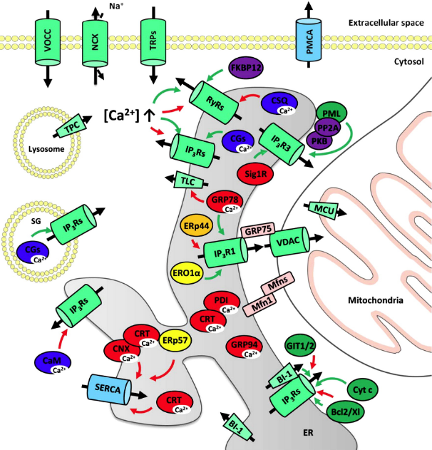
Fig. 1. Overview of the proteins involved in cellular Ca2 + handling. The ER and other membrane systems are represented with the main Ca2 +-transporting and Ca2 +-binding proteins, together with a number of regulatory proteins of importance for this review. Ca2 + channels and the Na +/Ca2 + exchanger are in light green, ATP-dependent Ca2 + pumps are in light blue, Ca2 +-binding proteins are in dark blue, chaperones, including Ca2 +-binding chaperones, are in red, oxidoreductases are in yellow, thioreductases are in orange, anti- and pro-apoptotic proteins are in dark green, regulatory proteins are in purple and linker proteins in salmon. Ca2 + fluxes are shown by the large black arrows, regulatory arrows are green (stimulatory effect) or red (inhibitory effect), while the flux of the counterion Na+ occurring at the level of the Na+/Ca2 + exchanger is represented by a small black arrow. Abbreviations are as defined in the text, except for the following ones that are only used in the figure: Bcl2/Xl, anti-apoptotic proteins of the Bcl-2 family; CaM, calmodulin; CGs, chromogranins; CNX, calnexin; CRT, calreticulin; Cyt c, cytochrome c; FKBP12, FK506-binding protein; GRP78, BiP/GRP78; MCU, mitochondrial Ca2 + uniporter; Mfn, mitofusin; NCX, Na+/Ca2 + exchanger; PKB, protein kinase B; PMCA, plasma-membrane Ca2 + ATPase; PP2A, protein phosphatase 2A; SG, secretory granule; Sig1R, sigma-1 receptor; TLC, translocon; TPC, two-pore channel; VDAC, voltage-dependent anion channel; VOCC, voltage-operated Ca2 + channels. For more information, see text.
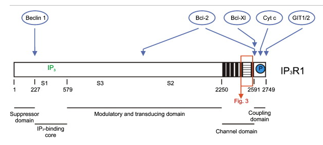
Fig. 2. Structure of the IP3R1 with indication of domains important in cell-fate regulation. The 5 functional domains of the IP3R1 are indicated, together with a number of crucial sites, like the IP3-binding site (IP3 in green) and the splice sites (S1, S2 and S3). Interaction sites of regulatory proteins related to apoptosis and autophagy (Beclin 1, Bcl-2, Bcl-Xl, cytochrome c, GIT1 and 2) are shown in blue and the protein kinase B-dependent phosphorylation site is represented by a blue circle. The 6 transmembrane helices are in black and the third luminal loop has a horizontal stripe pattern. This loop region is the interacting place for a number of important proteins discussed in detail in this review. This part of the IP3R1 (orange box) is therefore represented in detail in Fig. 3.
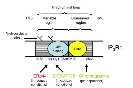
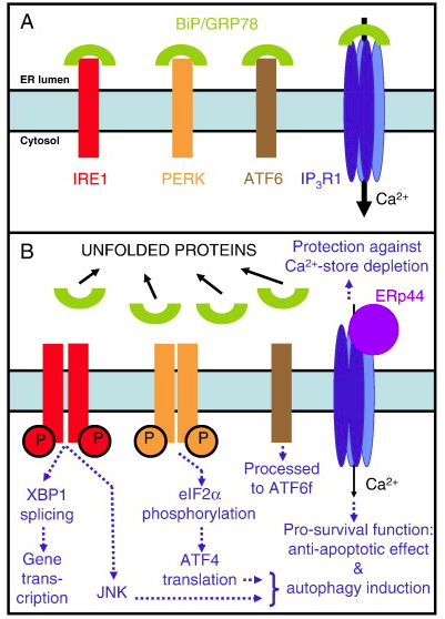
Fig. 4. The ER-stress sensors and the initial steps of the UPR. This new model includes not only the three canonical ER-stress sensors (IRE1, PERK and ATF6) but also the here proposed fourth ER-stress sensor, the IP3R1. Panel A represents the ER in normal, healthy cells. Panel B shows the activation of the ER-stress sensors during the adaptive phase of the UPR and the main steps involved in pro-survival signaling. The proteins are color-coded: BiP/GRP78 (green), IRE1 (red), PERK (orange), ATF6 (brown), IP3R1 (blue) and ERp44 (purple) are depicted. Dimerization and autophosphorylation (circled P) of IRE1 and PERK are shown. The processing of AFT6 is performed in the Golgi apparatus. Full black arrows indicate the magnitude of the Ca2 + fluxes. Dotted blue arrows indicate the cellular processes occurring after the activation of the ER-stress sensors. JNK denotes the activation of the c-Jun N-terminal kinase pathway. See text for further explanations.
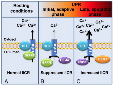
Fig. 5. Regulation of IP3R1 and IICR during ER stress and UPR progression. A. In resting conditions, the chaperone BiP/GRP78 (GRP78) stabilizes IP3R function and allows efficient Ca2 + channeling through the IP3R1. BI-1 can in these conditions interact with the IP3R and stimulate IICR. BI-1 is a pleiotropic regulator of cell function during ER stress, and a role in IP3R regulation during UPR is appealing. However, the role of its interaction with IP3R1 during ER stress was not yet ascertained. B. Induction of ER stress induces the UPR and both the dissociation of BiP/GRP78 and its replacement by ERp44 lead to a decreased IICR. C. Prolonged ER stress leads to hyperoxidation by ERO1α and displacement of ERp44. Subsequently IICR increases, leading to pro-apoptotic Ca2 + signaling.
ReviewRegulation of inositol 1,4,5-trisphosphate receptors during endoplasmic reticulum stress☆
Highlights
► ER stress is a condition of disturbed ER function. ► ER stress can lead to autophagy or apoptosis. ► Autophagy and apoptosis are affected by intracellular Ca2 + signaling. ► The activity of IP3Rs and other Ca2 +-handling proteins is modulated during ER stress. ► Ca2 + signals participate in cell-fate determination subsequently to ER stress.
Abbreviations
Keywords
1. Introduction
The endoplasmic reticulum (ER) forms an extensive intracellular network of tubules and cisterns, representing together the largest membrane system of animal cells [1]. The ER plays a crucial role in the synthesis, correct folding and sorting of proteins, but is also involved in many other functions like the synthesis of phospholipids, cholesterol and steroids, the degradation of glycogen, detoxification processes and, last but not least, intracellular Ca2 + signaling. Although the various functions are at least partly performed in different areas of the ER, they are not completely independent of each other [2], [3], [4], [5]. Importantly, several of these functions are coupled to the ER Ca2 + level [1], [3], [5]. A concerted regulation of the ER Ca2 +-uptake and the ER Ca2 +-release mechanisms is therefore essential for correct ER functioning [6], [7] and a decreased Ca2 + concentration in the ER can lead to a phenomenon called ER stress (see Part 3). Such an alteration in ER homeostasis is an upstream event in many pathological conditions [8], [9], [10], including many neurodegenerative diseases [11], [12].
The most ubiquitously expressed Ca2 +-release channel of the ER is the inositol 1,4,5-trisphosphate (IP3) receptor (IP3R). The IP3R and the IP3-induced Ca2 + release (IICR) resulting from IP3R activation have a central role in many cellular processes including the regulation of cell fate [13]. Their role in apoptosis and autophagy has been highlighted in a number of recent reviews [14], [15], [16], [17], [18], [19], [20]. The relation between the IP3R on the one hand and ER stress and the subsequent processes occurring in the cell, globally named unfolded protein response (UPR), on the other, has however, to the best of our knowledge, not yet been systematically reviewed.
In this review, we therefore will first discuss the function of the ER as a Ca2 + store and the various proteins hereby involved, including the IP3R in particular. We will then treat the phenomenon of ER stress and the subsequent UPR. Subsequently, we will go into further detail on how the IP3R and IICR are regulated during ER stress, and on how this will affect UPR progression and subsequent cell fate. The importance of the IP3R and IICR during the adaptive, pro-survival phase as well as, when the ER stress can not be alleviated, in cell death will be highlighted.
2. The ER as central player in cellular Ca2 + homeostasis
In mammalian cells, the ER forms the main intracellular Ca2 + reservoir. To function as a dynamic Ca2 + store, the ER basically contains three types of proteins (Fig. 1): Ca2 + pumps allowing active Ca2 + uptake, Ca2 +-binding proteins allowing the storage of significant amounts of Ca2 + in its lumen, and, last but not least, Ca2 + channels allowing a controlled release of Ca2 + into the cytosol in response to well-determined stimuli [2], [4], [13], [21], [22], [23]. Noteworthy, these proteins not only control the Ca2 +-loading level of the ER but are themselves often regulated positively or negatively by the luminal Ca2 + concentration [24].
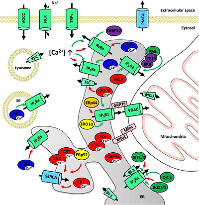
Fig. 1. Overview of the proteins involved in cellular Ca2 + handling. The ER and other membrane systems are represented with the main Ca2 +-transporting and Ca2 +-binding proteins, together with a number of regulatory proteins of importance for this review. Ca2 + channels and the Na +/Ca2 + exchanger are in light green, ATP-dependent Ca2 + pumps are in light blue, Ca2 +-binding proteins are in dark blue, chaperones, including Ca2 +-binding chaperones, are in red, oxidoreductases are in yellow, thioreductases are in orange, anti- and pro-apoptotic proteins are in dark green, regulatory proteins are in purple and linker proteins in salmon. Ca2 + fluxes are shown by the large black arrows, regulatory arrows are green (stimulatory effect) or red (inhibitory effect), while the flux of the counterion Na+ occurring at the level of the Na+/Ca2 + exchanger is represented by a small black arrow. Abbreviations are as defined in the text, except for the following ones that are only used in the figure: Bcl2/Xl, anti-apoptotic proteins of the Bcl-2 family; CaM, calmodulin; CGs, chromogranins; CNX, calnexin; CRT, calreticulin; Cyt c, cytochrome c; FKBP12, FK506-binding protein; GRP78, BiP/GRP78; MCU, mitochondrial Ca2 + uniporter; Mfn, mitofusin; NCX, Na+/Ca2 + exchanger; PKB, protein kinase B; PMCA, plasma-membrane Ca2 + ATPase; PP2A, protein phosphatase 2A; SG, secretory granule; Sig1R, sigma-1 receptor; TLC, translocon; TPC, two-pore channel; VDAC, voltage-dependent anion channel; VOCC, voltage-operated Ca2 + channels. For more information, see text.
2.1. Ca2 +-handling mechanisms of the ER
2.1.1. Ca2 + pumps
Active Ca2 + uptake in the ER is mediated by pumps, belonging to the sarco- and endoplasmic-reticulum Ca2 +-ATPase (SERCA) family (Fig. 1). Three different genes encode a SERCA pump (SERCA1, SERCA2, SERCA3) but the variety of Ca2 + pumps is increased by the existence of splice variants [25], [26], [27]. The crystal structure of SERCA1a was determined in various conditions, enabling the reconstitution of the almost complete reaction cycle at a structural level [28]. Structurally the SERCA pumps (molecular mass of ~ 110 kDa) are divided in the following domains: a nucleotide-binding domain, a phosphorylation domain, an actuator domain and transmembrane helices containing the Ca2 +-binding sites and joined by small luminal loops. SERCA2b is most widely expressed, and displays the highest Ca2 + affinity. In most cells Ca2 +-store loading therefore primarily depends on SERCA2b. All SERCA isoforms are characterized by a ratio of 2 Ca2 + ions transported per hydrolyzed ATP and are extremely sensitive to the inhibitor thapsigargin in the nanomolar range [25], [29], [30]. The latter is commonly used to induce ER stress, as it irreversibly blocks ER Ca2 + uptake and therefore leads to the complete depletion of the ER Ca2 + stores (see Part 3). Other specific, but reversible, inhibitors for the SERCA pumps are cyclopiazonic acid and 2,5-di-(t-butyl)-1,4-hydroquinone [25], [30].
2.1.2. Ca2 +-binding proteins
The Ca2 + ions taken up in the lumen of the ER are for the largest part bound to Ca2 +-binding proteins [31], [32] (Table 1 and Fig. 1). The ER in this way contains roughly 2 mM total Ca2 +, corresponding to a free Ca2 + concentration of about 500 μM, a concentration which is thus much higher than the free Ca2 + concentration in the cytosol [31], [33]. The luminal Ca2 +-binding proteins involved generally also function as chaperones. A prime example is calreticulin that accounts for most of the bound Ca2 + in the lumen of the ER. Calreticulin has a molecular mass of 46 kDa and is characterized by three functional domains: an N-domain, a centrally located P-domain and a C-domain [34]. All three domains interact in one way or another with various luminal ER proteins, including protein disulfide isomerase (PDI) and ERp57. The proline-rich P-domain has a high-affinity Ca2 +-binding site, while the C-domain contains > 20 low-affinity Ca2 +-binding sites (Table 1).
Table 1. ER Ca2 +-dependent chaperones determining luminal Ca2 + levels (see text and [32] for more explanations).
| Ca2 +-binding proteins (in alphabetical order) | Ca2 +-binding characteristics | References |
|---|---|---|
| BiP/GRP78 | 1 or 2 low-affinity sites | [35] |
| Calnexin | high capacity | [228] |
| Calreticulin | 1 high-affinity (Kd = 1 μM) site (P-domain) and 25 low-affinity (Kd = 2 mM) sites (C-domain) | [229] |
| GRP94 | 4 high-affinity (Kd = 2 μM) sites and 11 low-affinity (Kd = 0.6 mM) sites | [37] |
| PDI | 19 low-affinity (Kd = 4.7 mM) sites | [43] |
BiP/GRP78 is another important low-capacity, low-affinity Ca2 +-buffering protein, responsible for ~ 25% of the ER Ca2 + binding [35]. GRP94 is also an abundant ER Ca2 +-buffering protein, with 4 high-affinity and 11 low-affinity Ca2 +-binding sites [36]. These various proteins, as well as several members of the PDI family, are present in the IP3-sensitive Ca2 + stores [37], [38], [39], [40], [41], [42] where they participate to high-capacity, low-affinity ER Ca2 + binding [4], [43], [44], [45] (Table 1).
In addition to the Ca2 +-binding chaperones mentioned above, a number of other Ca2 +-binding proteins can be present in the ER. Calsequestrin, of which two isoforms (CSQ1 and CSQ2) exist, is a high-capacity (up to 80 mol Ca2 +/mol protein), low-affinity Ca2 +-binding protein predominantly present in skeletal and cardiac muscle, but also in some other tissues [32]. Calsequestrin changes extensively from conformation after Ca2 + binding, leading to its dimerization and polymerization. Finally, depending on the tissue, the Ca2 +-store levels, the polymerization and the phosphorylation level and the presence of other proteins like junctin and triadin, calsequestrin affects the function of the ryanodine receptor (RyR) (see Section 2.1.3) [32], [46].
Chromogranins A and B are two low-affinity, high-capacity Ca2 +-binding proteins of the granin family. Their Ca2 +-binding properties are pH-dependent with a maximal capacity between 50 and 100 mol Ca2 +/mol protein and a Kd in the 2–4 mM range [47]. These proteins are mostly found in the secretory granules but also in the ER of various types of (neuro)endocrine cells and neurons. Both chromogranins A and B interact with the various IP3R isoforms and thereby stimulate IICR [48], [49], [50]. Stimulation of the IP3R1 activity occurs by increasing channel open probability and mean open time [51], [52].
2.1.3. Ca2 +-release channels and the Ca2 +-leak mechanisms
Ca2 + ions can be released from the ER Ca2 + store in a controlled way upon cell stimulation [3], [22], [23]. The Ca2 + ions released by the members of 2 families of related intracellular Ca2 +-release channels, the IP3R and the RyR, form complex spatio-temporal signals in the cytosol, controlling a multitude of cell functions [13].
IP3Rs (Fig. 1) are ubiquitously expressed Ca2 +-release channels that are activated after phospholipase C activation and subsequent IP3 production [53], [54], [55], [56]. The resulting IICR leads to the formation of complex spatio-temporal Ca2 + signals regulating cell processes like fertilization, proliferation, differentiation, metabolism, secretion, contraction, synaptic plasticity, gene transcription and cell death [13]. These channels will be discussed in more detail in Section 2.2.
Three different genes encode a RyR. These proteins assemble to very large (> 2 MDa) tetrameric Ca2 +-release channels (RyR1, RyR2 and RyR3). RyR1 and RyR2 are expressed at a very high level in the sarcoplasmic reticulum of skeletal and cardiac muscle respectively [57], [58], [59], [60], [61]. RyRs are also expressed at significant levels in the ER (Fig. 1) of smooth muscle [62], [63], neurons (e.g. cerebellum and hippocampus) [64], [65], liver [66] and pancreatic acinar cells [67]. Although in most cell types the expression level of RyRs is much lower than that of IP3Rs, their physiological role can still be very important, as at each opening the RyRs release about 20 times more Ca2 + ions than IP3Rs [68].
In addition to the IP3R and RyR intracellular Ca2 +-release channels that are responsible for a controlled release of Ca2 + subsequent to a stimulus, the ER membrane displays an inherent Ca2 + leak. It is at this moment not yet clear which protein or proteins are responsible for this Ca2 + leak [6], [69], but several possible candidates have been proposed (Table 2 and Fig. 1), of which a number of them will be discussed later (see Section 3.4). Collectively, these channels are responsible, depending on the cell type, for the release of 20 to 200 μM Ca2 +/min, and together with the SERCA Ca2 +-uptake pumps, they thereby control the steady-state Ca2 + level in the ER under resting conditions [69].
Table 2. Proposed Ca2 +-leak mechanisms present in the ER. These proteins were all proposed to contribute to the basal Ca2 + leak out of the ER, but are not all discussed in the present review. In addition to the indicated proteins, the full-length endogenous ER Ca2 + channels or Ca2 + pumps may display an inherent Ca2 + leak and/or be activated in basal conditions and therefore participate in physiologic conditions to the basal Ca2 + leak [6], [69]. Please note that not all of the proteins listed below are necessarily expressed at significant levels in all cell types. Moreover, for some proteins contradictory results have been published, and cell-specific differences may explain some of them. For each candidate protein a number of pertinent references has therefore been selected.
2.2. Structure, function and regulation of the IP3R
2.2.1. IP3R structure
Three genes encode an IP3R leading to the expression of three distinct isoforms (IP3R1, IP3R2 and IP3R3) with additional splice variants. They form large tetrameric channels with a mass of about 1.2 MDa [53], [54], [55], [56]. The various IP3R isoforms have a similar structure but the latter was investigated to a greater depth for IP3R1. This includes high-resolution cryo-electron microscopy (the closed state of IP3R1 was recently resolved at a resolution of ~ 1 nm [70]) and X-ray crystallography. The latter could however until now only be performed on the N-terminal part of the receptor containing the IP3-binding site [71]. Recent studies performed at a resolution of 3.0–3.8 Å allowed the comparison of the apo and the IP3-bound form [72], [73].
At the functional level, the IP3R1 can be divided into five distinct domains (Fig. 2): an N-terminal suppressor domain (a.a. 1–225), an IP3-binding core (a.a. 226–578), a modulatory and transducing domain (a.a. 579–2275), a channel domain with 6 transmembrane helices (a.a. 2276–2589), and finally a C-terminal coupling domain (a.a. 2590–2749) [74]. Of particular interest for this review is the very small part of the IP3R that protrudes into the lumen of the ER. Basically, apart from the very short loops between the transmembrane helices 1 and 2, and 3 and 4, only the larger third loop of 106 a.a. located between transmembrane helices 5 and 6 is in contact with the ER lumen [75]. This loop (Fig. 3) consists of a variable region (66 a.a.) containing (in IP3R1) two N-glycosylation sites followed by a conserved region (40 a.a.) contributing to the channel pore.
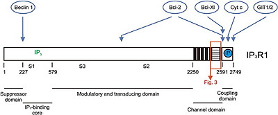
Fig. 2. Structure of the IP3R1 with indication of domains important in cell-fate regulation. The 5 functional domains of the IP3R1 are indicated, together with a number of crucial sites, like the IP3-binding site (IP3 in green) and the splice sites (S1, S2 and S3). Interaction sites of regulatory proteins related to apoptosis and autophagy (Beclin 1, Bcl-2, Bcl-Xl, cytochrome c, GIT1 and 2) are shown in blue and the protein kinase B-dependent phosphorylation site is represented by a blue circle. The 6 transmembrane helices are in black and the third luminal loop has a horizontal stripe pattern. This loop region is the interacting place for a number of important proteins discussed in detail in this review. This part of the IP3R1 (orange box) is therefore represented in detail in Fig. 3.
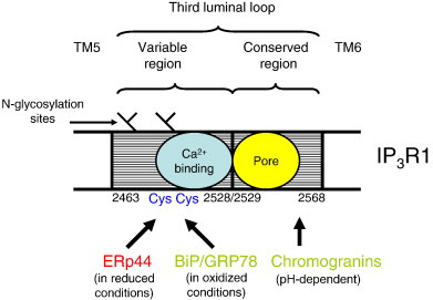
Fig. 3. Detailed structure of the third luminal loop of IP3R1, located between transmembrane helices 5 (TM5) and 6 (TM6). The variable and conserved regions are indicated, as well as the N-glycosylation sites [75], a region with a high density of negatively charged amino acids that can bind Ca2 + [176] and the pore domain. The interaction sites for ERp44 [112], BiP/GRP78 [113] and the chromogranins [111] are indicated. The latter interaction is pH-dependent and of particular importance in secretory granules. Please note that the interaction sites for ERp44 and BiP/GRP78 are specific for IP3R1, while chromogranins can interact with the three IP3R isoforms. The red color indicates an inhibitory action on IICR, and the green color a stimulatory effect. Cys2496 and Cys2504 that are important for ERp44 interaction are depicted (Cys).
2.2.2. Regulation of IP3R activity
The IP3R is activated by IP3 and the 3 isoforms are characterized by a different affinity for IP3, with IP3R2 having the highest and IP3R3 the lowest sensitivity [76], [77], [78], [79], [80]. In addition to IP3, cytosolic Ca2 + is considered to function as a co-agonist. Importantly, Ca2 + activates the IP3R at low concentrations, typically below 300 nM, while it inhibits the IP3R at higher concentrations [81], [82], [83], [84]. Also luminal Ca2 + is deemed important, and the depletion of the Ca2 + stores leads to a decreased IP3R sensitivity [85], [86], [87], [88].
Modulation of IP3R activity occurs by three main mechanisms: (1) the local environment, including pH, ATP and Mg2 + concentration and redox status; (2) its phosphorylation status, which depends on the activity of many different kinases and phosphatases, some of them forming a complex with the IP3R; (3) regulatory proteins that directly affect IP3R activity in either a stimulatory or an inhibitory way [53], [54], [56], [89].
With respect to the latter mode of modulation, several proteins regulate the IP3R in relation to cell-fate decisions. The regulation of the IP3R by anti-apoptotic B-cell lymphoma 2 (Bcl-2)-family members like Bcl-2 and Bcl-Xl (Fig. 2) is important for antagonizing apoptosis and has been extensively investigated [90], [91], [92], [93], [94], [95], [96], [97], [98], [99], [100]. In addition, other proteins interacting with the IP3R C-terminal coupling domain (Fig. 2) are involved in either protection against apoptosis or its stimulation. Regulation of the IP3R by GIT1 and GIT2 [101] or protein kinase B-mediated phosphorylation of the IP3R [102], [103], [104] has an anti-apoptotic effect. In contrast, cytochrome c stimulates the IP3R by suppressing its Ca2 +-mediated inhibition and helps driving the cell into apoptosis [105], [106]. Finally, the interaction of the N-terminal part of the IP3R with Beclin 1 is important for the regulation of the autophagy process [107], [108], [109], [110] (Fig. 2). With respect to ER stress (see Section 3), it is important to mention the luminal ER proteins interacting with the third luminal loop of the IP3R, i.e. the small part of the IP3R protruding into the ER lumen (Fig. 3). While all three IP3R isoforms can interact with the chromogranins A and B in this loop [50], [111], only the loop of IP3R1 is the target of ERp44 [112] and BiP/GRP78 [113], two proteins important during the UPR (see Section 3.3).
2.2.3. Subcellular localization of IP3Rs and their effect on cell fate
The role of IICR in cell function is very much dependent on the subcellular localization of the IP3R [114]. Very important in that respect is the existence of microdomains in the cell where the Ca2 + concentration is distinct from that of the bulk cytosol. This is particularly the case for the mitochondria-associated membranes (MAMs), which are areas of close contact between the ER and the mitochondria [115], [116], and where the local free Ca2 + concentration amounts to between 10 and 20 μM [117], [118], [119]. MAMs make up between 5 and 20% of the mitochondrial surface. MAMs form signaling platforms composed of many proteins that regulate the various types of functional interplay between the ER and the mitochondria including the flux of Ca2 + [16], [120], [121], [122]. The IP3Rs present in the MAMs are in close functional contact with the voltage-dependent anion channels located in the outer mitochondrial membrane, which allows an efficient transfer of Ca2 + from the ER to the mitochondria [123]. As it is important to avoid either a too low level of Ca2 + transfer or a Ca2 + overload of the mitochondria, tight regulation of the IP3R activity in the MAMs is needed [16], [124]. The amount of Ca2 + entering the mitochondria will indeed not only control mitochondrial metabolism, but most importantly will eventually determine cell fate [16], [19], [20], [125], [126], [127], [128]. If Ca2 + flux to the mitochondrial matrix is impaired, ATP production will decrease and an increase in the AMP/ATP ratio will lead to the activation of AMP-activated kinase, and ultimately to autophagy induction [19], [129]. In contrast, mitochondrial Ca2 + overload may provoke opening of the permeability transition pore and subsequent changes in the permeability of the outer mitochondrial membrane, provoking a release of pro-apoptotic factors like cytochrome c, ultimately leading to apoptosome formation, caspase activation and apoptosis induction [130], [131], [132].
3. Dysfunction of the ER resulting in the UPR
The steady-state ER Ca2 + level is in a state of dynamic equilibrium and is the result of the integrated activity of the various proteins involved in ER Ca2 + homeostasis (see Section 2.1). Any change in expression level, activity or regulation of either Ca2 +-uptake, Ca2 +-binding, or Ca2 +-release proteins will result in a change of the Ca2 + level in the ER. A small decrease in ER Ca2 + content may be beneficial for cell survival, as less Ca2 + can be transmitted to the mitochondria, thus avoiding mitochondrial Ca2 + overload and subsequent apoptosis [133], [134], [135], [136], [137]. A severe and/or chronic Ca2 + depletion of the ER leads however to a situation characterized by the accumulation of misfolded or unfolded proteins in the ER, which is known as ER stress. The basic reason for this effect is that ER-resident chaperones like calreticulin, BiP/GRP78, and GRP94 require a high Ca2 + concentration for their activity [138], [139]. Moreover, the interaction of calreticulin with chaperones like PDI and ERp57 is also depending on the luminal ER Ca2 + concentration [140]. Finally, the relation between ER stress and Ca2 + handling is more complex than is apparent at first sight, as not only the depletion of the Ca2 + stores leads to ER stress but ER stress also leads to a number of changes in ER Ca2 + handling and in IICR (see 3.3 Regulation of the IP, 3.4 Modulation of IICR during ER stress).
For the reasons mentioned above, ER stress can be provoked by treatment with agents like thapsigargin that inhibit SERCA activity and ultimately lead to Ca2 +-store depletion (see Section 2.1.1) or by an altered expression or activity of Ca2 +-handling proteins. ER stress can also be induced as a consequence of metabolic changes, glucose/energy deprivation, redox changes, viral infection overloading the ER with newly synthesized viral proteins, or pharmacologically by interfering with the normal pathway of protein synthesis and quality control by applying tunicamycin (an inhibitor of N-glycosylation), brefeldin A (an inhibitor of transport from the ER to the Golgi apparatus) or proteasome inhibitors [141], [142], [143]. All these processes lead to high levels of unassembled, incompletely oligomerized, misfolded and aggregated proteins in the ER and thus to ER stress [138], [144]. This in turn triggers an UPR, aiming to return to normal ER function or, if this is not possible, to eliminate the cell by apoptosis [10], [142]. Ca2 +-signaling mechanisms in the cytosol as well as the extent of the ER Ca2 + depletion may hereby control the pro-survival or pro-death outcome by regulating autophagy and apoptosis [145], [146].
3.1. BiP/GRP78, a link between ER quality control, ER Ca2 + levels and the UPR
One of the important links between ER quality-control mechanisms and ER Ca2 + homeostasis is the Ca2 +-binding chaperone BiP/GRP78, which requires high ER Ca2 + levels for its proper function. BiP/GRP78 mediates its chaperone function by binding and subsequently releasing unfolded proteins until they are properly folded and hydrophobic residues become inaccessible [147], [148], [149]. During ER stress, the balance between the protein synthesis and their further handling and processing is perturbed, leading to the accumulation of unfolded proteins. In healthy cells, the canonical ER-stress sensors (see Section 3.2) are kept in a dormant state by BiP/GRP78. The accumulation of unfolded proteins during ER stress acts as a sink for chaperones, including BiP/GRP78. This results in BiP/GRP78 dissociation from the ER-stress sensors, the subsequent activation of the latter and the initiation of the UPR [150], [151].
BiP/GRP78 levels themselves are very tightly related to ER Ca2 + levels. Depletion of the Ca2 + stores, e.g. by treatment with a low concentration of a Ca2 + ionophore, caused BiP/GRP78 upregulation, while phosphorylation of the eukaryotic initiaton factor 2α (eIF2α) required higher concentrations of Ca2 + ionophore [152]. Conversely, upregulation of BiP/GRP78 levels by treating cells with thapsigargin rendered them more tolerant to environmental stress [153]. The role of BiP/GRP78 in stabilizing ER Ca2 + homeostasis and suppressing oxidative stress has also been shown to protect neurons against excitotoxicity and apoptosis [154]. Similarly, other Ca2 +-binding chaperones, like calreticulin, are upregulated during ER stress [155], [156].
3.2. The canonical ER-stress sensors and the UPR
As mentioned above (see Section 3.1), the binding of BiP/GRP78 to the accumulating unfolded or misfolded proteins releases and activates the three canonical ER-stress sensors (Fig. 4), which are: inositol-requiring enzyme 1 (IRE1), protein kinase RNA-like ER kinase (PERK), and activating transcription factor 6 (ATF6). Their activation in turn leads to the start of the UPR [151].
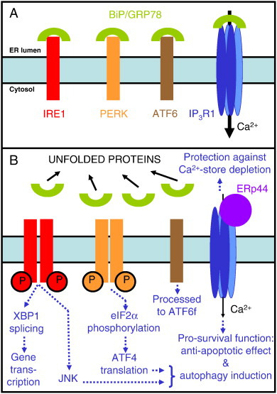
Fig. 4. The ER-stress sensors and the initial steps of the UPR. This new model includes not only the three canonical ER-stress sensors (IRE1, PERK and ATF6) but also the here proposed fourth ER-stress sensor, the IP3R1. Panel A represents the ER in normal, healthy cells. Panel B shows the activation of the ER-stress sensors during the adaptive phase of the UPR and the main steps involved in pro-survival signaling. The proteins are color-coded: BiP/GRP78 (green), IRE1 (red), PERK (orange), ATF6 (brown), IP3R1 (blue) and ERp44 (purple) are depicted. Dimerization and autophosphorylation (circled P) of IRE1 and PERK are shown. The processing of AFT6 is performed in the Golgi apparatus. Full black arrows indicate the magnitude of the Ca2 + fluxes. Dotted blue arrows indicate the cellular processes occurring after the activation of the ER-stress sensors. JNK denotes the activation of the c-Jun N-terminal kinase pathway. See text for further explanations.
The UPR consists in the first place of adaptive mechanisms leading to an enhanced translation of ER chaperones to increase the folding capacity in the ER, a reduced translation of other proteins, and the degradation of misfolded proteins. If the ER stress can not be resolved by these adaptive responses, the cell itself will be marked for elimination by apoptosis [10], [144], [157], [158], [159], [160]. In addition to being the cellular response to ER stress, the UPR can also integrate other signals and in this way affect basal cellular physiology independently of protein misfolding [161].
Each of the three ER-stress sensors activates a distinct set of mechanisms [10], [144], [157], [158], [159], [160]. In short, PERK phosphorylates eIF2α leading to the translation of ATF4. The RNase activity of IRE1 leads to the production of spliced X box-binding protein 1 (XBP1), that further activates a number of pathways, while ATF6 is processed to its active form, ATF6f (Fig. 4). The early responses involving PERK-dependent phosphorylation of eIF2α attenuate protein synthesis by inhibiting translation, while mRNA decay is regulated by IRE1. Moreover, damaged/unwanted proteins are degraded [162] and autophagy is triggered to remove either damaged ER (a process known as reticulophagy [163]) or abnormal proteins, particularly if the capacity of the proteasome pathway is exceeded. Autophagy can be induced by ER stress through various pathways, including IRE1 in conjunction with the c-Jun N-terminal kinase pathway, subsequently to PERK activation and downstream of intracellular Ca2 + signaling [20], [145], [146], [160], [164], [165]. A possible Ca2 +-dependent mediator for triggering autophagy induction after ER stress is protein kinase C θ [166], [167]. In a second wave of events, the transcription factors ATF4, ATF6f and spliced XBP1 promote further responses to restore ER function. More severe ER stress however induces apoptosis to eliminate the irreversibly damaged cells. Induction of apoptosis is largely due to the upregulation of the pro-apoptotic transcription factor C/EBP-homologous protein (CHOP), occurring subsequently to prolonged PERK activation. CHOP activation leads to downregulation of the anti-apoptotic protein Bcl-2 and upregulation of pro-apoptotic Bcl-2 homology 3 (BH3)-only proteins and of growth arrest and DNA damage-inducible 34 (GADD34), factors that lead to apoptosis [160], [168], [169], [170].
3.3. Regulation of the IP3R during ER stress
During ER stress induced by a decreased ER Ca2 + level, not only the driving force for Ca2 + release is diminished, but studies in various cell types have indicated that the IP3R becomes less sensitive to IP3 when the luminal Ca2 + concentration decreases [85], [86], [87], [171], [172]. Especially when Ca2 + stores are quite extensively depleted, the IP3R sensitivity markedly decreases [88], [173], [174], which fits with the idea of a protective mechanism against too low levels of Ca2 + in the lumen of the ER. The precise molecular mechanism involved was originally proposed to involve either Ca2 +-binding sites on the cytosolic [175] or luminal part of the IP3R [176] itself or regulation by luminal proteins [33], [177]. More recent work indicated that indeed several luminal proteins could regulate the IP3R, of which ERp44 [112] inhibited the IP3R1 at low levels of ER Ca2 + content. Its role will be further made explicit below in the context of ER stress.
In fact, during ER stress complex changes in IP3R regulation occur, leading to changes in Ca2 + homeostasis. A first observation in that respect was the disruption of signaling complexes in the ER-plasma-membrane microdomains involved in intracellular Ca2 + signaling in PERK-deficient cells having high levels of ER stress [178]. Furthermore, induction of ER stress in heterozygous IP3R1-knockout mice led to more cell damage than in wild-type mice [113]. Similarly, an increased level of ER stress-induced apoptosis was observed in HeLa cells in which IP3R1 was downregulated with siRNA. This phenomenon was however not due to changes in the three classical UPR pathways, but to a switch in the regulation of IICR by the chaperone BiP/GRP78. Under normal, unstressed conditions the latter binds to IP3R1, though not to IP3R2 and IP3R3 [113]. The BiP/GRP78-binding site of IP3R1 corresponds to the variable part (a.a. 2463–2528) of the large luminal loop of IP3R1 located between transmembrane helices 5 and 6 (Fig. 3). This BiP/GRP78-binding site exactly overlaps with the previously identified binding site for ERp44 [112], a member of the thioredoxin family [179]. As both proteins share the same binding site, ERp44 also only interacts with the IP3R1 isoform [112], and both proteins compete for binding to IP3R1. The binding of BiP/GRP78 to IP3R1 is ATP-dependent and stabilizes the assembly of IP3R1 into functional tetrameric channel complexes with consequently a higher level of IICR (Fig. 5A).
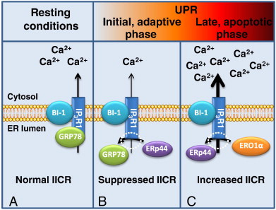
Fig. 5. Regulation of IP3R1 and IICR during ER stress and UPR progression. A. In resting conditions, the chaperone BiP/GRP78 (GRP78) stabilizes IP3R function and allows efficient Ca2 + channeling through the IP3R1. BI-1 can in these conditions interact with the IP3R and stimulate IICR. BI-1 is a pleiotropic regulator of cell function during ER stress, and a role in IP3R regulation during UPR is appealing. However, the role of its interaction with IP3R1 during ER stress was not yet ascertained. B. Induction of ER stress induces the UPR and both the dissociation of BiP/GRP78 and its replacement by ERp44 lead to a decreased IICR. C. Prolonged ER stress leads to hyperoxidation by ERO1α and displacement of ERp44. Subsequently IICR increases, leading to pro-apoptotic Ca2 + signaling.
During ER stress, BiP/GRP78 dissociates from IP3R1 in order to bind to unfolded or misfolded proteins in the ER lumen, leading to a decreased IICR (Fig. 5B). Similarly, after BiP/GRP78 knockdown, Ca2 + release through the IP3R1, but not through IP3R2 or IP3R3, was impaired [113]. This confirms that BiP/GRP78 binding to IP3R1 is important for IP3R1-channel activity. The decreased IICR activity on the one hand protects the ER from further depletion, but on the other hand can also decrease mitochondrial Ca2 + uptake below the threshold level needed for cell survival [113].
In addition, BiP/GRP78 can also indirectly influence IICR via binding to the sigma-1 receptor. The sigma-1 receptor is a chaperone that is enriched at the MAMs [180]. At normal ER Ca2 + levels BiP/GRP78 forms a complex with the sigma-1 receptor, in which both proteins display minimal activity. Low ER Ca2 + levels, like those that trigger ER stress, cause a rapid disassembly of the complex, leading to the activation of both chaperones, and a redistribution of the sigma-1 receptors from the MAMs to the bulk ER [180]. At the MAMs, sigma-1 receptors stabilize IP3R3s by protecting them from degradation. When released from BiP/GRP78, their action on IP3R3 promotes Ca2 + transfer to the mitochondria and favors cell survival. In contrast, the recently discovered shorter splice variant of the sigma-1 receptor (106 a.a. instead of 223 a.a.) does not interact with IP3Rs and acts antagonistically to its longer counterpart after ER stress [181]. Indeed, expression of the short variant leads to a decreased IICR towards the mitochondria. In ER-stress conditions an increased degradation of the IP3Rs is observed, which leads to a decreased ATP production, increased autophagy and eventually apoptosis, while the wild-type sigma-1 receptor favors Ca2 + transfer to the mitochondria, increased bioenergetics and cell survival [180], [181].
During ER stress, the promyelocytic leukemia (PML) protein forms at the MAMs a complex with IP3R3, protein phosphatase 2A and protein kinase B [182]. Protein phosphatase 2A hereby counteracts the inhibitory effect of protein kinase B on the IP3R [102], [103], [104], hereby promoting an increased, pro-apoptotic, Ca2 + transfer to the mitochondria [182].
During the UPR, the thioredoxin-family member ERp44 is upregulated [179]. The release of BiP/GRP78 from IP3R1 during ER stress therefore also leads to increased binding of ERp44 to the overlapping binding site, thereby further inhibiting IICR [112] (Fig. 5B). In agreement with these results, overexpression of ERp44 inhibited IICR, while its knockdown increased IICR. The binding of ERp44 to IP3R1 is dependent on Ca2 +, since the depletion of the Ca2 + stores increased IP3R1/ERp44-complex formation. Also luminal pH and the redox state of the ER influences the interaction of ERp44 to IP3R1. Mutation of cysteine residues (Cys2496 and Cys2504) in the third luminal loop of IP3R1 (Fig. 3) decreased ERp44 binding, indicating that free thiol groups are required [112]. Mutational analysis of ERp44 has indicated that it interacts with the IP3R1 through a.a. 236–285 [112], though it is presently not clear whether this region is responsible for IP3R inhibition or whether other regions and/or other proteins are involved [183]. In this respect, it is interesting to mention that ERp44 can also bind to ER oxidase 1α (ERO1α) [179], [184].
ERO1α, together with PDI, is a major catalyst of oxidative folding of secretory proteins, yielding H2O2 as a by-product. Specific and limited PDI oxidation by ERO1α is essential to avoid ER hyperoxidation. Under normal physiological conditions the ER forms an oxidizing environment and Ca2 + stores are filled, allowing proper function of the various chaperones. Not only the ER Ca2 + level, but also the ER redox state can be altered during ER stress. Moreover, the conformation of the third luminal loop of the IP3R1 depends on the oxidation state [185]. During ER stress, ERO1α is upregulated in a CHOP-dependent manner, leading to ER hyperoxidation [186], [187]. This hyperoxidation could disrupt the interaction between ERp44 and IP3Rs, causing IP3R hypersensitivity, increased IICR and induction of apoptosis [187] (Fig. 5C). More recent data support this model, as ERO1α appears to be enriched at the MAMs [188], [189]. The effect of ERO1α on IICR appears complex, as it not only can act by oxidizing the IP3R, but also by acting as a sink for ERp44. In addition it can affect mitochondrial Ca2 + uptake through the mitochondrial Ca2 + uniporter, either directly, via H2O2 production, or indirectly, e.g. via changes in the ER Ca2 + level [189].
3.4. Modulation of IICR during ER stress
The data shown above (Section 3.3) demonstrate that during ER stress the IP3R isoforms are modulated in a complex way. The final effect for the cell, including the choice between autophagy and apoptosis, will however also depend on other factors influencing either IICR directly or indirectly, and on the proximity between the ER and the mitochondria.
3.4.1. Truncated SERCA variants
Two short, C-terminally truncated SERCA1 variants (43 and 46 kDa, resp.) are expressed in various adult and fetal tissues, with the notable exception of skeletal and cardiac muscle [190]. These variants, named SERCA1T, localize to the ER but in contrast to the normal SERCA are unable to pump Ca2 +; instead they increase Ca2 + leakage out of the ER (Table 2), putatively by forming homodimers. ER-stress inducers cause the accumulation of SERCA1T [191]. Their induction occurs biphasically, first through PERK/eIF2α phosphorylation/ATF4 activation and subsequently also by a pathway dependent on ATF6 and CHOP. The induction of SERCA1T variants further amplifies the ER-stress responses, probably due to enhanced ER Ca2 +-store depletion. Interestingly, SERCA1T is especially expressed at the MAMs, thereby inhibiting mitochondrial movement and increasing their docking to the ER [191]. The expression of SERCA1T thus leads to increased apoptosis by the combined action of an increased ER leak in the neighborhood of the mitochondria, the consequently increased coupling between the ER and the mitochondria and the increased efficiency of IICR towards the mitochondria, all contributing to a Ca2 + overload of the mitochondria.
3.4.2. The regulatory role of calreticulin and calnexin
The Ca2 +-binding chaperones calreticulin and calnexin (Table 1) can regulate SERCA-mediated Ca2 + uptake either directly or indirectly. Calreticulin can interact with SERCA2b and with ERp57, a ubiquitous ER thiol-dependent oxidoreductase. At high ER Ca2 + levels, the N-domain of calreticulin targets glycosylated residues in the C-terminal tail of SERCA2b while recruiting ERp57 through its P-domain (Fig. 1). ERp57 promotes disulfide-bond formation between thiol groups in the fourth luminal loop of SERCA2b and it thereby reduces SERCA pumping activity [192]. Furthermore, under the same conditions, phosphorylated calnexin interacts with and inhibits SERCA2b [193]. At low ER Ca2 + levels, however, ERp57 dissociates from SERCA2b, while the increase in cytosolic Ca2 + concentration may concomitantly result in calnexin dephosphorylation, also causing its dissociation from SERCA2b. Both effects lead to increased SERCA activity that can counteract ER Ca2 + depletion. Indeed, during ER stress, the Ca2 +/calmodulin-dependent phosphatase calcineurin is upregulated and plays a double role to alleviate the stress situation: on the one hand it will associate with PERK and stimulate PERK-dependent phosphorylation of eIF2α to attenuate protein synthesis, but on the other hand calcineurin can dephosphorylate calnexin, relieving the SERCA inhibition and allowing recovery of ER stress by restoring ER Ca2 + levels [194]. Eventually, calcineurin will be phosphorylated by PERK, diminishing its activity, while its expression returns to resting levels.
3.4.3. The possible role of the translocon during ER stress
The translocon or SEC61 complex is a protein-conducting channel of the ER. It is formed by the association of Sec61α, β and γ, translocation-associated membrane protein, BiP/GRP78, calnexin, calreticulin, and ERp57. In addition to its function in translocation of the nascent polypeptide chain during protein translation, it was shown that in its protein-free form it could mediate Ca2 + leakage out of the ER [195], [196], [197], [198] (Fig. 1 and Table 2). Although it was never really questioned that the translocon could act as a Ca2 +-leak channel after treatment with puromycin, which opens the channel, it was not clear whether the translocon played a role in the basal Ca2 + leak occurring under physiological conditions [199]. It is however conceivable that the translocon channel is subject to intracellular regulation, as a permanent Ca2 + leakage out of the ER would indubitably lead to cellular malfunction. Interestingly, it was recently found that calmodulin could inhibit Ca2 + release through the translocon [200]. Even more interesting in the context of ER stress is that BiP/GRP78 itself binds to loop 7 of Sec61α and inhibits Ca2 + leakage through the translocon [201]. ER-stress induction or BiP/GRP78 silencing both increased Ca2 + leakage through the translocon. Parallel experiments performed in LNCaP cells led to identical results [202]. Moreover, the translocon inhibitor anisomycin not only blocked the Ca2 + leak, but also antagonized apoptosis, indicating that during ER stress the Ca2 + leakage out of the ER through the translocon contributes to cell death.
3.4.4. Bax inhibitor-1 (BI-1), a pleiotropic player in ER stress
BI-1, which is also named transmembrane Bax inhibitor motif-containing 6 (TMBIM-6), is an evolutionary highly conserved anti-apoptotic protein [203] that especially protects against ER stress and ischemia–reperfusion injury [204]. BI-1-deficient cells are hypersensitive to ER-stress inducers, while BI-1 overexpression protects against apoptosis induced by ER stress [205]. These cytoprotective properties of BI-1 correlate with its ability to reduce the ER Ca2 + level [206], [207], [208]. BI-1 can increase the Ca2 + leak out of the ER (Fig. 1 and Table 2), though at this moment the mechanism is not yet clear. It has been proposed that BI-1 has Ca2 +-channel properties [207], [209], but also that it can function as a Ca2 +/H+ antiporter [210] or as an IP3R-sensitizing protein [211], functions that moreover are not mutually exclusive. In addition to effects on Ca2 + handling, BI-1 prevents the accumulation of reactive oxygen species (ROS) that arises during ER-stress induction, mainly by upregulation of heme oxygenase 1 [212], but also by interfering with NADPH-dependent cytochrome P450 reductase [213]. In contrast to these protective functions, BI-1 also inhibits the ER-stress sensor IRE1α by binding to its C-terminus that contains the kinase and endoribonuclease domains [214]. The BI-1 domain responsible for IRE1α interaction is the C-terminal part, which is also essential for the Ca2 + permeability [209]. Finally, the effect of BI-1 on autophagy induction is controversial, as both a repression [215] and an enhancement [216] of autophagy were reported. The latter phenomenon was dependent on the presence of IP3R channels and may relate to the fact that IP3Rs are sensitized by BI-1 [211], leading to basal Ca2 + release and a decreased Ca2 + level in the ER, which would then result in insufficient Ca2 + transfer to the mitochondria [216]. BI-1 seems therefore to function as a stress integrator controlling a variety of homeostatic processes, including the adaptive ER-stress response, ER-stress dependent apoptosis and pro-survival signaling through autophagy, dependently on its interaction partners. Other members of the BI-1 family may also be involved. For example, GRINA (glutamate receptor, ionotropic NMDA-associated protein 1, also termed TMBIM-3) protects cells against ER stress-induced apoptosis [217]. During ER stress it is strongly upregulated in a PERK- and ATF4-dependent way, and may act synergistically with BI-1 in the modulation of ER Ca2 + homeostasis and apoptosis, at least in part by binding to and regulating IP3R channels.
3.4.5. Regulation of ER-mitochondrial contacts during ER stress
ER-mitochondria contact points, the so-called MAMs, are very important cellular microdomains, not only for Ca2 + handling, but also for lipid synthesis and for the control of mitochondrial dynamics [122]. With respect to Ca2 + transfer, the proximity between the ER and mitochondria is a key element, controlling mitochondrial Ca2 + uptake. It is conceivable that the modulation of the distance or of the area of interaction allows a precise control of the ER-to-mitochondria Ca2 + transfer. In addition to Ca2 +, ATP and ROS are other molecules that can be exchanged between the ER and the mitochondria, and that can affect processes in both organelles, like e.g. protein folding and Ca2 + transport. Their eventual effects will therefore also depend on the level of interaction between the two organelles [16], [218], [219].
Mitochondrial motility is determined by the microtubular network and ER-mitochondria contacts can be reversibly regulated by changes in the cytosolic Ca2 + concentration [220], [221], [222]. Mitochondria appear to be trapped in the neighborhood of ER Ca2 +-release sites, to allow a more efficient transfer of Ca2 +. Moreover, ER-mitochondria contacts appear to be strengthened under ER stress and/or apoptotic conditions [223], [224]. This may lead in the late phases of ER stress to increased apoptosis [191], but in the early phases it will lead to increased respiration and ATP production, and thus have a pro-survival role [225], [226].
In PERK-deficient cells a distended and fragmented ER was observed that was disconnected from contacts with the plasma membrane [178]. PERK did not directly affect IP3Rs, but since IP3 is produced at the plasma membrane the distance between the plasma membrane and the ER is important for efficient IP3R activation. PERK-deficient cells were indeed found to display decreased rates of agonist-induced Ca2 + release [178]. In addition, PERK activity was shown to be stimulated during ER stress in a Ca2 +-dependent way by the Ca2 +/calmodulin-dependent phosphatase calcineurin [194]. Finally, very recent work has demonstrated that PERK is enriched at the MAMs, and is thereby involved in ER-mitochondria tethering [227]. PERK-deficient cells therefore not only display aberrant Ca2 + signaling consecutively to changes in the ER-plasma-membrane contact sites, but also due to changes in the interaction of the ER with the mitochondria. These changes will not only affect the Ca2 + transmission between the two organelles, but also the transmission of ROS-mediated signals, and therefore the occurrence of apoptosis subsequently to ROS-induced ER stress.
IP3-induced Ca2 + signals arising during ER stress and the UPR therefore not only depend on the IP3R but also on the presence and activity of various other proteins involved in Ca2 + binding or transport and on the ER-mitochondria interactions. The latter form signaling hubs containing crucial proteins participating in cell-fate decisions subsequent to ER stress.
3.5. IP3R1 as a fourth ER-stress sensor
From the preceding, it is clear that the IP3R and IICR play a central role in the Ca2 + signaling needed for the adequate progression through the UPR. Moreover, by influencing the expression and the function of IP3R-modulating proteins in the ER lumen (e.g. BiP/GPR78, ERp44, ERO1α), the UPR itself contributes to the modulation of IP3R activity and thus ultimately to its own outcome. Changes in Ca2 + signaling will determine whether or not autophagy or apoptosis will be activated [17], [18]. Importantly, the cytosolic and the mitochondrial Ca2 + concentration should each be kept within strict limits, and conditions leading to Ca2 + signals that are either too large or too small, have detrimental effects for the cell.
The UPR is initiated by the recruitment of BiP/GRP78 to the excess of misfolded and unfolded proteins in the lumen of the ER (Fig. 4). It is well established that BiP/GRP78 thereby dissociates from the canonical ER-stress sensors, IRE1, PERK and ATF6, leading to their activation [151].
In addition, BiP/GRP78 is also recruited from other binding partners like the IP3R1, the sigma-1 receptor and the translocon, leading also to changes in their activity. In particular, after dissociation from the IP3R1, BiP/GRP78 will no longer assist in its assembly to functional tetrameric channels [113]. Moreover, the disassembly of the complex between BiP/GRP78 and IP3R1 will allow ERp44 to interact now with IP3R1, which will further reduce IICR [112]. The latter will contribute to the adaptive pro-survival ER-stress response by limiting the decrease in ER Ca2 +-store loading. This will promote re-establishing ER homeostasis and proper ER-protein folding capacities. These favorable conditions for cell survival are further supported by the dissociation of BiP/GRP78 from the sigma-1 receptors [180], leading to the stabilization of IP3R3 activity in the MAMs and sustained ATP production by the mitochondria. ATP is an essential co-factor for chaperones and will further help to restore protein-folding processes in the ER and/or regulate UPR-related gene expression. Since in healthy cells BiP/GRP78 is bound to IP3R1 where it is required for proper IP3R1 function and becomes dissociated as an early event in ER stress, leading to a decrease in activity which on the one hand supports reestablishment of ER homeostasis, and at the other hand can contribute to pro-survival signaling, we propose that IP3R1 should be seen as a fourth ER-stress sensor in addition to the three canonical ER-stress sensors IRE1, PERK and ATF6 (Fig. 4).
If during prolonged ER stress IP3R activity decreases up to the point that ATP production by the mitochondria is no longer supported, autophagy can be triggered [129]. If, on the other hand, the combined UPR and autophagic response do not sufficiently alleviate ER stress, upregulation of ERO1α will lead to a hypersensitization of IP3R1, an excessive Ca2 + transfer to the mitochondria and finally cell death by apoptosis [187]. This outcome is further supported by other changes in the Ca2 +-handling proteins, including the dissociation of BiP/GRP78 from the translocon, which activates a Ca2 +-leak pathway leading to further Ca2 +-store depletion and apoptosis [201], [202].
4. Conclusions
The relation between ER stress and Ca2 + homeostasis appears complex. A decreased ER Ca2 + load induces ER stress, during which ER Ca2 +-binding proteins are upregulated as a protective mechanism. Moreover, during ER stress, changes occur in the regulation of the IP3R leading to changes in IICR and in Ca2 + signaling. These changes in Ca2 + signaling in turn impact on the process of the UPR and on the progression of the cell towards autophagy or apoptosis. The activity of the IP3R1 is decreased in the initial phase of ER stress and this contributes to the UPR. It is therefore appropriate to consider the IP3R1 as a fourth ER-stress sensor, besides the three canonical ER-stress sensors IRE1, PERK and ATF6. However, although at low levels of ER stress Ca2 + release is adjusted to promote survival, under persistent stress the IP3R will participate in the eventual demise of the cell.
Acknowledgements
Work performed in the laboratory of the authors in this area was supported by the Research Council of the KU Leuven (Collaborative grant BIL/LA/10/09, Concerted Action 09/012 and research grant STRT1/10/044) and by the Research Foundation Flanders (research grants G.0604.07, G.0731.09N, G.0724.09N and G.0634.13).
References
- [1]
- O. Baumann, B. WalzEndoplasmic reticulum of animal cells and its organization into structural and functional domainsInt. Rev. Cytol., 205 (2001), pp. 149-214
- [2]
- J. Meldolesi, T. PozzanThe heterogeneity of ER Ca2 + stores has a key role in nonmuscle cell signaling and functionJ. Cell Biol., 142 (1998), pp. 1395-1398
- [3]
- M.J. BerridgeThe endoplasmic reticulum: a multifunctional signaling organelleCell Calcium, 32 (2002), pp. 235-249
- [4]
- S. Papp, E. Dziak, M. Michalak, M. OpasIs all of the endoplasmic reticulum created equal? The effects of the heterogeneous distribution of endoplasmic reticulum Ca2 +-handling proteinsJ. Cell Biol., 160 (2003), pp. 475-479
- [5]
- A. Görlach, P. Klappa, T. KietzmannThe endoplasmic reticulum: folding, calcium homeostasis, signaling, and redox controlAntioxid. Redox Signal., 8 (2006), pp. 1391-1418
- [6]
- E. Sammels, J.B. Parys, L. Missiaen, H. De Smedt, G. BultynckIntracellular Ca2 + storage in health and disease: a dynamic equilibriumCell Calcium, 47 (2010), pp. 297-314
- [7]
- T. Vervliet, S. Kiviluoto, G. BultynckER stress and UPR through dysregulated ER Ca2 + homeostasis and signalingP. Agostinis, A. Samali (Eds.), Endoplasmic Reticulum Stress in Health and Disease, Springer Science + Business Media, Dordrecht (2012), pp. 107-142
- [8]
- D. Mekahli, G. Bultynck, J.B. Parys, H. De Smedt, L. MissiaenEndoplasmic-reticulum calcium depletion and diseaseCold Spring Harb. Perspect. Biol., 3 (2011), p. a004317
- [9]
- S. Wang, R.J. KaufmanThe impact of the unfolded protein response on human diseaseJ. Cell Biol., 197 (2012), pp. 857-867
- [10]
- G.C. Shore, F.R. Papa, S.A. OakesSignaling cell death from the endoplasmic reticulum stress responseCurr. Opin. Cell Biol., 23 (2011), pp. 143-149
- [11]
- S. Matus, L.H. Glimcher, C. HetzProtein folding stress in neurodegenerative diseases: a glimpse into the ERCurr. Opin. Cell Biol., 23 (2011), pp. 239-252
- [12]
- K.M. Doyle, D. Kennedy, A.M. Gorman, S. Gupta, S.J. Healy, A. SamaliUnfolded proteins and endoplasmic reticulum stress in neurodegenerative disordersJ. Cell. Mol. Med., 15 (2011), pp. 2025-2039
- [13]
- M.J. Berridge, P. Lipp, M.D. BootmanThe versatility and universality of calcium signallingNat. Rev. Mol. Cell Biol., 1 (2000), pp. 11-21
- [14]
- S.K. Joseph, G. HajnoczkyIP3 receptors in cell survival and apoptosis: Ca2 + release and beyondApoptosis, 12 (2007), pp. 951-968
- [15]
- M.W. Harr, C.W. DistelhorstApoptosis and autophagy: decoding calcium signals that mediate life or deathCold Spring Harb. Perspect. Biol., 2 (2010), p. a005579
- [16]
- J.P. Decuypere, G. Monaco, G. Bultynck, L. Missiaen, H. De Smedt, J.B. ParysThe IP3 receptor-mitochondria connection in apoptosis and autophagyBiochim. Biophys. Acta, 1813 (2011), pp. 1003-1013
- [17]
- S. Kiviluoto, H. Akl, T. Vervliet, G. Bultynck, J.B. Parys, L. Missiaen, H. De SmedtIP3 receptor-binding partners in cell-death mechanismsWIREs Membr. Transp. Signal., 1 (2012), pp. 201-210
- [18]
- J.P. Decuypere, G. Bultynck, J.B. ParysA dual role for Ca2 + in autophagy regulationCell Calcium, 50 (2011), pp. 242-250
- [19]
- C. Cárdenas, J.K. FoskettMitochondrial Ca2 + signals in autophagyCell Calcium, 52 (2012), pp. 44-51
- [20]
- J.B. Parys, J.P. Decuypere, G. BultynckRole of the inositol 1,4,5-trisphosphate receptor/Ca2 +-release channel in autophagyCell Commun. Signal, 10 (2012), p. 17
- [21]
- P. Volpe, T. Pozzan, J. MeldolesiRapidly exchanging Ca2 + stores of non-muscle cellsSemin. Cell Biol., 1 (1990), pp. 297-304
- [22]
- T. Pozzan, R. Rizzuto, P. Volpe, J. MeldolesiMolecular and cellular physiology of intracellular calcium storesPhysiol. Rev., 74 (1994), pp. 595-636
- [23]
- M.J. Berridge, M.D. Bootman, H.L. RoderickCalcium signalling: dynamics, homeostasis and remodellingNat. Rev. Mol. Cell Biol., 4 (2003), pp. 517-529
- [24]
- D. Burdakov, O.H. Petersen, A. VerkhratskyIntraluminal calcium as a primary regulator of endoplasmic reticulum functionCell Calcium, 38 (2005), pp. 303-310
- [25]
- P. Vangheluwe, M.R. Sepulveda, L. Missiaen, L. Raeymaekers, F. Wuytack, J. VanoevelenIntracellular Ca2 +- and Mn2 +-transport ATPasesChem. Rev., 109 (2009), pp. 4733-4759
- [26]
- M. Brini, E. CarafoliCalcium pumps in health and diseasePhysiol. Rev., 89 (2009), pp. 1341-1378
- [27]
- I. Vandecaetsbeek, P. Vangheluwe, L. Raeymaekers, F. Wuytack, J. VanoevelenThe Ca2 + pumps of the endoplasmic reticulum and Golgi apparatusCold Spring Harb. Perspect. Biol., 3 (2011), p. a004184
- [28]
- C. ToyoshimaHow Ca2 +-ATPase pumps ions across the sarcoplasmic reticulum membraneBiochim. Biophys. Acta, 1793 (2009), pp. 941-946
- [29]
- O. Thastrup, P.J. Cullen, B.K. Drobak, M.R. Hanley, A.P. DawsonThapsigargin, a tumor promoter, discharges intracellular Ca2 + stores by specific inhibition of the endoplasmic reticulum Ca2 +-ATPaseProc. Natl. Acad. Sci. U. S. A., 87 (1990), pp. 2466-2470
- [30]
- F. Michelangeli, J.M. EastA diversity of SERCA Ca2 + pump inhibitorsBiochem. Soc. Trans., 39 (2011), pp. 789-797
- [31]
- J. Meldolesi, T. PozzanThe endoplasmic reticulum Ca2 + store: a view from the lumenTrends Biochem. Sci., 23 (1998), pp. 10-14