beyond reason
https://www.cell.com/immunity/pdf/S1074-7613(19)30198-0.pdf
Nearly every tissue in the body undergoes routine turnover of cells as part of normal healthy living. The majority of these cells undergoing turnover die via apoptosis, and then are rapidly removed by phagocytes by the process of efferocytosis that is anti-inflammatory. However, a number of pathologies have recently been linked to defective clearance of apoptotic cells. Perturbed clearance arises for many reasons, including overwhelming of the clearance machinery, disruptions at different stages of efferocytosis, and responses of phagocytes during efferocytosis, all of which can alter the homeostatic tissue environment. This review covers linkages of molecules involved in the different phases of efferocytosis to disease pathologies that can arise due to their loss or altered function.
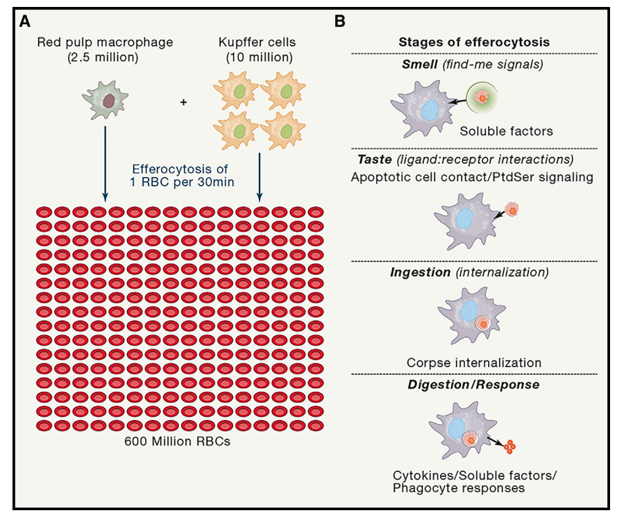
Figure 1. A Game of Numbers—Erythrocyte Clearance in Mice and the Phases of Efferocytosis
(A) Every symbol represents 2.5 3 106 cells of the respective population. Red blood cells (RBC) are depicted in red, Kupffer cells (KC) are depicted in yellow, and red pulp macrophages (RPM) are depicted in blue. Every day, 600 3 106 RBC are cleared, resulting in a clearance rate of 7,000 RBC per second. This task is performed by either 10 3 106 KC, 2.5 3 106 macrophages, or both, resulting in the necessity of each phagocyte to engulf one RBC on average every 24 min, 6min, or 30 min, respectively.
(B) Efferocytosis is carried out via four distinct steps, and potential disturbance in one or more of these steps could result in defective apoptotic cell clearance and the associated pathologies. The first step in engulfment of apoptotic cells is the ‘‘Smell phase’’ that involves communication of dying cells with nearby phagocytes through findme signals that are released in the early stages of apoptosis. The second step is the ‘‘Taste phase’’ that involves recognition of the ligands of apoptotic cells, termed ‘‘eat-me signals’’ by the phagocytes through specific efferocytotic receptors. The third step of the process is the ‘‘Ingestion phase,’’ where signaling in the phagocyte downstream of engagement of apoptotic cells by phagocytic receptors leads to corpse internalization. The fourth step is the ‘‘Digestion and response phase’’ that involves processing of the corpses and the production of anti-inflammatory mediators by the efferocytic phagocytes.
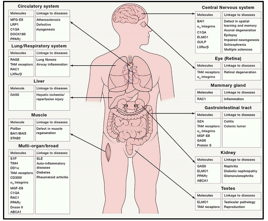
Figure 2. Molecules Involved in Efferocytosis and Their Disease Linkage in Different Tissues In many tissues of the body, clearance of apoptotic cells is performed by the professional phagocytes and non-professional phagocytes. Pathologies associated with either natural or induced deletions or mutations of the specific molecules and their linkage t0 diseases or the different tissues are shown.
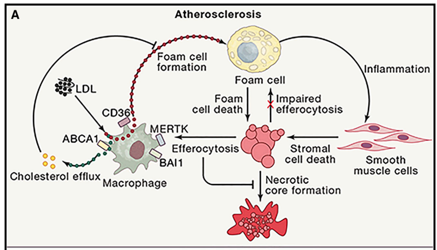
Apoptotic Cell Clearance in Atherosclerosis and Gut Inflammation
(A) Monocyte-derived macrophages in atherosclerotic plaques take up LDL via CD36, and the free cholesterol is often released via ABCA1 to HDL (part of reverse cholesterol transport to the liver). The macrophages express phosphatidylserine receptors such as MERTK and BAI1, limiting the accumulation of dead cells and preventing necrotic core formation. Further, BAI1- mediated efferocytosis increases the capacity of macrophages to perform reverse cholesterol efflux via upregulation of ABCA1. If the load of LDL is too high, efferocytosis is impaired, or the reverse cholesterol efflux is reduced, then macrophages accumulate cholesterol and develop into foam cells. Foam cells show inflammatory characteristics, impaired efferocytosis, and are poised to undergo cell death. Dying foam cells and inflammation-induced cell death of plaqueassociated cells contribute to necrotic core formation.
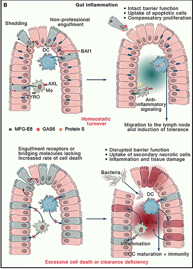
(B) During homeostasis, apoptotic cells in the
epithelial lining are taken up by nearby healthy
epithelial cells, by professional phagocytes of the
lamina propria (such as dendritic cells), or shed into
the lumen. Under these circumstances, the epithelial barrier function is maintained, and cell clearance
establishes a tolerogenic, anti-inflammatory environment. If apoptotic cell clearance is impaired or
cell death exceeds the clearance capacity of the
intestine, apoptotic cells progress into secondary
necrosis. Secondary necrotic cells either release
pro-inflammatory mediators themselves or induce
an inflammatory signature in the phagocytes. The
disruptions of epithelial lining allow bacterial passage through the barrier, further contributing to the
overall inflammatory environment associated with
increased cell death and clearance deficiency