1. 주관절의 구조
가. 동영상 : https://www.youtube.com/watch?feature=player_embedded&v=3l3-5Ij3JZ8
나. 구조 개요 : http://cafe.naver.com/panicbird/131
다. 촉진 : http://cafe.naver.com/panicbird/132
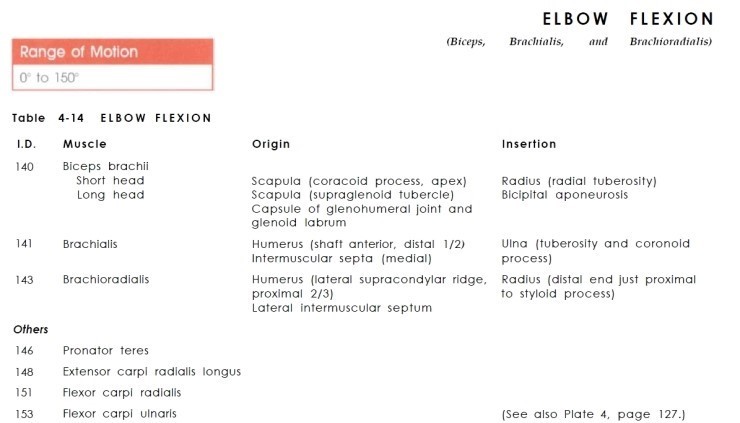
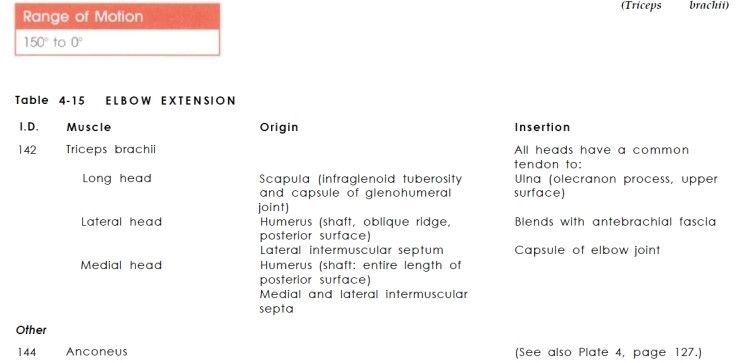
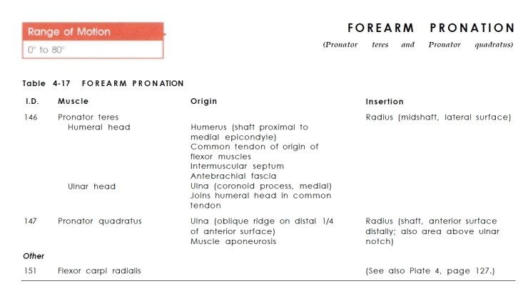
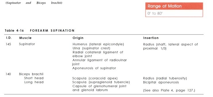
2. 주관절의 운동
가. 팔꿈치 굴곡 150도
나. 신전 0도
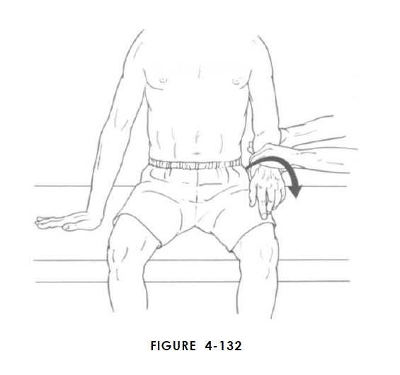
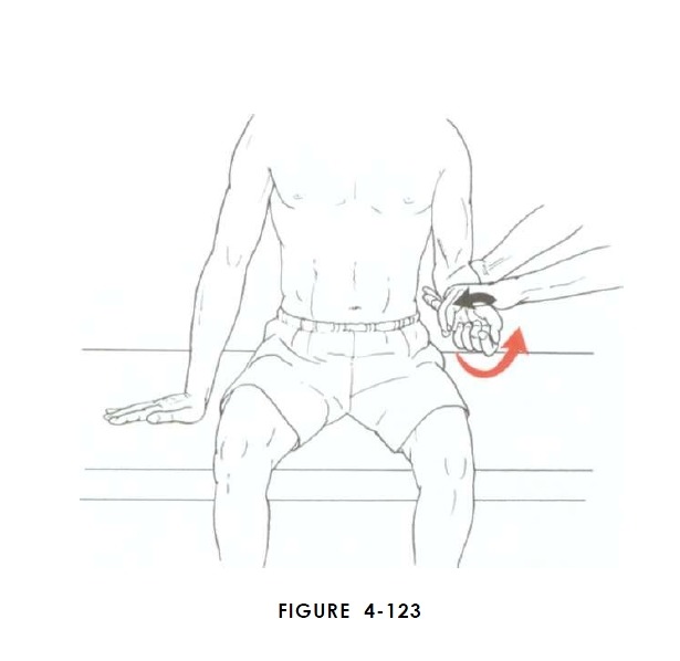
다. forearm 회내 80도
라. 회외 80도
3. 주관절 주변의 근육
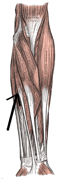
가. anterior superficial
- Biceps brachii
| Biceps brachii | |
|---|---|
|
Location of biceps. Two different colors represent two different bundles which compose biceps.
Short head
Long head
| |
| Details | |
| Latin | musculus biceps brachii |
| Origin | Short head: coracoid process of the scapula. Long head: supraglenoid tubercle |
| Insertion | Radial tuberosity and bicipital aponeurosis into deep fascia on medial part of forearm |
| Brachial artery | |
| Musculocutaneous nerve (C5–C6) | |
| Actions | Flexes elbow, flexes and abducts shoulder and supinates radioulnar joint in the forearm |
| Antagonist | Triceps brachii muscle |


- Pronator teres muscle
| Pronator teres muscle | |
|---|---|

Anterior view of the left forearm. Superficial muscles. (Pronator teres colored at center.) | |
| Details | |
| Latin | Musculus pronator teres |
| Origin | humeral head: medial supracondylar ridge of humerus (common flexor tendon) ulnar head: coronoid process of ulna |
| Insertion | Middle of the lateral surface of the body of the radius |
| ulnar artery and radial artery | |
| median nerve | |
| Actions | pronation of forearm, flexeselbow |
| Antagonist | Supinator muscle |
- Palmaris longus muscle
| Palmaris longus muscle | |
|---|---|

Front of right upper extremity. (Palmaris longus labeled at bottom, second from left.) | |
 | |
| Details | |
| Latin | musculus palmaris longus |
| Greek | Μακρύς παλαμικός μυς |
| Origin | medial epicondyle of humerus(common flexor tendon) |
| Insertion | palmar aponeurosis |
| ulnar artery | |
| median nerve | |
| Actions | wrist flexor |
| Antagonist | Extensor carpi radialis brevis,Extensor carpi radialis longus,Extensor carpi ulnaris |
-Flexor carpi radialis muscle
| Flexor carpi radialis muscle | |
|---|---|

Anterior view of the left forearm. Superficial muscles. (Flexor carpi radialis and its tendon visible in blue.) | |
|
Anterior view of right upper extremity. (Flex. carp. rad. labeled at upper left.) | |
| Details | |
| Latin | musculus flexor carpi radialis |
| Origin | medial epicondyle of humerus(common flexor tendon) |
| Insertion | Bases of second and third metacarpal bones |
| Radial Artery | |
| Median nerve | |
| Actions | Flexion and abduction at wrist |
| Antagonist | Extensor carpi ulnaris muscle |
- Flexor carpi ulnaris
| Flexor carpi ulnaris | |
|---|---|

Dorsal (left) and ventral (right) views of deep muscles of the forearm. FCU is visible in blue. | |
| Details | |
| Latin | musculus flexor carpi ulnaris |
| Origin | medial epicondyle (common flexor tendon) and medial margin on olecranon of ulna |
| Insertion | pisiform, hook of the hamate,base of the fifth metacarpal bone |
| ulnar artery | |
| muscular branches of ulnar nerve | |
| Actions | flexion and adduction of wrist |
| Antagonist | Extensor carpi radialis brevis muscle, Extensor carpi radialis longus muscle |
- Flexor digitorum superficialis
| Flexor digitorum superficialis | |
|---|---|

| |
| Details | |
| Latin | musculus flexor digitorum superficialis |
| Origin | medial epicondyle of the humerus (common flexor tendon) as well as parts of theradius and ulna. |
| Insertion | anterior margins on the bases of the middle phalanges of the four fingers |
| ulnar artery | |
| median nerve | |
| Actions | flexor of fingers (primarily atproximal interphalangeal joints) |
| Antagonist | Extensor digitorum muscle |
나. anterior deep
- Pronator quadratus muscle
| Pronator quadratus muscle | |
|---|---|

Anterior view of left forearm. Deep muscles. (Pronator quadratus visible at bottom-center right.) | |
| Details | |
| Latin | Musculus pronator quadratus |
| Origin | medial, anterior surface of the ulna |
| Insertion | lateral, anterior surface of the radius |
| anterior interosseous artery | |
| median nerve (anterior interosseous nerve) | |
| Actions | pronates the forearm |
| Antagonist | Supinator muscle |
-Flexor digitorum profundus muscle
| Flexor digitorum profundus muscle | |
|---|---|

Ventral view of the deep muscles of the forearm. FDP is shown in blue. | |
| Details | |
| Latin | Musculus flexor digitorum profundus |
| Origin | upper 3/4 of the volar and medial surfaces of the body of the ulna, interosseous membrane and deep fascia of the forearm |
| Insertion | base of the distal phalanges of the fingers |
| anterior interosseous artery | |
| median (anterior interosseous),muscular branches of ulnar | |
| Actions | flex hand and bothinterphalangeal joints |
| Antagonist | Extensor digitorum muscle |
- Flexor pollicis longus muscle
| Flexor pollicis longus muscle | |
|---|---|

Front of the left forearm. Deep muscles. (Flexor pollicis longus is shown in blue) | |
| Details | |
| Latin | Musculus flexor pollicis longus |
| Origin | The middle 2/4 of the volarsurface of the radius and the adjacent interosseus membrane. |
| Insertion | The base of the distal phalanxof the thumb |
| Anterior interosseus artery | |
| Anterior interosseous nerve(branch of median nerve) (C8, T1) | |
| Actions | Flexion of the thumb. |
| Antagonist | Extensor pollicis longus muscle,Extensor pollicis brevis muscle |
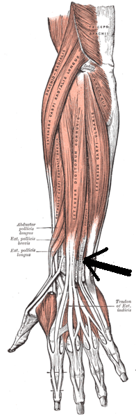
다. posterior superficial
_ Triceps Brachii
Triceps Brachii


Details
Latin
Musculus triceps brachii
Origin
Long head: infraglenoid tubercle of scapula
Lateral head: above the radial sulcus
Medial head: below the radial sulcus
Insertion
Olecranon process of ulna
Deep brachial artery
Radial nerve and axillary nerve (long head)
Actions
Extends forearm, long head extends,adducts arm, Extends shoulder
Antagonist
Biceps brachii muscle
- Brachioradialis muscle
| Brachioradialis muscle | |
|---|---|

Anterior view of muscles of the left forearm with brachioradialis shown in blue | |

Cross-section through the middle of the forearm. Brachioradialis labeled at center left, sixth from the top. | |
| Details | |
| Latin | musculus brachioradialis |
| Origin | Lateral supracondylar ridge of thehumerus |
| Insertion | Distal radius (radial styloid process) |
| Radial recurrent artery | |
| Radial nerve | |
| Actions | Flexion of elbow and supination |
- Extensor carpi radialis longus
| Extensor carpi radialis longus | |
|---|---|

Superficial muscles of the forearm. Extensor carpi radialis longus visible in blue. | |

Transverse section across the wrist and digits. (Ext. carp. rad. long. labeled at center left.) | |
| Details | |
| Latin | musculus extensor carpi radialis longus |
| Origin | lateral supracondylar ridge |
| Insertion | 2nd metacarpal |
| radial artery | |
| radial nerve | |
| Actions | extensor at the wrist joint,abducts the hand at the wrist |
| Antagonist | Flexor carpi ulnaris muscle |
- Extensor carpi radialis brevis
| Extensor carpi radialis brevis | |
|---|---|

Posterior surface of the left forearm. Superficial muscles. The partially obscured extensor carpi radialis brevis is shown in blue. | |
| Details | |
| Latin | musculus extensor carpi radialis brevis |
| Origin | humerus at the anterior oflateral epicondyle (common extensor tendon)[1] |
| Insertion | Posterior base of the 3rd metacarpal[1] |
| radial artery | |
| deep branch of the radial nerve | |
| Actions | extensor and abductor of thehand at the wrist joint[1] |
| Antagonist | Flexor carpi ulnaris muscle |
- Extensor digitorum muscle
| Extensor digitorum muscle | |
|---|---|

Posterior surface of the forearm. Superficial muscles. Extensor digitorum muscle is labeled in purple. | |
|
Transverse section across the wrist and digits. (Ext. dig. communis labeled at bottom center.) | |
| Details | |
| Latin | musculus extensor digitorum |
| Origin | lateral epicondyle (common extensor tendon) |
| Insertion | extensor expansion of middle and distal phalanges of the 2nd, 3rd, 4th, and 5th fingers[1] |
| posterior interosseous artery | |
| radial nerve | |
| Actions | extension of hand, wrist andfingers |
| Antagonist | Flexor digitorum superficialis muscle, Flexor digitorum profundus muscle |
- Extensor digitorum muscle
| Extensor digiti minimi | |
|---|---|
 | |
| Details | |
| Latin | musculus extensor digiti minimi |
| Origin | the anterior portion of thelateral epicondyle of thehumerus (common extensor tendon) |
| Insertion | at the extensor expansion, located at the base of theproximal phalanx of digit V on the dorsal side |
| posterior interosseous artery | |
| posterior interosseous nerve (C7, 8) | |
| Actions | extends the wrist and the little finger at all joints |
| Antagonist | Flexor digiti minimi brevis |
- Extensor carpi ulnaris
| Extensor carpi ulnaris | |
|---|---|

Posterior surface of the forearm. Extensor carpi ulnaris labeled in purple at center right. | |
| Details | |
| Latin | musculus extensor carpi ulnaris |
| Origin | Common extensor tendon(lateral epicondyle), ulna |
| Insertion | 5th metacarpal |
| ulnar artery | |
| Deep branch of the radial nerve(C7, C8) | |
| Actions | extends and adducts the wrist |
| Antagonist | Flexor carpi radialis |
- Anconeus muscle
| Anconeus muscle | |
|---|---|

Back of right upper extremity. (Anconeus labeled at bottom center.) | |

Posterior surface of the forearm. Superficial muscles. (Anconeus visible at center right.) | |
| Details | |
| Latin | Musculus anconeus, musculus anconaeus |
| Origin | lateral epicondyle of the humerusproximally |
| Insertion | lateral surface of the olecranonprocess and the superior part of the posterior ulna distally |
| deep brachial artery, recurrent interosseous artery | |
| radial nerve (C7, C8, and T1) | |
| Actions | It is partly blended in with thetriceps, which it assists inextension of the forearm. It also stabilizes the elbow duringpronation and supination and pulls slack out of the elbow joint capsule during extension to prevent impingement. |
라. posterior deep
- Supinator muscle
| Supinator muscle | |
|---|---|

Posterior view of the supinator. (Right arm.) | |
| Details | |
| Latin | musculus supinator |
| Origin | Lateral epicondyle of humerus,supinator crest of ulna, radial collateral ligament, annular ligament |
| Insertion | Lateral proximal radial shaft |
| Radial recurrent artery | |
| Deep radial nerve | |
| Actions | Supinates forearm |
| Antagonist | Pronator teres, pronator quadratus |
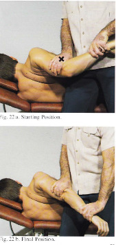
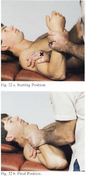
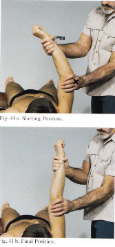
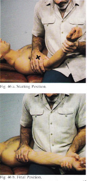
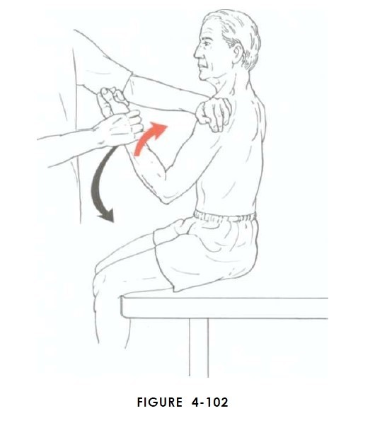
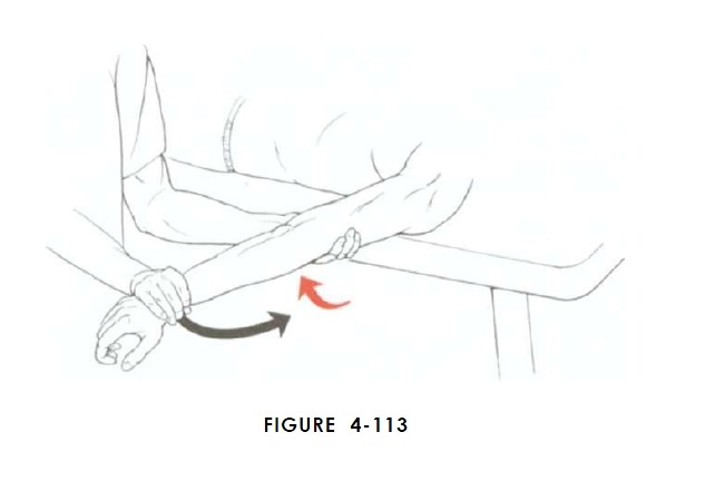
4. 주관절의 흔한 질환 http://cafe.naver.com/panicbird/134 5. 근막이완 1) 롤핑 http://cafe.daum.net/panicbird/OJs0/281 2) CMP건 - http://cafe.daum.net/panicbird/Qibc/88 - http://cafe.daum.net/panicbird/Qibc/89 3) 폼롤러를 이용한 근막이완..... 6. 주요 근육 스트레칭 1) 스트레칭 기본개념 *** 각 동작은 5초씩 3회가 기본(7초가 넘어가면 보상기전이 발생하므로) *** 모든 동작은 다음 동작을 위한 예비동작 임, 건너 뛰지 말 것 http://cafe.daum.net/panicbird/QtNi/299 2) biceps brachii - 능동 - 수동 long head short head 3) triceps brachii - 능동 - 수동 4) extensor, flexor muscle - 능동
- 수동 http://cafe.daum.net/panicbird/RUDq/79 5) supinator - 능동
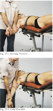
-수동 6) pronator -능동
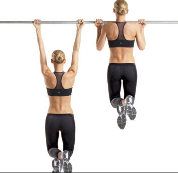
- 수동 7. 관절가동 1) 총괄 http://cafe.daum.net/panicbird/OUdz/3 2) radiohumeral mobilization, compression, distraction https://www.youtube.com/watch?v=mxkGPEyEYC4&feature=player_embedded https://www.youtube.com/watch?v=EuMgwnPXlEw&feature=player_embedded https://www.youtube.com/watch?v=ELrw7cmiU8s&feature=player_embedded 3) humeroulnar distraction, mobilization https://www.youtube.com/watch?v=r6DOE1xDTjo&feature=player_embedded https://www.youtube.com/watch?v=8IH6njiDlC4&feature=player_embedded 4) proximal radioulnar joint mobilization https://www.youtube.com/watch?v=IiBJPVgO0zA&feature=player_embedded 5) 기타 http://cafe.daum.net/panicbird/QzIa/54 8. 인대 약침, 가열식 화침 http://cafe.daum.net/panicbird/RNu8/21 9. stabilizing ex. 가. biceps brachii 1) chin-up 2) biceps curl
nabil 동영상 : https://www.youtube.com/watch?v=DRywmb1ueOI&feature=player_embedded


나.triceps brachii
http://cafe.daum.net/panicbird/RUDq/36


다. flexor, extensor

라. pronator, supinator

10. 근력 불균형 검사
1)근력 검사의 고려사항
http://cafe.daum.net/panicbird/QzZ4/15
2) 굴곡 검사
3) 신전 검사
4) 회내 검사 5) 회외 검사
11. isolation ex.
stabilizing ex. 참조
12. functional ex.


13. 정렬운동
여러 운동 참조......
14. 행동수정
충분한 휴식
자가 스트레칭
아이스팩
손목 신전이나 굴곡 제한(물건을 들 때 손 바닥을 위로하여 굴곡하며 들기 등) 등