Cervical Spine Radiographs
Radiology Cases in Pediatric Emergency Medicine
Volume 5, Case 2
Tai-Chuen Lin, Medical Student
Loren G. Yamamoto, MD, MPH
Kapiolani Medical Center For Women And Children
University of Hawaii John A. Burns School of Medicine
Introductory Notes
Most spinal cord injuries are attributed to trauma.
Absence of radiographic findings does not exclude a
spinal cord injury. A substantial portion of spinal cord
injuries in children (25% to 50%) have no radiographic
abnormalities--SCIWORA (spinal cord injury without
radiographic abnormalities). Some patients with
cervical spine injury may also have thoracolumbar
lesions. In the younger child, injuries to the cervical
spine often involve the upper three vertebrae.
Pediatric Considerations
Pediatric anatomy differs from the adult in several
important ways, particularly in the ossification pattern of
the cervicocranium (occiput-atlas-axis) and the normal
laxity of the developing soft tissue structures of the
cervicocranium. These differences can lead to false
positive interpretation as fractures, subluxations, and/or
tumors etc. (Refer to Case 5 of Volume 1, Cervical
Spine Malalignment - True or Pseudo Subluxation?,
and Case 1 of Volume 5, Fever With Neck Stiffness . . .
Rule Out Meningitis).
Clinical Aspects
The assessment of cervical spine injuries must first
be a clinical evaluation. Clinical and radiographic data
should be interpreted together to yield the most
accurate assessment.
Diagnostic strategies depend on whether the patient
is conscious and can freely move his or her neck.
Unconscious or poorly conscious patients should be
examined radiographically while maintaining cervical
spine immobilization since history and examination will
be unreliable.
A conscious patient with a significant cervical spine
injury will complain of pain. A significant cervical spine
injury is not likely to be present in a patient without neck
pain who is alert, not intoxicated, and lacks other painful
injuries (that may distract neck pain). Normal cervical
range of motion is consistent with the absence of a
cervical spine injury and such patients generally do not
need any radiographs.
Anatomy
In order to properly evaluate the radiographic
images of the cervical spine, an understanding of the
cervical spine anatomy is necessary to appreciate the
structural organization that lends to spinal stability. The
vertebrae are bony building blocks connected by
ligamentous and muscular structures. This resulting
stable skeleton provides the scaffold for the soft tissue
structures that communicate between the head and the
thorax, the spinal cord being one of the most delicate
and important.
The cervical spine is made up of seven sequentially
numbered cervical vertebrae, C1 through C7.
Superiorly, C1 is connected to the occiput of the
cranium. Inferiorly, C7 is connected to the first thoracic
vertebrae, T1. The upper portion of the cervical spine,
C1 and C2, together with the occiput is also referred to
as the cervicocranium. All vertebrae share many
common features. These will be reviewed along with
features unique to the cervical vertebrae. C1 and C2
are atypical cervical vertebrae and will be treated
separately.
Vertebral Body
The anterior and most easily identifiable structure of
a vertebra is the vertebral body, also known as the
centrum. The body is the largest and appropriately the
main weight-bearing structure of a vertebra. The back
of the body also forms the anterior border of the spinal
canal.
View C4.
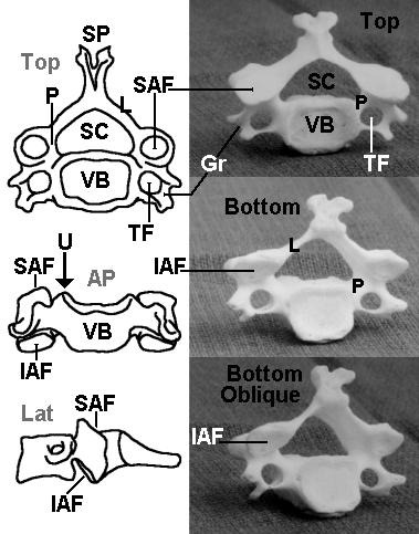 The three line diagrams on the left from top to
bottom include an axial view, viewed from the top (Top),
an anterior view (AP), and a lateral view (Lat). The
three photographs of C4 on the right from top to bottom
include a view from the top (Top), a view from the
bottom (Bottom), and an oblique view from the bottom
(Bottom oblique).
Identify the following structures on these diagrams
and photos:
SP - spinous process
L - lamina (forms roof of the neural arch)
P - pedicle (forms supports of the neural arch)
SC - spinal canal
VB - vertebral body
SAF - superior articular facet
IAF - inferior articular facet
TF - transverse foramen
Gr - groove for spinal nerve (transverse process)
U - uncinate process
The neural arch is formed by the laminae, the base
of the spinous process and the pedicles. The pedicles
are very short in the cervical spine. The facet joints are
formed by the inferior and superior facets such that the
C4-C5 facet joint is formed by the inferior articular facet
of C4 and the superior articular facet of C5.
On a lateral film, the body is a rhomboid with the
posterior portion slightly taller than the anterior portion.
View lateral C-spine view.
The three line diagrams on the left from top to
bottom include an axial view, viewed from the top (Top),
an anterior view (AP), and a lateral view (Lat). The
three photographs of C4 on the right from top to bottom
include a view from the top (Top), a view from the
bottom (Bottom), and an oblique view from the bottom
(Bottom oblique).
Identify the following structures on these diagrams
and photos:
SP - spinous process
L - lamina (forms roof of the neural arch)
P - pedicle (forms supports of the neural arch)
SC - spinal canal
VB - vertebral body
SAF - superior articular facet
IAF - inferior articular facet
TF - transverse foramen
Gr - groove for spinal nerve (transverse process)
U - uncinate process
The neural arch is formed by the laminae, the base
of the spinous process and the pedicles. The pedicles
are very short in the cervical spine. The facet joints are
formed by the inferior and superior facets such that the
C4-C5 facet joint is formed by the inferior articular facet
of C4 and the superior articular facet of C5.
On a lateral film, the body is a rhomboid with the
posterior portion slightly taller than the anterior portion.
View lateral C-spine view.
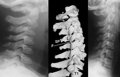 The lateral view of a very young child is shown on
the left compared to the lateral view of a teenager on
the right. Alignment is assessed by the integrity of lines
drawn along: 1) the anterior borders of the vertebral
bodies, 2) the posterior borders of the vertebral bodies
and 3) the anterior borders of the vertebral arch's apex
(spinolaminal line). The facet joints should be clearly
visible.
View identifying landmarks.
The lateral view of a very young child is shown on
the left compared to the lateral view of a teenager on
the right. Alignment is assessed by the integrity of lines
drawn along: 1) the anterior borders of the vertebral
bodies, 2) the posterior borders of the vertebral bodies
and 3) the anterior borders of the vertebral arch's apex
(spinolaminal line). The facet joints should be clearly
visible.
View identifying landmarks.
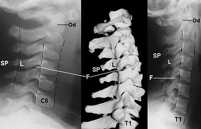 The contour lines of alignment are shown. Identify
the following areas on the radiographs:
F - facet joint
SP - spinous process
L - lamina
Od - odontoid
On an AP view, the lateral superior edges of the
body form bilateral ridges, called the uncinate
processes (U).
View AP C-spine view.
The contour lines of alignment are shown. Identify
the following areas on the radiographs:
F - facet joint
SP - spinous process
L - lamina
Od - odontoid
On an AP view, the lateral superior edges of the
body form bilateral ridges, called the uncinate
processes (U).
View AP C-spine view.
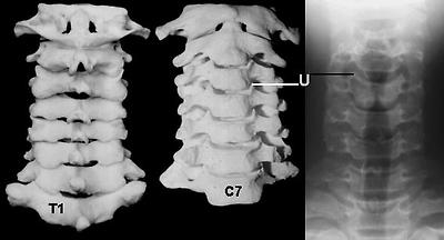 A posterior view of the cervical spine is shown on
the left. An anterior view is shown in the center.
Axial compression can result in compression
fractures which can lead to decreased vertebral body
height or a burst fracture that fragments the vertebral
body. A strong lateral force can cause a shearing
action and create fractures of an uncinate process.
Hyperflexion and hyperextension may also result in
teardrop fractures of the anterior superior or inferior
corner of the body.
Between the vertebral bodies are the intervertebral
disks. These function as shock absorbers. As in the
lumbar region, rupture of the annulus can lead to
encroachment into the spinal canal. The vertebral body
also serves as the attachment site of the anterior and
posterior longitudinal ligaments. Tears in these
ligamentous structures can result from displacement or
extensive fractures of the vertebral body. Without
these ligamentous connections, the vertebral column is
unstable.
Neural Arch
Posterior to the vertebral body is the neural arch
(vertebral arch covering the spinal canal). The neural
arch refers to all the structures dorsal to the body. The
arch serves to protect the spinal cord, provide
attachment sites for ligaments and muscles, and forms
synovial joints that facilitate movement of the vertebral
column. The major structures that make up the arch
include: 1) the pedicles, 2) the laminae, 3) the spinous
process, 4) the articular processes and facets, and 5)
the transverse processes.
View C4.
Pedicles
The pedicles ("little feet") form the supports of the
neural arch as it is attached to the vertebral body. In
the cervical spine, the pedicles are short. They project
posteriorly (dorsally) from the body and form the lateral
borders of the spinal canal. Superior and slightly larger
inferior vertebral notches above and below the pedicles
form intervertebral foramina in the articulated vertebral
column. Through these foramina pass the cervical
spinal nerves.
View lateral.
On a lateral film, the pedicles appear as small
connections between the body and the articular
processes (see below). On the AP view, the pedicles
appear as small doughnut densities on the lateral upper
portion of the vertebral body, just below the uncinate
processes. Fractures in this region can disrupt the
spinal nerves or the spinal cord itself.
View AP.
Laminae
The laminae (meaning "layers") form a roof over
the neural arch, supported by the pedicles. In addition
to the obvious protective function, the laminae also
serve as the site of attachment for the ligamentum
flavum. Because the laminae are thinner in the C-spine
compared to other vertebrae, their relative radiolucency
appears as an apparent gap between the posterior
cortex of the articular facets and the anterior cortex of
the spinous process (posterior aspect of the neural
arch) on the lateral view. In general, the laminae (L)
are not easily appreciable on an AP view.
View lateral.
Spinous Process
The spinous process projects dorsoinferiorly from
the point of union of the laminae. Unique to the typical
cervical vertebrae, the spinous processes of C3 through
C6 are typically bifid at the tips. The spine of C7 is an
easily visible surface landmark called the vertebra
prominens. The spinous processes are the site of
attachment for a number of ligamentous and tendinous
structures. The major ligaments associated with the
spine include the interspinous and supraspinous
ligaments as well as the ligamentum nuchae. A number
of intrinsic muscles of the spine as well as large back
muscles such as the trapezius, the levator scapularis,
and the rhomboids are attached to the cervical spinous
processes. Excessive load on these muscles may
result in avulsion of the spinous processes of C6 and
C7, commonly known as the clay shoveller's fracture.
This fracture is more commonly found in adults.
View lateral.
On a lateral view, the spinous processes appear as
triangular extensions. The anterior border with the
laminae (spinolaminal line) is an easily visible feature
marking the posterior border of the vertebral canal
(spinal canal). On an AP view, the spinous processes
appear as a midline density superimposed on the
vertebral body. The bifid nature of some of the cervical
spines can be easily appreciated in this view.
View AP.
Articular Processes and Articular Facets
The articular processes are cylindrical structures at
the junction of the pedicles and the laminae. Like the
pedicles, articular processes also delimit the lateral
margins of the spinal canal. The articular facets, the
oblique elliptical ends of the cylinders, are higher
anteriorly and lower posteriorly.
View articular facets of C4.
Capsular articular ligaments join adjacent inferior
and superior articular facets of successive vertebrae to
form synovial joints. Strong rotary forces can stretch or
tear these ligaments resulting in unilateral or bilateral
dislocated facets.
View lateral.
On a lateral view, the articular processes are
rhomboidal in shape and superimposed upon one
another. Unlike the vertebral body which slopes
downward anteriorly, the articular processes slope
sharply downward posteriorly. They appear
superimposed on the spinal canal.
View oblique view.
A posterior view of the cervical spine is shown on
the left. An anterior view is shown in the center.
Axial compression can result in compression
fractures which can lead to decreased vertebral body
height or a burst fracture that fragments the vertebral
body. A strong lateral force can cause a shearing
action and create fractures of an uncinate process.
Hyperflexion and hyperextension may also result in
teardrop fractures of the anterior superior or inferior
corner of the body.
Between the vertebral bodies are the intervertebral
disks. These function as shock absorbers. As in the
lumbar region, rupture of the annulus can lead to
encroachment into the spinal canal. The vertebral body
also serves as the attachment site of the anterior and
posterior longitudinal ligaments. Tears in these
ligamentous structures can result from displacement or
extensive fractures of the vertebral body. Without
these ligamentous connections, the vertebral column is
unstable.
Neural Arch
Posterior to the vertebral body is the neural arch
(vertebral arch covering the spinal canal). The neural
arch refers to all the structures dorsal to the body. The
arch serves to protect the spinal cord, provide
attachment sites for ligaments and muscles, and forms
synovial joints that facilitate movement of the vertebral
column. The major structures that make up the arch
include: 1) the pedicles, 2) the laminae, 3) the spinous
process, 4) the articular processes and facets, and 5)
the transverse processes.
View C4.
Pedicles
The pedicles ("little feet") form the supports of the
neural arch as it is attached to the vertebral body. In
the cervical spine, the pedicles are short. They project
posteriorly (dorsally) from the body and form the lateral
borders of the spinal canal. Superior and slightly larger
inferior vertebral notches above and below the pedicles
form intervertebral foramina in the articulated vertebral
column. Through these foramina pass the cervical
spinal nerves.
View lateral.
On a lateral film, the pedicles appear as small
connections between the body and the articular
processes (see below). On the AP view, the pedicles
appear as small doughnut densities on the lateral upper
portion of the vertebral body, just below the uncinate
processes. Fractures in this region can disrupt the
spinal nerves or the spinal cord itself.
View AP.
Laminae
The laminae (meaning "layers") form a roof over
the neural arch, supported by the pedicles. In addition
to the obvious protective function, the laminae also
serve as the site of attachment for the ligamentum
flavum. Because the laminae are thinner in the C-spine
compared to other vertebrae, their relative radiolucency
appears as an apparent gap between the posterior
cortex of the articular facets and the anterior cortex of
the spinous process (posterior aspect of the neural
arch) on the lateral view. In general, the laminae (L)
are not easily appreciable on an AP view.
View lateral.
Spinous Process
The spinous process projects dorsoinferiorly from
the point of union of the laminae. Unique to the typical
cervical vertebrae, the spinous processes of C3 through
C6 are typically bifid at the tips. The spine of C7 is an
easily visible surface landmark called the vertebra
prominens. The spinous processes are the site of
attachment for a number of ligamentous and tendinous
structures. The major ligaments associated with the
spine include the interspinous and supraspinous
ligaments as well as the ligamentum nuchae. A number
of intrinsic muscles of the spine as well as large back
muscles such as the trapezius, the levator scapularis,
and the rhomboids are attached to the cervical spinous
processes. Excessive load on these muscles may
result in avulsion of the spinous processes of C6 and
C7, commonly known as the clay shoveller's fracture.
This fracture is more commonly found in adults.
View lateral.
On a lateral view, the spinous processes appear as
triangular extensions. The anterior border with the
laminae (spinolaminal line) is an easily visible feature
marking the posterior border of the vertebral canal
(spinal canal). On an AP view, the spinous processes
appear as a midline density superimposed on the
vertebral body. The bifid nature of some of the cervical
spines can be easily appreciated in this view.
View AP.
Articular Processes and Articular Facets
The articular processes are cylindrical structures at
the junction of the pedicles and the laminae. Like the
pedicles, articular processes also delimit the lateral
margins of the spinal canal. The articular facets, the
oblique elliptical ends of the cylinders, are higher
anteriorly and lower posteriorly.
View articular facets of C4.
Capsular articular ligaments join adjacent inferior
and superior articular facets of successive vertebrae to
form synovial joints. Strong rotary forces can stretch or
tear these ligaments resulting in unilateral or bilateral
dislocated facets.
View lateral.
On a lateral view, the articular processes are
rhomboidal in shape and superimposed upon one
another. Unlike the vertebral body which slopes
downward anteriorly, the articular processes slope
sharply downward posteriorly. They appear
superimposed on the spinal canal.
View oblique view.
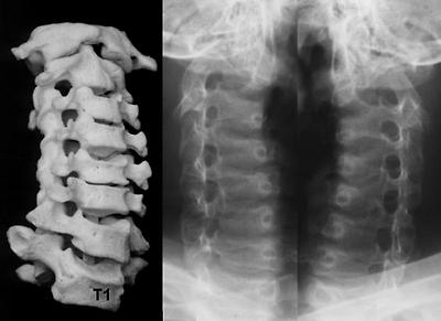 The skeletal model on the left shows variability in
the intervertebral disk spacing due to poor positioning of
the model bones during photography. This oblique view
shows the intervertebral foramina formed by the inferior
notch of the pedicle of the vertebrae above and the
superior notch of the pedicle of the vertebrae below.
Transverse Processes
The transverse processes project outward
anteroinferiorly from the pedicles like half-cylindrical
scoops.
View C4.
Along the grooved portion of the transverse
process pass the ventral rami of the cervical nerves.
The dorsal rami pass more posteriorly. In the middle
of the transverse process is a foramen for the vertebral
artery as it courses upward toward the foramen
magnum. Lesions in this region can damage the
nerves of cervical and brachial plexi as well as
compromise the arterial supply of the posterior brain.
The Cervicocranium
The articulations between the occiput, the atlas
(C1), and the axis (C2) are highly specialized to allow
the extensive range of motion of the head upon the
neck. As such, C1 and C2 differ sufficiently from the
typical vertebrae that they deserve special mention.
View odontoid view.
The skeletal model on the left shows variability in
the intervertebral disk spacing due to poor positioning of
the model bones during photography. This oblique view
shows the intervertebral foramina formed by the inferior
notch of the pedicle of the vertebrae above and the
superior notch of the pedicle of the vertebrae below.
Transverse Processes
The transverse processes project outward
anteroinferiorly from the pedicles like half-cylindrical
scoops.
View C4.
Along the grooved portion of the transverse
process pass the ventral rami of the cervical nerves.
The dorsal rami pass more posteriorly. In the middle
of the transverse process is a foramen for the vertebral
artery as it courses upward toward the foramen
magnum. Lesions in this region can damage the
nerves of cervical and brachial plexi as well as
compromise the arterial supply of the posterior brain.
The Cervicocranium
The articulations between the occiput, the atlas
(C1), and the axis (C2) are highly specialized to allow
the extensive range of motion of the head upon the
neck. As such, C1 and C2 differ sufficiently from the
typical vertebrae that they deserve special mention.
View odontoid view.
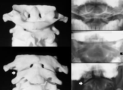 The atlas (C1) articulates superiorly with the
occipital bone. The occipital bone forms the base of the
cranium, and articulation with the cervical spine is via
the pair of large convex occipital condyles situated on
either side of the anterior half of the foramen magnum.
The brain stem becomes the spinal cord as it leaves the
cranium through the foramen magnum. Anterior to the
foramen magnum is an upward incline to the dorsum
sellae called the clivus. The posterior aspect of the
foramen magnum is in-line with the posterior arch of C1
and C2 (the spinolaminar line).
Lacking a body, the atlas is essentially a ring with
prominent articular processes that are appropriately
called lateral masses. The lateral masses divide the
ring into a smaller anterior and a larger posterior arch.
View C1-C2 cross section CT scan.
The atlas (C1) articulates superiorly with the
occipital bone. The occipital bone forms the base of the
cranium, and articulation with the cervical spine is via
the pair of large convex occipital condyles situated on
either side of the anterior half of the foramen magnum.
The brain stem becomes the spinal cord as it leaves the
cranium through the foramen magnum. Anterior to the
foramen magnum is an upward incline to the dorsum
sellae called the clivus. The posterior aspect of the
foramen magnum is in-line with the posterior arch of C1
and C2 (the spinolaminar line).
Lacking a body, the atlas is essentially a ring with
prominent articular processes that are appropriately
called lateral masses. The lateral masses divide the
ring into a smaller anterior and a larger posterior arch.
View C1-C2 cross section CT scan.
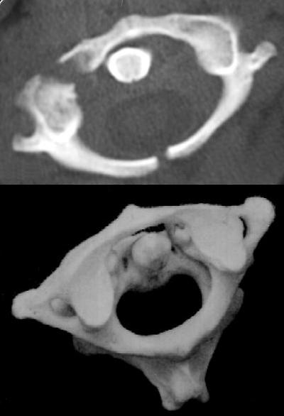 On the inner aspects of the lateral masses are
tubercles for the transverse ligament that run between
these tubercles. The concave superior facets articulate
with the convex condyles of the occipital bone, while the
larger inferior facets (of C1) articulate with C2.
Because the atlas lacks a body, the lateral masses are
the major weight bearing structures, and a compression
force (axial load) can result in a bursting fracture of the
ring of C1. The upper CT image shows such fracture of
the C1 ring.
View odontoid view.
On an open-mouth odontoid view, the lateral
masses are easily visible as trapezoidal wedges. The
anterior and posterior arches are superimposed over
the odontoid process.
Radiographically, the surfaces of the anterior
atlantoaxial gap are parallel to each other and the
distance is less than 5 mm in a child. Widening of this
space can be a result of a transverse ligament tear,
allowing unstable motion between the two bones.
View lateral.
On a lateral view, the atlas is a simple ring structure
seen edge on. The lateral masses are superimposed
on the odontoid process of C2 and are difficult to
identify. The inner aspect of the anterior arch can be
easily appreciated, as can the inner aspect of the
posterior arch. Note that the anterior arch articulates
with the anterior aspect of the odontoid, while the
posterior arch forms the very first posterior border of
the vertebral canal.
The most prominent feature of C2 (axis) is the
odontoid process, also called the dens. Both names
refer to its resemblance to a tooth. The odontoid
process projects superiorly from the body of the axis.
Articular processes centered around the odontoid have
smooth superior facets that facilitate rotational
articulation within the atlas. The inferior facets are
more posterior and in-line with the articular processes
of the rest of the cervical vertebrae area. On a lateral
view, the axis appears much like a typical cervical
vertebra, however, it is easily identified by the odontoid
process projecting vertically from the body and the large
and wide spinous process.
On the inner aspects of the lateral masses are
tubercles for the transverse ligament that run between
these tubercles. The concave superior facets articulate
with the convex condyles of the occipital bone, while the
larger inferior facets (of C1) articulate with C2.
Because the atlas lacks a body, the lateral masses are
the major weight bearing structures, and a compression
force (axial load) can result in a bursting fracture of the
ring of C1. The upper CT image shows such fracture of
the C1 ring.
View odontoid view.
On an open-mouth odontoid view, the lateral
masses are easily visible as trapezoidal wedges. The
anterior and posterior arches are superimposed over
the odontoid process.
Radiographically, the surfaces of the anterior
atlantoaxial gap are parallel to each other and the
distance is less than 5 mm in a child. Widening of this
space can be a result of a transverse ligament tear,
allowing unstable motion between the two bones.
View lateral.
On a lateral view, the atlas is a simple ring structure
seen edge on. The lateral masses are superimposed
on the odontoid process of C2 and are difficult to
identify. The inner aspect of the anterior arch can be
easily appreciated, as can the inner aspect of the
posterior arch. Note that the anterior arch articulates
with the anterior aspect of the odontoid, while the
posterior arch forms the very first posterior border of
the vertebral canal.
The most prominent feature of C2 (axis) is the
odontoid process, also called the dens. Both names
refer to its resemblance to a tooth. The odontoid
process projects superiorly from the body of the axis.
Articular processes centered around the odontoid have
smooth superior facets that facilitate rotational
articulation within the atlas. The inferior facets are
more posterior and in-line with the articular processes
of the rest of the cervical vertebrae area. On a lateral
view, the axis appears much like a typical cervical
vertebra, however, it is easily identified by the odontoid
process projecting vertically from the body and the large
and wide spinous process.
다음검색