muscle recruitment pattern의 개념에 대한 새로운 눈을 뜨고 있다.
움직임의 ABC인 muscle recruitment pattern
자전거는 upstroke 와 downstroke의 반복.
트레이닝은 downstrke동안 고관절, 무릎, 발목의 강력하고 리드미컬한 신장과 협응력으로 이루어짐
The most common sites for overuse injury were the neck (48.8%) followed by the knee (41.7%).
panic bird...
cycling cycle
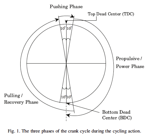
Pushing phase - 20도의 top dead center - power phase(downstroke) - bottom dead center - pulling phase - recovery(upstroke) phase

.

멋지다. 인간의 몸으로 할 수 있는 멋진 운동. 협응력 트레이닝 최고의 방법이다!!
![]() Muscle recruitment pattern in cycling. 리뷰논문.pdf
Muscle recruitment pattern in cycling. 리뷰논문.pdf
Abstract
Studies have indicated that the muscles work in a systematic and coordinated way to generate and direct power from the human body to the crank during cycling. Understanding of the muscle involvement or recruitment pattern during cycling will be useful for developing specific and effective muscle training and rehabilitation programs for cyclists. Moreover, it will also facilitate the use of the cycling ergometer for therapeutic purpose.
- 자전거를 타는 동안 인체로부터 생성되는 직접적인 힘과 힘생성의 시스템적 그리고 협응적 방법에 있어서 근육작업에 대한 탐구
- 자전거를 타는 동안 근육동원의 이해는 자전거 선수를 위한 재활프로그램과 효과적인 근육트레이닝 개발을 위해 유용함. 게다가 치료적 목적을 위한 자전거 작업계사용을 촉진할 수 있음.
This paper reviews the current literature on muscle recruitment pattern during cycling and the effects of muscle fatigue,
cadence, riding posture and seat height on this recruitment pattern. In the power phase or ‘downstroke’, the hip, knee and ankle joints extend simultaneously for the pushing action, whilst in the recovery phase or ‘upstroke’, they flex together to pull the pedal back to the top dead center of the crank cycle.
- 이 논문은 자전거를 타는 동안 근육동원에 대한 문헌과 근피로, 리듬, 자전거타는 자세 그리고 의자높의 효과를 리뷰함. 페달을 내밟는 순간(downstroke) 고관절, 무릎 그리고 발목관절은 미는 동작을 위하 동시에 신장되고, upstroke동안 그것들은 페달을 위로당겨 함께 굴곡함.
Recent studies have indicated that in this repeated sequence, the mono-articular muscles are mainly involved in the
generation of positive work whereas the biarticular muscles are responsible for regulating force transmission. Some muscles co-activate during cycling to provide synergistic actions and other functional needs.
- 최신논문은 이 반복적인 연속동안, 단관절 근육이 힘생성하는 방법을 주로 연구함. 반면에 다관절 근육이 힘생성을 조절함. 어떤 근육은 자전거를 타는 동안 협력작용과 다른 기능적 요구를 제공하는 동시수축을 함.
Muscle fatigue is an important factor affecting cycling performance. It has been reported that muscle fatigue in the lower body would alter the cycling motion and muscle activation pattern. Therefore, studying the change of muscle activation pattern during cycling at the fatigued level may shed light on the sequel of local muscle fatigue.
- 근피로는 자전거를 타는 동안 중요한 요소임. 그것은 하체에서 근피로는 자전거 동작을 변경할 수 있고, 근육활성패턴을 바꿀 수있다고 보고함. 그래서 자전거를 타는동안 근피로 레벨에서 근활성 패턴 변화 연구는 국소적 근피로의 결과에 초점을 맞출수 있음.
A muscle training program specifically for cycling can then be designed accordingly. Additionally, the change of cadence during cycling will affect the muscle recruitment pattern. There is a unique cadence that minimizes the muscle activation level at a specific level of power output. This cadence will increase as the power output increases. The change of riding posture from sitting to standing renders the pelvis unsupported and the body weight will assist the power phase of pedalling. Similarly, changes to the seat height will alter the posture which will affect the directions of muscle actions to the crank, thus changing the muscle recruitment pattern.
- 자전거를 위한 특별한 근육트레이닝 프로그램은 동시에 디자인 될 수 있음. 게다가 자전거 동안 리듬의 변화는 근육 동원패턴에 영향을 줄 수 있음. power output의 특별한 레벨에서 근수축을 최소화하는 유일한 리듬이 있음. 이 리듬은 power output이 증가함에 따라 증가할 수 있음. 앉기에서 서기로 자전거 타는 자세 변화는 골반을 지지하지 않게하고, 인체무게는 power phase의 페달링에 도움을 줄 수있음. 유사하게 의자 높이 변화는 자세를 바꿔 손잡이에 근육행위의 변화에 영향을 주고, 근육동원패턴을 바꿀 수 있음.
1. Introduction
Cycling is a one of the most popular sports in the world. Cyclists may race, tour or simply exercise to keep fit. As more people become involved in cycling, the incidence of its associated injuries has increased. Cycling is non-weight bearing and the action is smooth and non-paring, making injuries less likely.
- 자전거는 세상에서 가장 유행하는 스포츠. 자전거타는 사람은 경주, 여행, 또는 단순 운동을 유지할수 있음. 사람들이 자전거에 관여할수록 자전거 관련손상을 늘어남. 자전거는 비체중부하 운동으로 움직임은 부드럽고 비쌍으로 움직이어서 손상위험이 적음.
However, due to considerably longer periods spent in training and racing compared to many other sports, there are patterns of overuse injury unique to cycling (MacAuley, 1995). Wilber, Holland, Madison, and Loy (1995) reported that among all the recreational cyclists who responded to their questionnaire, 85% reported one or more overuse injuries, with 30% requiring medical treatment. The most common sites for overuse injury were the neck (48.8%) followed by the knee (41.7%). Holmes, Pruitt, and Whalen (1994) stated that knee pain is the most common lower extremity overuse problem in cyclists which, ironically, is caused by strong knee extensors. If only the knee extensors are strengthened, the patella will be overstrained because most of the energy in the power phase is transmitted through the patella (Sanner & O’Halloran, 2000).
- 하지만 자전거를 오래타면 자전거만의 과사용패턴이 있음. 자전거를 타는 사람에게 가장 흔한 손상부위는 경추 48.8%, 무릎 41.7%임.
This problem can result in decreased performance, participation and enjoyment for cyclists at all levels. The knee extensor muscle group is the prime mover to generate energy to the crank in the downstroke phase of cycling (Raasch, Zajac, Ma, & Levine, 1997) and many serious cyclists emphasize their muscle training on the knee extensor muscles for performance enhancement. However, this may increase their risk of getting knee injuries. Therefore, an understanding of the muscle involvement or recruitment pattern during cycling is essential for developing a specific and effective muscle training program for cyclists for performance enhancement and injury prevention. Moreover, it also helps sport physiotherapists to determine which muscles need specific training to recondition elite cyclists after injury.
- 무릎 신전근육은 사이클링의 downstroke 기간동안 주동근이고, 많은 자전거선수는 무릎신전근 트레이닝을 위해 자전거운동임. 자전거를 타는동안 근육동원을 잘 알아야 부상을 방지할 수 있음.
Cycling has been recognized as a recreational and sporting activity that has many therapeutic qualities. Fleming et al. (1998) suggested that stationary cycling at the appropriate cadence and resistance is an effective rehabilitation exercise for patients with anterior cruciate
ligament injury to increase muscle activity without subjecting the ligament to undue strain. With an understanding of the normal muscle recruitment pattern of this exercise, sports physiotherapists can focus on a particular phase of the cycling action to train a particular muscle group.
- 사이클링을 하는 동안 정상근육사용패턴으 이해는 특별한 근육트레이닝에 중요한 역할을 함.
Even though cycling is generally regarded as a closed-kinetic chain exercise, the joint position and loading in the kinetic chain may not always be predicted at a particular sequence. de Groot et al. (1994) indicated that muscular work depends on the length–tension, force–velocity power relationships of the involved muscles.
- 사이클링은 닫힌사슬 운동으로 간주될지라도, 관절위치와 부하는 틀별한 연속성은 항상 예측할 수 없을 수 있음.
- 그루트는 사이클링은 근육 길이-긴장관계, 힘-속도 관계에 의존한다고 밝힘.
The effectiveness of force production is affected by joint angles, muscle lengths and muscle moment arms. These variables in turn are altered by changes in the rate of pedaling, position and orientation of the body and changes in seat height. In addition to this, Asplund and St Pierre (2004) stated that muscle fatigue during cycling might induce alternations in pedaling technique leading to stress and symptoms in other parts of the kinetic chain.
- 힘 생성의 효과성은 관절, 근육길이, 근육지레팔에 의해 영향을 받음. 이것들의 변수는 차례로 페달링 비율, 신체의 위치와 방향 그리고 의자의 높이에 따라서 변화함. 피에르는 다음과 같이 말함. 사이클링 동안 근피로는 페달링테크닉이 운동사슬의 서로다른 일부에서 스트레스와 증상을 야기하는 변화를 유도함.
Therefore, knowledge of the muscle fatigue pattern in the major muscles during cycling will not just enable a better prescription of muscle training programs, it may also reduce the risk of overuse injury. This article provides a review for clinicians to appreciate the complexity of cycling so that they can modify the exercise to achieve the purpose of training on a particular muscle or range that they aim for.
- 그래서 사이클링 동안 주요근육 근피로에 대한 지식은 더 나은 근육 트레이닝 프로그램을 처방할 뿐만 아니라 과사용 손상을줄일 수 있음. 이 논문은 그 목적을 위해 쓰여짐.
2. Classical view on muscle recruitment during cycling
Schmidt (1994) , an experienced scientific cycling coach, stated that the most important muscle for cycling is the quadriceps, whilst pulling on the pedals depends on the hip flexors. The lower leg muscles power the pedaling motion with the calf muscles being active during the entire pedal revolution and the muscles of the anterior tibial compartment are only responsible for fixing the foot in place whilst pulling back on the pedals. Schmidt (1994) also stressed the importance of the smooth pedal stroke for the bicycle racer, which refers to the even distribution of power to the pedals during the course of the entire pedal revolution.
- 슈미트는 과학적으로 훈련된 사이클링 코치인데, 다음과 같이 언급함. 사이클링동안 가장 중요한 근육은 대퇴사두근, 페달을밟는데는 고관절 신전근(대둔근)에 의존함. 하지 근육들은 종아리근육과 함께 페달링 동작에 힘을 부여함. 그리고 전경골근 등 근육들은 페달을 끌어올리는 동안 고정하는 역할을 함. 슈미트는 ....
The use of racing pedals that restrain the foot on the pedal enable power transmission from top to bottom during pushing, pressing and pulling. This enables the road racers to use all the leg and hip muscles during the entire crank revolution cycle. Muscles in the trunk and arms provide a counterbalancing force to the lower limbs during the pedaling motion. The hand, arm, shoulder, abdomen and back form a muscular sling, which rhythmically moves back and forth in supporting the trunk and pelvis (Schmidt, 1994 ). The ipsilateral arm and other structures provide the support to counteract the leg extension force. Nearly all the major skeletal muscle groups are utilized. A general description of the function of the major muscle groups during cycling has been developed but no detailed description of the level of force and working time/phase of each muscle group with respect to the pedal cycle is available.
- 레이싱 페달의 사용은 페달의 누름과 당김동안 힘을 이동함. 이것은 사이클 동안 모든 하지, 엉덩이 근육 사용을 가능케 함. 몸통과 팔근육은 하지에 역균형을 제공함. 손, 팔, 어깨, 복부 그리고 등 근육은 리드미컬하게 움직여 몸통과 골반을 지지하는 동안 앞뒤로 움직임. 동통의 팔과 다른 구조는 하지 신전힘을 지지함. 거의 모든 근육이 사용됨.
3. Muscle recruitment during cycling based on electromyopraphic (EMG) pattern
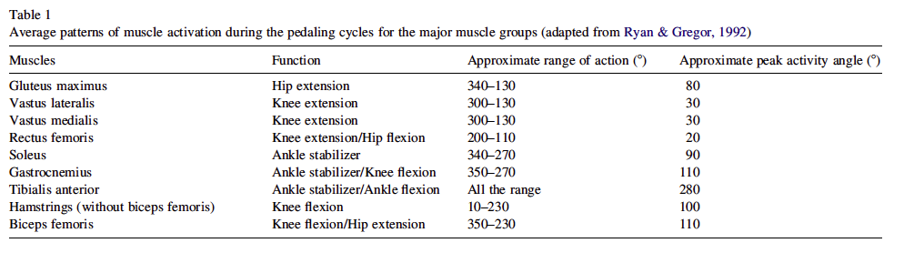
Before discussing the muscle recruitment in cycling, a brief description of the crank cycle is essential. The crank cycle can be broken down into three phases (Fig. 1 ):
1. The propulsive/power/downstroke phase.
2. The pulling/recovery/upstroke phase.
3. The pushing phase, in which the foot is pushed forward at the top dead center (TDC).
.
Gregor and Rugg (1986) studied the activity pattern of eight muscles by electromyographic monitoring in the leg whilst cycling at 85 rpm against a moderate load in 10 competitive male cyclists riding at their own personal comfort level. Six muscles (vastus medialis, vastus lateralis, rectus femoris, tibialis anterior, biceps femoris and gluteus maximus) had more than 50% of their respective maximum activity during the first half of propulsion (08 TDC to 908 ). The quadriceps (knee extensors) became less active, whilst the hamstrings, gastrocnemius and gluteus maximus maintained their activities until the bottom dead center (BDC) in order to complete the propulsive phase. This would lead to significant hip and knee extension and ankle plantar flexion in the propulsion phase.
- 그레고는 85 회전수로 사이클링 동안 하지에서 근전도를 이용하여 8개 근육의 활성패턴을 연구함. 6개의 근육(내측광근, 외측광근, 대퇴직근, 전경골근, 대퇴이두근, 대둔근)은 ... - 대퇴사두근(무릎 신전근)은 덜 확성회됨. 햄스트링, 비복근, 대둔근은 bottom dead center에서 완전한 propulsive단계에 까지 활성을 유지함. 이는 고관절과 무릎 신전, 발목 배굴을 이끌어냄. ...
In a later report, Pruitt (1988) also described a similar muscle activation pattern. In a more recent study, Gregor and Conconi (2000)
presented the muscle recruitment pattern during cycling, with more detailed description of the muscle activity, especially during the recovery phase. Both medial and lateral vasti muscles, being single-joint knee extensors, exhibited a rapid onset and cessation with relatively constant activity in-between during the downstroke phase.
- 내외측광근은 무릎 신전근으로 페달누름 기간동안 지속적으로 활성화되면서 rapid onset and cessation을 보임.
Conversely, the rectus femoris, which is a hip flexor and knee extensor, demonstrated a more gradual rise and decline. The soleus, being a single-joint ankle plantar flexor, was recruited just before the gastrocnemius. The semimembranosus and semitendinosus were recruited after TDC and their peak activities occurred at, or slightly after, 908 from TDC. Peak activity in the semitendinosus occurred slightly after that of the semimembranosus, whereas biceps femoris was the most variable among the hamstring muscles.
- 반면에 대퇴직근은 고관절 굴곡근, 무릎신전근으로 좀더 점차적인 rise and decline을 보임. 가자미근은 발목 저굴을 위한 단관절 근육으로 비복근이 활성화되기 전에 동원됨. 반막양근과 반건양근은 TDC후에 동원되고, 그들의 최고 활성은 TDC후에 일어남. 반건양근 최고활성은 반막양근보다 약간 후에 일어남. 반면에 대퇴이두근은 햄스트링 중에서 가장 변수가 많음.
The authors stated that during the recovery phase, from 180 to 360 , the lower extremity flexed actively and that this served two functions: firstly, it reduced the resistance on the crank to the propulsive phase on the contralateral limb; and secondly, it provided enough flexion to rotate the crank and assisted the contralateral limb in propulsion. A summary of the muscle activity pattern is described in Table 1 .

From the above descriptions, an optimal pedaling technique could provide effective force on the pedals during both the propulsive and recovery phases. The hip and knee flexors work to drive the pedal rearward near the bottom dead center and to lift the pedal during a large portion of recovery (Broker & Gregor, 1996 ).
- 위의 분석을 보면 최적의 페달링 테크닉은 propulsive and recovery phase동안 효과적인 힘을 제공함. 고관절과 무릎굴곡근은 함께 작용하여 BOD에서 페달을 들어올리는데 큰 역할을 함.
The importance of the upper extremity and trunk muscles in cycling was stressed by Gregor and Conconi (2000) but no detailed description was stated. There has been no scientific study done to evaluate the upper body and trunk muscle activity pattern during cycling. Only the coach manual has illustrated the upper body muscle work in cycling (Schmidt, 1994 ). There is a need to evaluate the extent of upper body and trunk muscle activity level during cycling in order to guide the optimal strength-training program for the cyclists.
- 상체와 몸통근육에 대한 분석은 더 탐구되어야 함.
4. Muscle recruitment during cycling based on biomechanical modelling
Because the relationship between EMG and force is uncertain during non-isometric cyclic contractions, the precise muscle coordination in the delivery of energy to the crank cannot be clearly described. Also, some potentially important muscles, which are situated deep inside the body, are difficult to study with surface EMG.
- 근전도와 힘사이의 관계때문에 비등척성 사이클 수축동안 분명하지 않음. 정확한 근육 협응성은 에너지를 전달하는데 있어서 분명하게 기술할 수 없음. 또한 어떤 중요한 근육은 몸의 깊은 부위에 위치하여 표면근전도로 연구하기 힘듬.
Therefore, studying how energy transfer happens from the body to the bike is a means to better understand the muscle activation pattern. Raasch et al. (1997) reported that based on a simulation study, the net hip extension torque primarily delivered energy to the limb segment, which was then transferred to the crank by net ankle torque. Furthermore, Raasch and Zajac (1999) suggested a model utilizing three pairs of alternating muscle groups in excitation to describe the function of each muscle group in the torque transference.
- 그래서 몸에서 자전거로 에너지가 어떻게 전달되는가에 대한 탐구는 근육활성 패턴의 좀더 나은 이해를 의미함. 라쉬는 다음과 같이 보고함. ...
The uniarticular hip extensors—gluteus maximus and knee extensors—vastus lateralis and medialis (i.e. the extensor group, or E group) co-excited during cycling and worked alternately with the uniarticular hip flexors—iliacus and psoas and the knee flexor—short head of biceps femoris (i.e. the flexor group, or F group). These formed the E–F pair. A second pair consisted of the biarticular rectus femoris (RF) and tibialis anterior (TA), which alternated with the posterior thigh muscles—medial hamstrings and long head of biceps femoris (HAM) and ankle plantar flexors including the soleus and gastrocnemius (SG), forming the final pair.
- 단일 관절 고관절 신전근(대둔근)과 무릎신전근(내외측광근)이 사이클링 동안 동시 흥분하고, 단일관절 고관절 굴곡근(장골근과 대요근)과 무릎굴곡근(대퇴이두근 단두)가 교대로 작용함. 이것들은 신전-굴곡 쌍을 이룸. 두번째 쌍은 이중관절인 대퇴직근과 전경골근이 만들고, 이는 대퇴후근(내측햄스트링과 대퇴이두근 장두)과 발목족저굴곡근(가자미근과 비복근)이 마지막 쌍을 이룸.
The E/F groups produced energy to propel the crank whilst the RF/TA and HAM/SG groups acted as rigid links to facilitate the energy transfer. The RF/TA pair provided energy to propel the crank toward the end of flexion and during the flexion-to-extension transition; whereas the HAM/SG pair were active near the end of extension and during the extension-to-flexion transition (Zajac & Gordon, 1989; Raasch et al., 1997 ).
- 신전/굴곡 그룹은 ...
In this pattern of activity, Raasch and Zajac (1999) stressed that the ankle must be stabilized to ensure complete transfer of the extension energy to the crank. Raasch et al. (1997) highlighted that if the plantar flexors did not co-excite during downstroke, energy from extensors would go towards accelerating the limbs by dorsiflexing the ankle and hyperextending the knee rather than the crank.
For the ankle movement, Raasch and Zajac (1999) stated that the ankle could employ different strategies or pedaling movements during cycling. First, the ankle could have more motion before the pedal passed through the BDC by dorsiflexion to store the kinetic energy and then plantar flexion just before the BDC to force the pedal to go through the BDC. Second, the stiffness and stability of the ankle is maintained by activating the tibialis anterior and soleus earlier during downstroke. These two muscles would also be activated slightly through the upstroke phase to transfer the power generated by joint flexion to the crank. Third, the ankle could be in a plantar flexed position for much of the cycling motion with prolonged soleus and gastrocnemius excitation.
Therefore, muscles work in a coordinated way; some work together whilst others work in a coordinated sequence in order to maximize energy transfer from the cyclist to the bike. In this coordinated recruitment pattern, two major areas can be discussed further:
(1) Single-joint muscle activity pattern (e.g. soleus) is different from the multi-joint muscles (e.g.gastrocnemius);
(2) Co-activation is not restricted to the synergistic muscles, the agonist and antagonist muscles (e.g. knee extensors and flexors) also co-activate.
5. Activation patterns for single- and multi-joint muscles
The single-joint muscles were chiefly concerned with the generation of positive work, whereas multi-joint muscles were responsible for the fine regulation of the net moments about the joints (Jacobs, Bobbert, & van Ingen Schenau, 1993 ). Moreover, the multi-joint muscles served different purposes at different phases of cycling. For example, the rectus femoris assisted flexion of the hip during the recovery phase, but it also assisted knee extension in the propulsive phase (Eisner, Bode, Nyland, & Caborn, 1999 ). A similar pattern was also observed for the gastrocnemius; during the recovery phase, it functioned as a knee flexor, but during the propulsive phase, it functioned as an ankle stabilizer (Burke, 1995 ).
Rectus femoris and biceps femoris exhibited double bursts of activity at higher cadence. Gregor, Broker, and Ryan (1991) explained that the secondary bursts in these muscles were attributed to their biarticular function.
As the effective ‘downstroke’ only happens within a limited range of crank angles (about 10–1708 ), activities of the single-joint muscles were consistent across subjects to produce power at a similar portion of the crank cycle. In contrast, the activity patterns in multi-joint muscles were much more variable as they controlled the direction of pedal force produced by the single-joint muscles to adjacent joints; hence their activities were affected by the actions of other muscles (Gregor & Conconi, 2000 ).
The multi-joint muscles were also involved in the transition phase from downstroke to upstroke and vice versa (Mileva & Turner, 2003 ). Moreover, there may be other factors affecting their activations; for example, the different roles of biceps femoris (hip extensor and/or knee flexor) might be related to subject-specific pedaling techniques and also dependent upon the competitive level.
Relative muscle strength may be another factor, as stronger one-joint hip extensors might be associated with increased rectus femoris activity for power transportation; whereas weaker one-joint hip extensors might need help from bicep femoris to forcefully extend the hip joint (Li & Caldwell, 1998 ). Increased strength of the single-joint muscles will relieve the load on the synergistic multi-joint muscles, but increase the load on the antagonistic multi-joint muscles. Therefore, the force generated by the single-joint muscle requires the multi-joint muscles to facilitate the energy transfer, and the relative strength of such muscle couples could affect the total energy transferred to the pedals. Therefore, it is very important to balance muscle development when designing muscle training and rehabilitation programs for cyclists
6. Muscle co-activation pattern
During the propulsive phase, several agonist and antagonist muscle pairs activate together to enhance the power acting on the pedal. The plantar flexors (gastrocnemius and soleus) and dorsiflexor (tibialis anterior) could activate together to stabilize the ankle during propulsion and force transmission. Moreover, together with the quadriceps, the hamstring muscles were also active as hip extensors (Jorge & Hull, 1986; Gregor et al., 1991; Gregor & Conconi, 2000 ).
With more detailed studies, a feature of muscle coordination at the hip, knee, and ankle was identified, which was co-activation of one and two-joint muscles, agonist and antagonist pairs (e.g. gluteus maximus vs. rectus femoris) and the two-joint rectus femoris and gastrocnemius. A functional consequence of this coordination is the transfer of mechanical energy between joints. The one- and two-joint synergists were often co-activated but with a phase shift and time difference in peak occurrence. This occurs because activation of two-joint synergists depends on moment demands at both joints, whereas the activity of one-joint synergists is influenced primarily by the moment demand at only one joint (Prilutsky, 2000 ). Smith (1981) stated that the co-activation of agonist and antagonist muscles appeared to be an inherent component in many types of joint movements.
Therefore, in addition to just enhancing power output in the propulsive phase, there could be some other functions for the agonist and antagonist muscle co-activation. Aagaard et al. (2000) proposed that during isokinetic knee extension, a neural pathway existed to co-activate the flexors of the knee. This co-activation mechanism may be important for the stability of knee joint as it potentially prevents anterior tibial displacement and assists the mechanical and proprioceptive roles of the anterior cruciate ligament.
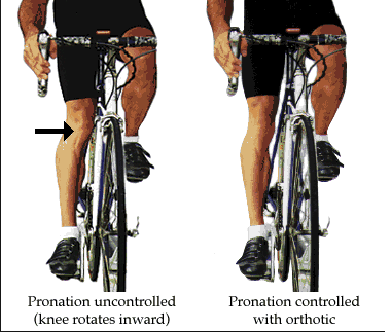
Sanderson, Hennig, and Black (2000) noted that if pedal forces were high and cadence was low, eversion of the foot with inward rotation of the tibia through the cycle would lead to stress in the knee. Co-activation of the hamstring muscles may help to relieve this stress. Therefore, coactivation of the agonist and antagonist pairs may serve the purpose of protecting the joints that they act upon. Hunter, St Clair Gibson, Lambert, and Noakes (2002) agreed by stating that the antagonistic activity of biceps femoris would progressively increase to stabilize the knee joint.
However, Raasch et al. (1997) stated that it was the plantar flexors that stabilized the knee in late downstroke whilst the hamstrings accelerated the knee towards extension. Therefore, the function of these muscles at different phases of the cycling stroke requires further study.
Muscles activated in a defined pattern are not just for optimizing the energy transfer from the human body to the machine but also provide protection to the major joints, e.g. knees. If any particular muscle becomes too strong or too weak, such optimized muscle activation link might be jeopardized. Hence, identification of weak components in these muscle links and strengthening them with specific muscle training may improve the cycling performance and reduce the risk of injury.
7. Effects of fatigue on muscle recruitment pattern
Muscle fatigue has been defined as the failure of the muscle to maintain the target force (Edwards, 1981 ). There is, however, considerable controversy about how fatigue is defined and measured. Some experts argue that before this failure point is reached, the muscle is already fatigued (Bigland-Ritchie & Woods, 1984 ; De Luca, 1984, 1993 ). Fatigue, from this perspective, is an ongoing process that begins from the start of a muscle contraction. Some people argue that muscle fatigue may be better defined as a time-dependent process with regard to physiological and biochemical changes (De Luca, 1984, 1993 ).
Electromyographic analysis during fatiguing contractions has been performed extensively on various muscles (De Luca, 1984 ). During a fatiguing contraction, the EMG amplitude has been found to increase in sub-maximal contractions (De Luca, 1984; Moritani, Muro, & Nagata, 1986; Krogh-Lund & Jorgenson, 1991 ). At the same time, changes in the frequency components of the EMG signals can also be observed (De Luca, 1984 ). There is an increase in the lower frequency components of the EMG spectrum during sustained contractions, and this frequency shift is commonly termed the ‘frequency/spectral shift’ or ‘frequency/spectral compression’ toward lower frequency (De Luca, 1984; Ha¨gg, 1992; Ng, Richardson, Kippers, Parnianpour, & Bui, 1996 ).
This frequency shift has been used as an indicator of muscle fatigue (De Luca, 1984, 1993 ). Muscle fatigue would decrease the rate of voluntary force development in cycling (Bentley, Smith, Davie, & Zhou, 2000; Lepers, Millet, & Maffiuletti, 2001 ). Paavolainen, Nummela, Rusko, and Hakkinen (1999) reported that fatigue produced by prolonged and repetitive high-intensity short-duration stretch and shortening cycle exercises could induce acute decrease in force production due to reduced neural input to the muscle and the efficiency of the contractile mechanism. van Diee¨n, van der Burg, Raaijmakers, and Toussaint (1998) stated that in the dynamic pattern-generation approach, muscles were viewed not as simple effectors operating under neural command but as an integral part of a mutually influencing network with the nervous system.
The nervous system adjusted the motor programs in order to compensate for changes of the effector organ characteristics. Such changes could be reduced force output of a particular muscle group due to fatigue. Hence the muscle recruitment in cycling would change due to fatigue.
In many studies on cycling, the fatigue process was registered by real-time or continuous monitoring of the rate of decline of pedal power output during maximal cycling workout. With a more detailed study to evaluate the muscular activities at different positions within the pedal revolution, von Tscharner (2002) adopted a time–frequency (wavelet analysis) analysis of EMG signals and found that shifting of the frequency components that occurred with fatigue was very specific for certain periods during the revolution. Some periods had the frequency shift to the relatively high end, while in other periods, the frequency shifted to the lower end. Thus the spectral changes are likely to reflect a systematic change of motor unit recruitment pattern with pedal position and with fatigue.
In order to generate the high force levels accompanying intense activity, maximal or near maximal activation of all the synergistic muscles is a fundamental requirement. It has been found that seven different synergistic muscles were recruited in cycling, their activation pattern being highly ordered and dependent on the limb and pedal positions (Green & Patla, 1992 ). Muscle activity patterns (i.e. timing) were not significantly affected by changes in pedaling conditions (load, seat height and shoe type) while the only change was the muscle activity levels result from different pedaling conditions (Jorge & Hull, 1986 ).
On the other hand, Kay et al. (2001) hypothesized that the lower limb inter-muscular recruitment strategies could be altered. During high-intensity cycling exercise, the activities of vastus lateralis and rectus femoris had both increased but vastus lateralis increased more. This could imply a shift of the synergistic muscle recruitment. Moreover, during prolonged exercise at a constant intensity, there were signs that changes occurred in the neuromuscular system as any progress in exercise might influence the efficacy or the pattern with which the contractile machinery was activated (Lepers, Maffiuletti, Rochette, Brugniaux, & Millet, 2002 ).
Therefore, fatigue could affect the pattern of recruitment of the multi-joint muscles. Kay et al. (2001) also found that activation of rectus femoris disappeared after five high-intensity short-sprint cycling strokes. They concluded that although qualitative, their data indicated an alteration in the coordination pattern in cycling movement in conjunction with the development of fatigue.
Hautier et al. (2000) found that muscle coordination in cycling could be efficiently adapted to the loss of the contractile power in the muscles due to local muscle fatigue. For instance, the lower activation of the antagonists after fatigue appeared to be an efficient adaptation of the inter-muscular coordination to modulate the net force generated by the fatigued agonist to the pedals for maintaining the force applied to the cycle. With a dynamic cycling study, Prilutsky and Gregor (2000) reported that the fatigue and perceived exertion might be reduced by preferential activation of two joint muscles during phases of movement where they could contribute to the moments at both joints and by reciprocal activation of one-joint antagonists such as soleus or tibialis anterior and two-joint antagonists such as rectus femoris and hamstrings.
Another feature of muscle coordination in cycling, which might potentially reduce fatigue and perceived effort was co-activation of one-joint muscles with their two-joint antagonists. The scenario may be further complicated with the addition of endurance training as Hawley and Stepto (2001) postulated that the number of years of training might improve the patterns of neuromuscular recruitment thus
adaptation in response to sport specific overload patterns. Therefore, studies to evaluate the effect of endurance training on the change of muscle recruitment due to fatigue are warranted.
8. Effects of cadence on muscle recruitment pattern
MacIntosh, Neptune, and Horton (2000) concluded that muscle activation at a given power output is minimized at a unique cadence and that cadence increases in unison with power output. As the cadence increased, the time for the recovery phase will decrease and the pedaling leg has to push the recovering leg back to the TDC with a higher force (Sanderson et al., 2000). This will demand more positive
force from the pedaling leg thus affecting the muscle recruitment pattern during cycling.
Timmer (1991) and Ericson, Nisell, Arborelius, and Ekholm (1985) noted that increasing the pedaling rate was associated with increased activitiy in the gluteus maximus, gluteus medius, vastus medialis, medial hamstrings, and the calf muscles. In knee extension/flexion, Miller, Croce, and Hutchins (2000) found that biceps femoris would be co-activating more as an antagonist with increasing movement
speed. Therefore, with an increasing cadence, the activity of the antagonists may also increase.
Recently, Sarre, Lepers, Maffiuletti, Millet, and Martin (2003) reported no cadence effect on the root mean square EMG value of vastus lateralis and vastus medialis whilst that of rectus femoris was significantly greater at the lowest cadence tested. They attributed this to the increased demand on the rectus femoris to propel the pedal crank anteriorly through the TDC at low cadence. In addition, Gregor et al.
(1991) stated that the activities of rectus and biceps femoris progressed earlier in the pedaling cycle as cadence increased, each exhibiting a double burst of activity at higher cadence.
Coyle et al. (1991) demonstrated that a difference in pedaling skill exists even amongst highly trained cyclists. There were large differences in the peak torque during the ‘downstroke’ when a given work output at the same pedaling rate was performed. This phenomenon was also reported by Takaishi, Yasuda, Ono, and Moritani (1996) who found that the pedaling rate of minimal neuromuscular fatigue at 80 or 90 rpm for non-cyclists was slightly lower than the pedaling rate predicted for experienced cyclists.
9. Effects of posture on muscle recruitment pattern
Standing out of the saddle causes a change in muscle recruitment pattern. The change of body position results in increased muscle activities from 340 to 1808 and a cessation of activities from 180 to 2808 (Pruitt, 1988 ). Li and Caldwell (1998) gave a detailed description of the muscle activities during cycling with standing posture. As the pelvis is not supported by the saddle during stand-cycling, gluteus
maximus would increase its activity to stabilize the pelvis just before the TDC and this would last well into the latter part of the ‘downstroke’.
Brown, Kautz, and Dairaghi (1996) also noted that joint
torques and muscle activation patterns observed during
pedaling changed systematically with body orientation.
Activation patterns of the ankle muscles changed significantly
in order to maintain a relatively consistent position of
the ankle even late in the upstroke, both net dorsiflexor
ankle torque and tibialis anterior activity increased as body
orientation became more vertical. However, in this study,
the body weight of the subjects was supported by the saddle
in all the assessed body orientations. Hence, body weight
would not contribute to the pushing force during ‘downdownstroke’.
The muscle recruitment pattern described in
this study may not be applicable to reflect the real picture in
stand-cycling.
94 R.C.H. So et al. / Physical Therapy in Sport 6 (2005) 89–96
Miller, Peach, and Keller (2001) examined the muscular
activation pattern produced while riding a stepper cycle
during sit- and stand-riding. Although the ergometer used in
this study was not a normal bicycle, the kinematics of riding
the stepper and racing bicycle is similar. Therefore, the
information gathered may help to understand the muscle
recruitment in stand-cycling. Miller et al. (2001) stated that
body weight helps in power production whilst riding in a
standing position. The body weight advantage would be lost
at low power levels, presumably because the muscular
requirements for balance and posture are larger when
riding in standing than sitting. From the existing literature,
there is no consensus on the optimal power and cadence
for stand-cycling.
10. Effects of seat height on muscle recruitment pattern
Seat height or saddle height is the distance between the top of the saddle and the center of the pedal axle measured when the pedal is down and the crank arm is in line with the seat tube. A change of seat height will alter the range of the legs and the muscle length, which would subsequently affect the muscle activity pattern during cycling.
Burke and Pruitt (2003) reported that oxygen consumption
in cycling is minimized at approximately 100% of
trochanteric height (from the greater trochanter of the femur
to the floor whilst standing barefoot), or 106–109% of pubic
symphysis height (measured from the ground to the pubic
symphysis). However, there is no general agreement on the
optimum seat height based on the relationship of muscle
activity pattern, joint force and moment patterns, and
pedaling effectiveness. Gregor et al. (1991) stated that
researchers generally agree that leg muscle activity
increases as seat height decreases. This increase appears
to be more prevalent in the quadriceps and hamstring
muscle groups. Similarly, MacAuley (1995) noted that
changes in seat height can alter the balance of the muscle
activity. Rectus femoris was found to be more active at a
lower seat height of 95% leg length, whilst raising the seat
height to 105% leg length increased the activity of the
sartorius muscle. Mellion (1991) summarized that
for serious cyclists, there are several scientific and quasiscientific
methods for determining the seat height of a
bicycle. The more scientific methods are the in seam
method, bone length method, and estimation from leg
length. All seat height formulae are estimates. After an
initial setting is made, the cyclist may fine-tune the seat
according to their comfort.
11. Conclusion
There is a well-defined muscle recruitment pattern in cycling. Most studies have focused on the lower extremities and indicated that the hip, knee and ankle generated the power during most of the power phase when all three joints extend. During the recovery phase, these joints flex to pull the pedal back to the top dead center. Mono- and biarticular muscle pairs, agonist and antagonist muscle pairs perform specific functions during cycling, such as diverting the power to the pedal and stabilizing the joints. The muscle recruitment patterns can be altered by muscle fatigue, cadence change and posture. More specific information of the above points will be needed to guide the strength training and rehabilitation programs for cyclists and an optimal specific training module can be developed accordingly.
댓글
댓글 리스트-
작성자tommyjung 작성시간 13.01.14 자전거 뿐만 아니라 모든 반복 운동 또는 자세 유지는 근육동원을 하는데, 물리학적으로 "가장 효율적인 것을 가장 먼저 동원하고 점차 효율성이 떨어지는 것을 사용한다" 가 핵심인 듯 하다.
그러면 가장 효율적인 근육을 강화시키는 것이 가장 효율적이라는 아주 단순무식한 결론을 도출해서는 안된다.
실제로 몸도 이런 합리성에 길들여 있어서 다들 그렇게 하려고 하지만 중요한 것은 전혀 상관없을 것 같은, 효율성이 가장 없는 것을 트레이닝 시켜야 한다.
그것은 반복적이고 의지적인 훈련이 필요하다.
반드시 누워서 다리를 올려보라.
그러면서 하는 행동이 목에도 힘을 준다.
이유가 뭘까!
-
작성자tommyjung 작성시간 13.01.14 허리는 해부학적 명칭 또는 위치성 때문에 뒤쪽만 생각한다.
하지만 허리는 위 아래 앞과 뒤를 명칭하는 통(barrel)이다.