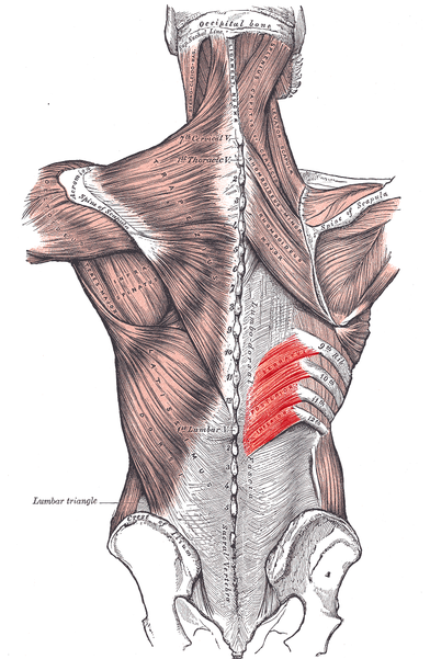
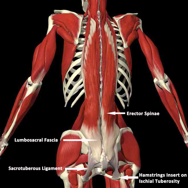
1. sacrotuberous ligament에서 기시하여 천장관절, 요추, 흉추, 경추, 두개골까지 연결되는 긴 근육군
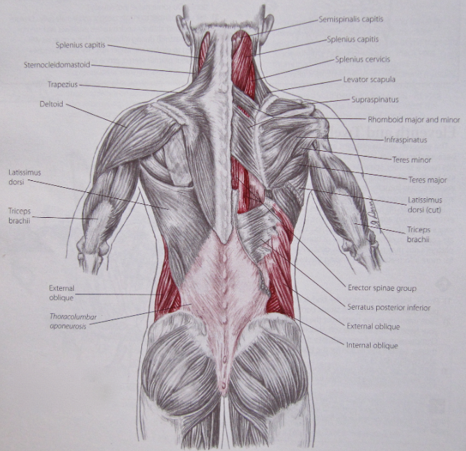
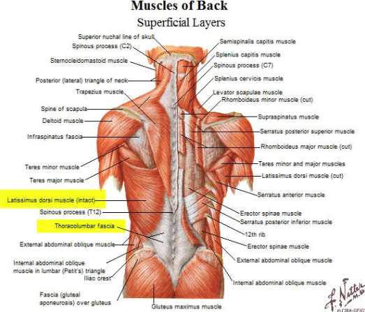
2. 흉요근막-광배근 - 척추기립근 - 다열근 - Interspinalis, intertransversarii
3. 하부승모근, 하후거근 - 척추기립근
4. 중부승모근, 능형근, 상후거근 - 척추기립근
5. 상부승모근, semispinlis capitus, splenius capitus, levator scapular, longissimus capitis, SCM
6. 꼭 천부, 심부 근육의 특성을 살펴서 기능적연결 문제를 해결해야 함.
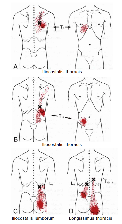
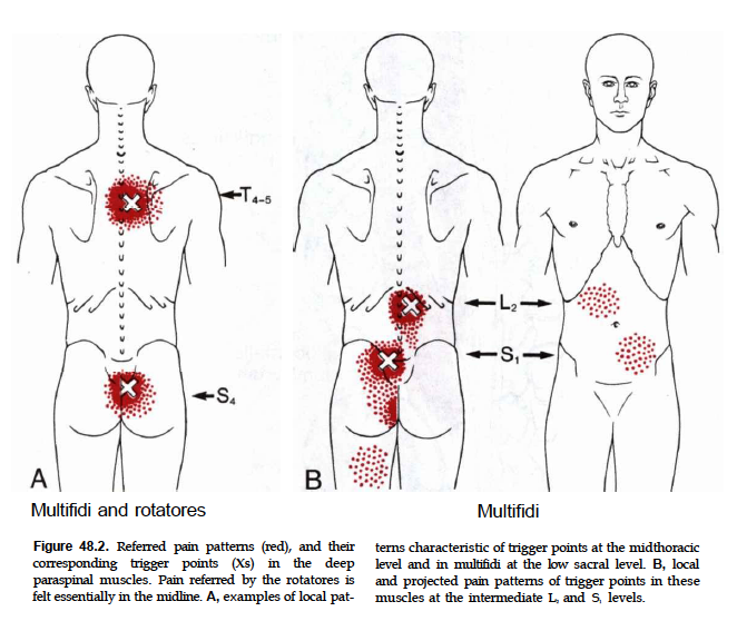
7. 엉치뼈, 꼬리뼈 부위 통증은 최장근, 다열근의 연관통부위
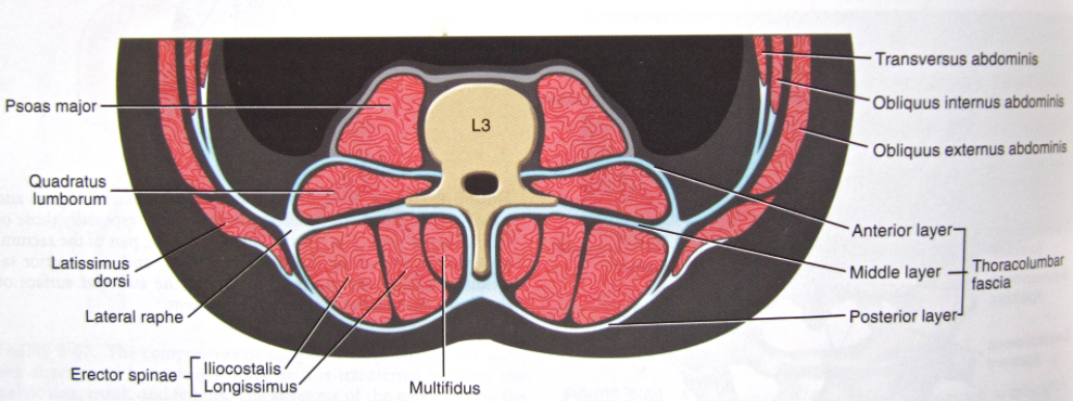
참고) 흉요근막은 3개의 근막층으로 구성되고, 요방형근, 다열근, 최장근, 장늑근(iliocostalis) 를 포함함.
꼭 끝까지 봐야 할 동영상
흉요근막의 구조를 보자
하후거근과 외복사근이 장력관계로 연결되어 있음을 보자.
척추기립근
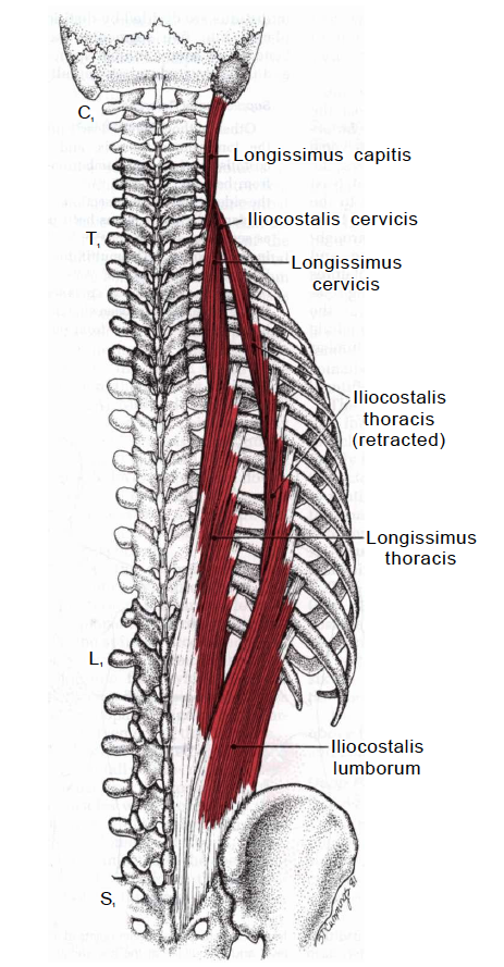
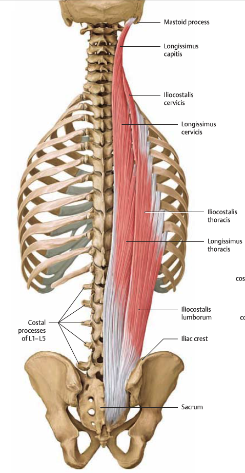
1. 두최장근(longissimus capitis)
기시 ; 흉추 1-3번 횡돌기, 경추 4-7번 후관절과 횡돌기
종지 : 유양돌기
2. 경최장근(longissimus cervicis)
기시 : 흉추 1-6번 횡돌기
종지 : 경추 2-5번 횡돌기
3. 흉최장근(longissimus thoracis)
기시 : sacrum, iliac crest, lumbar spinous process, lower thoracic 횡돌기
종지 : 2-12번째 rib, 흉추 횡돌기
4. 요장늑근(iliocostalis lumborum)
기시 : 천골, 장골능, 흉요근막
종지 : 6-12번째 늑골, 흉요근막의 deep layer, 상부요추의 횡돌기
5. 흉장늑근(iliocostalis thoracis)
기시 : 7-12번째 늑골
종지 : 1-6번 늑골
6. 경장늑근(iliocostalis cervicis)
기시 : 3-7번 늑골
종지 : 경추 4-6번 횡돌기
The erector spinæ (/ˌɨˈrɛktər ˈspiːniː/ ə-rek-tər spee-nee)[1] is a muscle group of the back in humans and other animals, which extends the vertebral column (bending the spine such that the head moves posteriorly while the chest protrudes anteriorly). It is also known as sacrospinalis in older texts. A more modern term is extensor spinae,[2] though this is not in widespread use.
Structure[edit]
The erector spinæ is not just one muscle, but a bundle of muscles and tendons. It is paired and runs more or less vertically. It extends throughout the lumbar, thoracic and cervical regions, and lies in the groove to the side of the vertebral column. Erector spinæ is covered in the lumbar and thoracic regions by the thoracolumbar fascia, and in the cervical region by the nuchal ligament.
This large muscular and tendinous mass varies in size and structure at different parts of the vertebral column. In the sacral region, it is narrow and pointed, and at its origin chiefly tendinous in structure. In the lumbar region, it is larger, and forms a thick fleshy mass. Further up, it is subdivided into three columns. These gradually diminish in size as they ascend to be inserted into the vertebræ and ribs.
The erector spinæ arises from the anterior surface of a broad and thick tendon. It is attached to the medial crest of the sacrum, to the spinous processes of the lumbar and the eleventh and twelfth thoracic vertebræ and the supraspinous ligament, to the back part of the inner lip of the iliac crests, and to the lateral crests of the sacrum, where it blends with the sacrotuberous and posterior sacroiliac ligaments.
Some of its fibers are continuous with the fibers of origin of the gluteus maximus. The muscular fibers form a large fleshy mass that splits, in the upper lumbar region, into three columns, viz., a lateral (Iliocostalis), an intermediate (Longissimus), and a medial (Spinalis). Each of these consists of three parts, inferior to superior, as follows:
Iliocostalis[edit]
The iliocostalis originates from the sacrum, erector spinae aponeurosis and iliac crest. The iliocostalis has three different insertions according to the parts:
- iliocostalis lumborum has the lumbar part(where its insertion is in the 12th to 7th ribs)
- iliocostalis thoracis where its insertion runs from the last 6 ribs to the first 6 ribs.
- iliocostalis cervicis which runs from the first 6 ribs to the posterior tubercle of the transverse process of C6-C4.
Longissimus[edit]
The longissimus muscle has three parts with different origin and insertion:
- longissimus thoracis originates from the sacrum, spinous processes of the lumbar vertebrae and transverse process of the last thoracic vertebra and inserts in the transverse processes of the lumbar vertebrae, erector spinae aponeurosis, ribs and costal processes of the thoracic vertebrae.
- longissimus cervicis originates from the transverse processes of T6-T1 and inserts in the transverse processes of C7-C2.
- longissimus capitis originates from the transverse processes of T3-T1 runs through C7-C3 and inserts in the mastoid process of the temporal bone.
Spinalis[edit]
The spinalis muscle has three parts:
- spinalis thoracis which originates from the spinous process of L3-T10 and inserts in the spinous process of T8-T2.
- spinalis cervicis originates from the spinous process of T2-C6 and inserts in the spinous process of C4-C2.
- spinalis capitis is an inconstant muscles fibres that runs from the cervical and upper thoracic that then inserts in the external occipital protuberance.
| Insertion | Lateral Column Iliocostalis | Intermediate Column Longissimus | Medial Column Spinalis |
| Lower thoracic vertebrae and ribs | I. lumborum | ||
| Upper thoracic vertebrae and ribs | I. thoracis | L. thoracis | S. thoracis |
| Cervical vertebrae | I. cervicis | L. cervicis | S. cervicis |
| Skull | L. capitis | S. capitis |
From lateral to medial, the erector spinæ muscles can be remembered using the mnemonic, "I Long for Spinach": Illiocostalis, Longissimus and Spinalis.[3]
Training[edit]
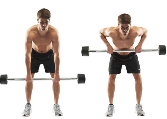
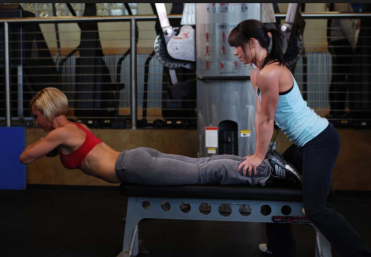
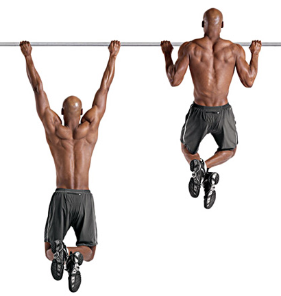
Examples of exercises by which the erector spinae can be strengthened for therapeutic or athletic purposes include, but are not limited to:
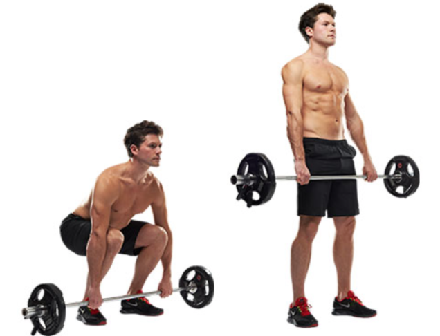
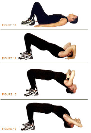
- Hyperextension exercise
- Good-morning exercise
- Rowing exercise
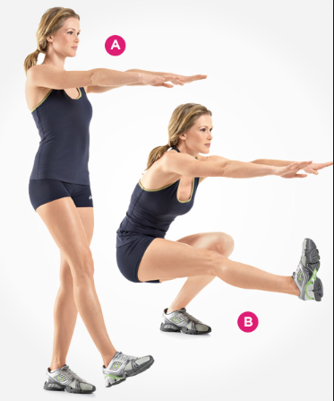
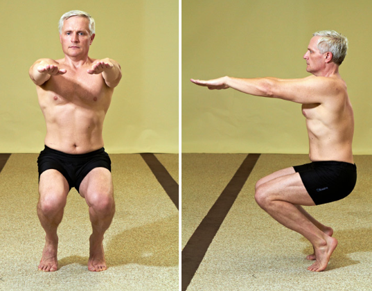
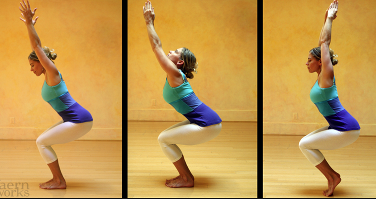
one leg squat
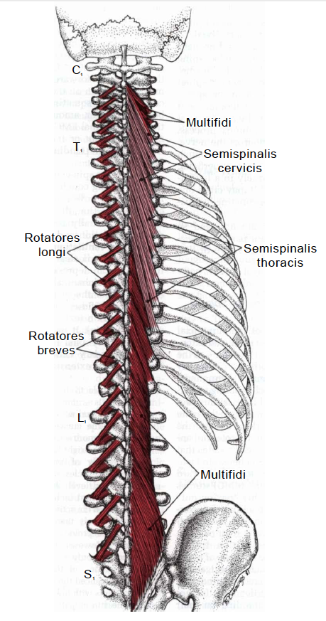
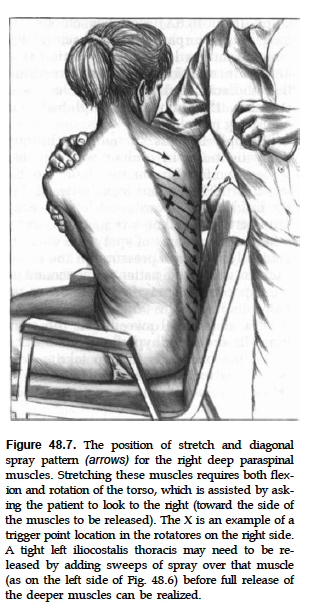
척추기립근 안쪽으로 더 들어가면서 보이는 근육
1. multifidi
2. semispinalis cervicis
3. semispinalis thoracis
4. rotators longi and breves
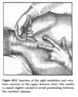
The multifidus (multifidus spinae : pl. multifidi ) muscle consists of a number of fleshy and tendinous fasciculi, which fill up the groove on either side of the spinous processes of the vertebrae, from the sacrum to the axis. The multifidus is a very thin muscle.
Deep in the spine, it spans three joint segments, and works to stabilize the joints at each segmental level.
The stiffness and stability makes each vertebra work more effectively, and reduces the degeneration of the joint structures.
These fasciculi arise:
- in the sacral region: from the back of the sacrum, as low as the fourth sacral foramen, from the aponeurosis of origin of the Sacrospinalis, from the medial surface of the posterior superior iliac spine, and from the posterior sacroiliac ligaments.
- in the lumbar region: from all the mamillary processes.
- in the thoracic region: from all the transverse processes.
- in the cervical region: from the articular processes of the lower four vertebrae.
Each fasciculus, passing obliquely upward and medialward, is inserted into the whole length of the spinous process of one of the vertebræ above. These fasciculi vary in length: the most superficial, the longest, pass from one vertebra to the third or fourth above; those next in order run from one vertebra to the second or third above; while the deepest connect two contiguous vertebrae.
The multifidus lies deep relative to the Spinal Erectors, Transverse Abdominus, Abdominal internal oblique muscle and Abdominal external oblique muscle.
다열근 활성화 방법
정리중...
